. 2020 Oct 9;48(18):10397-10412.
doi: 10.1093/nar/gkaa739.

A ribosomal RNA fragment with 2',3'-cyclic phosphate and GTP-binding activity acts as RIG-I ligand

Affiliations

A ribosomal RNA fragment with 2',3'-cyclic phosphate and GTP-binding activity acts as RIG-I ligand

Stephanie Jung et al. Nucleic Acids Res. .

Abstract

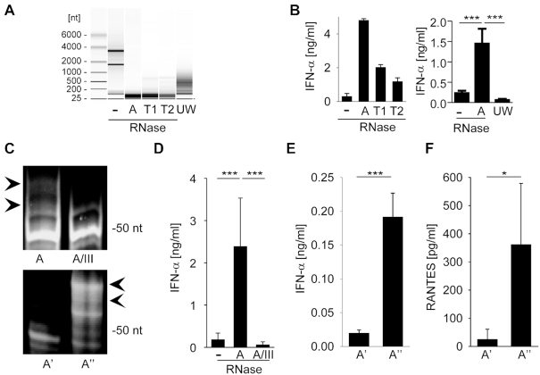
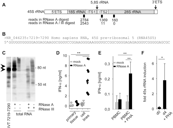
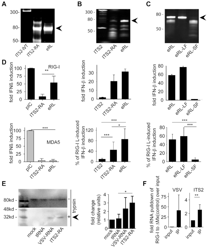
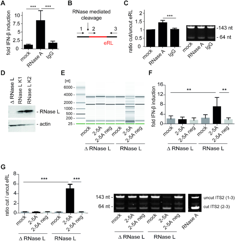
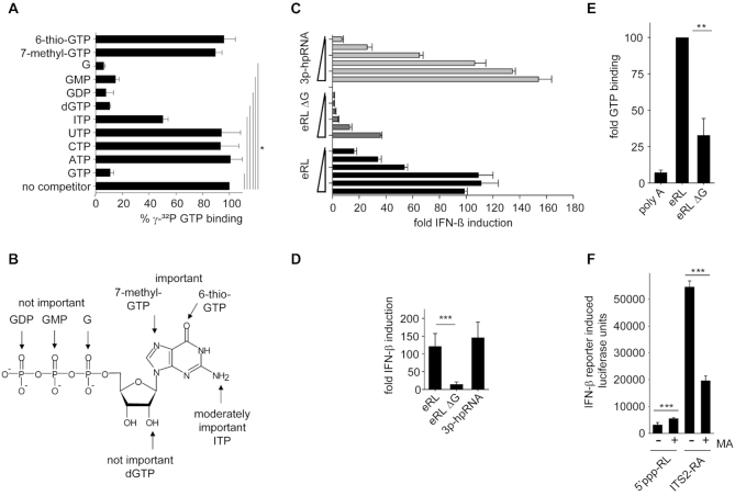
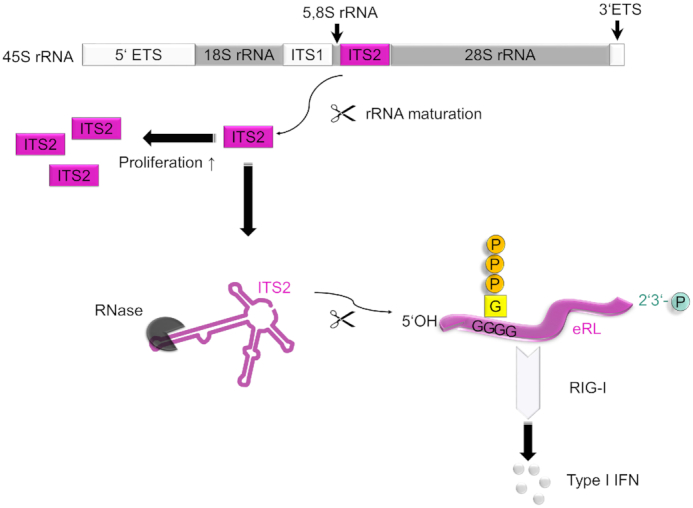
The RNA helicase RIG-I plays a key role in sensing pathogen-derived RNA. Double-stranded RNA structures bearing 5'-tri- or diphosphates are commonly referred to as activating RIG-I ligands. However, endogenous RNA fragments generated during viral infection via RNase L also activate RIG-I. Of note, RNase-digested RNA fragments bear a 5'-hydroxyl group and a 2',3'-cyclic phosphate. How endogenous RNA fragments activate RIG-I despite the lack of 5'-phosphorylation has not been elucidated. Here we describe an endogenous RIG-I ligand (eRL) that is derived from the internal transcribed spacer 2 region (ITS2) of the 45S ribosomal RNA after partial RNase A digestion in vitro, RNase A protein transfection or RNase L activation. The immunostimulatory property of the eRL is dependent on 2',3'-cyclic phosphate and its sequence is characterized by a G-quadruplex containing sequence motif mediating guanosine-5'-triphosphate (GTP) binding. In summary, RNase generated self-RNA fragments with 2',3'-cyclic phosphate function as nucleotide-5'-triphosphate binding aptamers activating RIG-I.


Figures

Figure 1.
Figure 1.
RNA digestion by single-strand specific RNases generates immune-stimulatory fragments. (A) Agilent bioanalyzer mediated RNA analysis of undigested HEK293 RNA (–), RNase-digested HEK293 RNA (A, T1 and T2) and ultrasonic waves (UW) fragmented HEK293 RNA. (B) RNAs visualized in panel A were complexed to DOTAP or Lipofectamine 2000 and incubated with human PBMCs at 2 μg/ml. After a 20 h incubation IFN-α levels were detected by ELISA. Left panel shows one individual experiment, right panel combines three independent experiments each in biological duplicates (six measurements per data point ± S.D.), *** P < 0.001. (C) Polyacrylamide gel electrophoresis of RNase A (A) and RNase A / RNase III (A/III) treated HEK293 RNA (upper panel). RNase A-digested RNA was size-fractionated by anionic exchange chromatography into fraction with lower (A') and higher (A'') molecular RNA size (lower panel). (D) PBMCs or human monocytes were stimulated with undigested, RNase A- or RNase A / RNase III treated RNA at 0.4 μg/ml and IFN-α levels were detected by ELISA. Graph combines six independent experiments each in biological duplicates (twelve measurements per data point ± S.D.), *** P < 0.001. (E and F) Human monocytes (E) or A549 cells (F) were stimulated with size fractionated RNA fragments (A', A'') for 20 h at 2 or 0.4 μg/ml, respectively. IFN-α or RANTES levels were determined by ELISA. Graph combines three independent experiments each in biological duplicates (six measurements per data point ± S.D.), *** P < 0.001, * P < 0.05.
Figure 2.
Figure 2.
Identification of an ITS2-derived immunostimulatory RNA fragment. (A) Schematic illustration of 45S rRNA, consisting of 5.8S, 18S and 28S as well as internal and external transcribed spacers (ETS, ITS). Positions and number of sequence reads in RNase A and RNase A / RNase III digested and size-fractionated samples A' and A'' (see Figure 1C) are depicted. (B) Sequence of most common read specifically found in the RNase A-digested but not in the RNase A / RNase III-digested sample. (C) Northern blot analysis of undigested, RNase A, RNase III or RNase A/RNase III digested HEK293 RNA with a specific probe for 45S rRNA 7219–7290. IVT-transcribed fragment 7219–7290 served as positive control. (D) Human PBMCs were stimulated for 20 h with 2 μg/ml mock-treated (grey symbols) or RNase A-digested RNA (black symbols) from primary tissue such as human PBMCs (five samples, circle), murine liver (one sample, square) or tumor cell lines such as HeLa (one sample, triangle up), Vero (two samples, hexagonal), MDCK (two samples, triangle down) and HEK293 (five samples, diamond). IFN-α was measured by ELISA. *** P < 0.001. (E) Human PBMCs and monocytes were stimulated with 2 μg/ml mock-treated (grey bars) or RNase A-digested RNA (black bars) from PBMCs, or with RNA from PBMCs activated with PHA for 4 days. IFN-α was measured by ELISA. Graph combines three independent experiments each in biological duplicates (six measurements per data point ± S.D.), ** P < 0.01. (F) 45S rRNA levels of unstimulated or PHA stimulated PBMCs were determined by RT-PCR targeting the 5.8S/ITS2 region. Three combined independent experiments are shown each in biological duplicates (six measurements per data point ± S.D.), * P < 0.05.
Figure 3.
Figure 3.
The eRL is single stranded and located in the ITS2 region. (A) Non-denaturing PAGE analysis of various RNA samples: a 358 nucleotide ITS2 RNA fragment (ITS2-IVT), RNase A-digested ITS2-IVT (ITS2-RA) and gel purified band (eRL, endogenous RIG-I ligand). (B) HEK293-RIG-I-IFN-ß reporter cells were stimulated with 0.5 μg/ml ITS2-IVT, ITS2-RA and eRL and fold IFN-ß induction was determined after 12 h. (C) HEK293-RIG-I-IFN-ß reporter cells were stimulated with 0.5 μg/ml eRL, large fragment (eRL-LF) and small fragment (eRL-SF) and fold IFN-ß induction was determined after 12 h. Integrity and composition of RNA samples used for stimulation in (B) and (C) were monitored by denaturing PAGE analysis with subsequent SYBR-Gold staining (upper row). Fold IFN-ß induction is shown for one individual experiment (middle row) and a diagram combining three independent experiments each in biological triplicates with data adjusted to % of pIC induced IFN-β activation (lower row; nine measurements per data point ± S.D.). * P < 0.05, *** P < 0.001. (D) Huh7.5 cells expressing RIG-I or MDA5 were stimulated with 1 μg/ml pIC and 0.4 μg/ml ITS2, ITS2-RA or eRL for 6 h and IFN-β upregulation was determined by RT-PCR. Graph combines three independent experiments each in biological duplicates (six measurements per data point ± S.D.), *** P < 0.001, ** P < 0.01, * P < 0.05. (E) A549 cells were transfected with controls and eRL at 200 ng/ml for 1 h. RIG-I activation was marked by a remaining 30 kDa fragment after limited trypsin digestion in conformational switch assay. Western blot shows one representative result out of three independent experiments. Diagram combines three independent experiments with data adjusted to % of mock-induced conformational switch (three measurements per data point ± S.D.), * P < 0.05. (F) HEK293 ΔRIG-I were transfected with flag-tagged RIG-I or ΔMx and stimulated with 250 ng VSV-RNA or eRL 24 h later. Immunoprecipitation of flag-tagged proteins was performed 6 h after stimulation and RNA binding was determined in qRT-PCR. First panel shows pulldown of VSV-RNA as a positive control, last panel shows pulldown of eRL. Diagram combines three independent experiments each in biological duplicates (six measurements per data point ± S.D), * P < 0.05.
Figure 4.
Figure 4.
The eRL is generated intracellularly and induces immune activation. (A) HEK293-RIG-I-IFN-ß reporter cells were mock-treated or transfected with 3.5 μg/ml RNase A and 3.5 μg/ml murine IgG using SAINT-Protein transfection reagent and interferon reporter activity was measured 24 h after transfection. Graph combines three independent experiments each in biological quadruplicates (12 measurements per data point ± S.D.), *** P < 0.001. (B) Schematic illustration of the RT-PCR strategy using three primers (depicted as 1, 2 and 3, see Materials and Methods) in one reaction to analyze uncut or cut ITS2 fragments (eRL). (C) Cleavage of eRL was determined in qRT-PCR after 24 h. Left panel combines three independent experiments each in technical duplicates (six measurements per data point ± S.D). *** P < 0.001. Right panel depicts a representative fragment analysis in a 12% PAA gel stained with SYBRGold. One individual experiment of three independent experiments is shown. (D) Western blot of HEK293-RIG-I-IFN-ß reporter cells devoid of or reconstituted with RNase L. ß-actin served as loading control. (E) RNase L-deficient (△ RNase L) and RNase L-competent (RNase L K2) HEK cell lysates were untreated (mock) or incubated with 2′-5′-oligoadenylate (2–5A) containing lysate or control lysate (2–5A neg). After incubation of 1 h at 37°C, RNA was purified and RNA integrity investigated utilizing Pico RNA chips on an Agilent bioanalyzer. One individual representative experiment of three independent experiment is shown. (F) RNAs depicted in (E) were transfected into HEK293-RIG-I-IFN-β reporter cells at 5 μg/ml RNA each. Fold IFN-ß induction was determined after 12–16 h. Graph combines three independent experiments each in biological duplicates (6 measurements per data point ± S.D.), ** P < 0.01. (G) RNAs depicted in (E) were analyzed for generation of eRL by RT-PCR. Left panel combines three independent experiments each in technical duplicates (six measurements per data point ± S.D), *** P < 0.001. Right panel depicts a representative fragment analysis in a 12% PAA gel stained with SYBRGold. One individual experiment of three independent experiments is shown. In vitro RNase A-digested HEK293 RNA served as positive control.
Figure 5.
Figure 5.
The activity of eRL depends on a 2′,3′-cyclic phosphate. (A and B) HEK293-RIG-I-IFN-ß reporter cells were stimulated with 2 μg/ml 5'-triphosphate RNA (5′ppp-RL), RNase A-digested HEK293 RNA (dig. RNA), 1 μg/ml pIC, and eRL-LF (untreated or treated with 10 mM HCl for 30 min) and luciferase activity was determined 12 to 20 h later. One individual experiment (A) and a diagram combining three independent experiments each in biological duplicates or triplicates with data adjusted to the ratio of IFN-β activation with and without HCl treatment (B) (6 to 8 measurements per data point ± S.D.) are shown, *** P < 0.001, ** P < 0.01. (C) 3′-phosphorylated RNA40 (RNA40–3′-P) and RNA40 with a 2′-3′-cyclic phosphate (generated with rtcA) (RNA40>P) were incubated with 10 mM HCL, SAP, PNK or in combinations as indicated. RNAs were analyzed at 50°C on a 12% polyacrylamide gel containing 8 M urea and stained with SyBRGold. (D) HEK293-RIG-I-IFN-ß reporter cells were stimulated with 0.0625 μg/ml eRL that was mock treated or incubated with SAP and PNK. The commercially available RIG-I activator 3p-hpRNA served as control. Graph combines six independent experiments each in biological duplicates (twelve measurements per data point ± S.D.), *** P < 0.001.
Figure 6.
Figure 6.
The eRL functions as an aptamer and binds GTP. (A) Competitive binding studies with IVT-eRL, γ-32P GTP and indicated unlabeled competitors. Percentage γ-32P GTP binding = [(amount of radiolabeled GTP bound in the presence of unlabeled competitor) / (amount of radiolabeled GTP bound in the absence of indicated competitor)] × 100. Graph combines two independent experiments each in duplicates (four measurements per data point ± S.D.), * P < 0.05. (B) Data from competitive binding experiments mapped to the structure of GTP. (C, D) HEK293-RIG-I-IFN-ß reporter cells were stimulated with eRL, eRL ΔG or 3p-hpRNA at 0.25; 0.063, 0.016, 0.0039 or 0.00098 μg/ml. Luciferase activity was measured 12 h after ligand stimulation. (C) One representative experiment is shown and (D) a statistical analysis at the concentration of 0.063 μg/ml combining three independent experiments each in biological duplicates (6 measurements per data point ± S.D.) is depicted, *** P < 0.001. (E) GTP binding studies utilizing 0,1 nmol eRL, eRL ΔG or poly A and γ-32P GTP. GTP binding by eRL is set to 100. Graph combines three independent experiments each in duplicates (six measurements per data point ± S.D.), * P < 0.05. (F) Stimulation of HEK293-RIG-I-IFN-ß reporter cells that where mock-treated or pre-incubated with 10 μM mycophenolic acid (MA) for 24 h and subsequently incubated with 0.5 μg/ml 5′ppp-RL or 1 μg/ml ITS2-RA. Luciferase activity was measured 12 h after ligand stimulation. One individual experiment is shown (first panel) and a diagram combining three independent experiments each in triplicates with data adjusted to the ratio of IFN-β activation without and with MA treatment (last panel; nine measurements per data point ± S.D.), *** P < 0.001.
Figure 7.
Figure 7.
Mechanism of RIG-I signaling activation by endogenous RNA. The endogenous RIG-I ligand (eRL) is derived from the internal transcribed spacer 2 (ITS2) region of polycistronic 45S ribosomal RNA. ITS2 is excised by processing of 45S precursor rRNA to mature ribosomal RNA species and more abundant in proliferating than in resting cells. RNase treatment exposes eRL as a specific sequence derived from ITS2 region. This stimulatory RNA fragment binds GTP by internal G-quadruplexes and bears a 2′,3′-cyclic phosphate. Both these features are crucial for eRL dependent RIG-I activation.

Similar articles

Cited by

References

    1. Blasius A.L., Beutler B.. Intracellular toll-like receptors. Immunity. 2010; 32:305–315. - PubMed
    1. Yoneyama M., Kikuchi M., Natsukawa T., Shinobu N., Imaizumi T., Miyagishi M., Taira K., Akira S., Fujita T.. The RNA helicase RIG-I has an essential function in double-stranded RNA-induced innate antiviral responses. Nat. Immunol. 2004; 5:730–737. - PubMed
    1. Kato H., Takeuchi O., Sato S., Yoneyama M., Yamamoto M., Matsui K., Uematsu S., Jung A., Kawai T., Ishii K.J. et al. .. Differential roles of MDA5 and RIG-I helicases in the recognition of RNA viruses. Nature. 2006; 441:101–105. - PubMed
    1. Runge S., Sparrer K.M.J., Lässig C., Hembach K., Baum A., García-Sastre A., Söding J., Conzelmann K.-K., Hopfner K.-P.. In vivo ligands of MDA5 and RIG-I in measles virus-infected cells. PLoS Pathog. 2014; 10:e1004081. - PMC - PubMed
    1. Kato H., Takeuchi O., Mikamo-Satoh E., Hirai R., Kawai T., Matsushita K., Hiiragi A., Dermody T.S., Fujita T., Akira S.. Length-dependent recognition of double-stranded ribonucleic acids by retinoic acid-inducible gene-I and melanoma differentiation-associated gene 5. J. Exp. Med. 2008; 205:1601–1610. - PMC - PubMed

Publication types