. 2020 Nov:183:104932.
doi: 10.1016/j.antiviral.2020.104932. Epub 2020 Sep 15.

A biaryl sulfonamide derivative as a novel inhibitor of filovirus infection

Affiliations

A biaryl sulfonamide derivative as a novel inhibitor of filovirus infection

Mao Isono et al. Antiviral Res. 2020 Nov.

Abstract

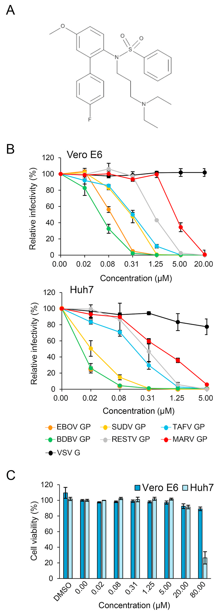
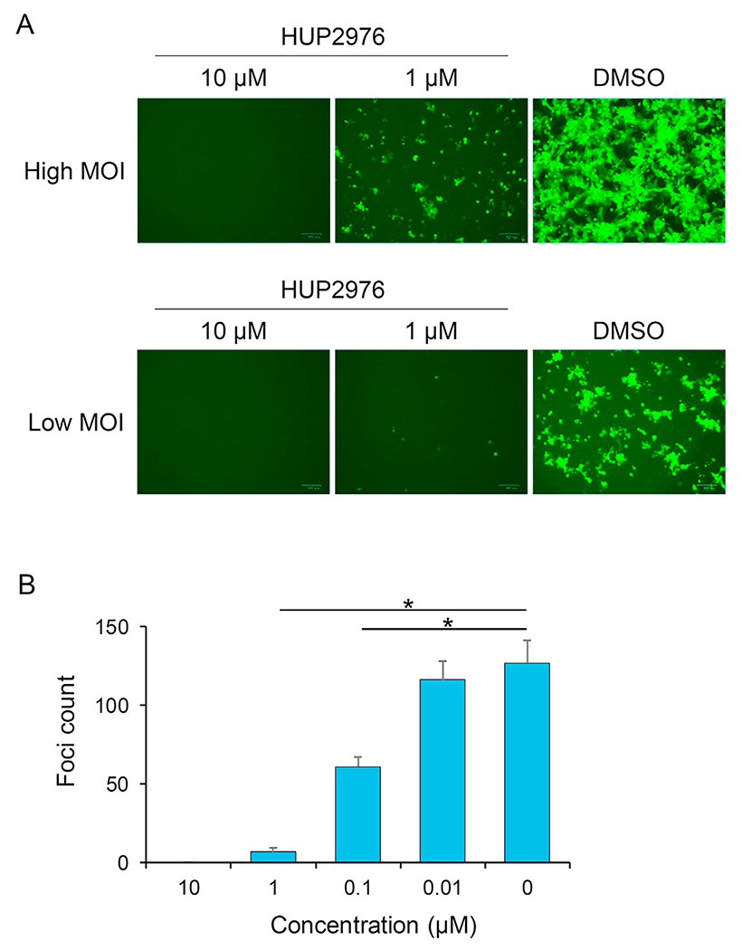
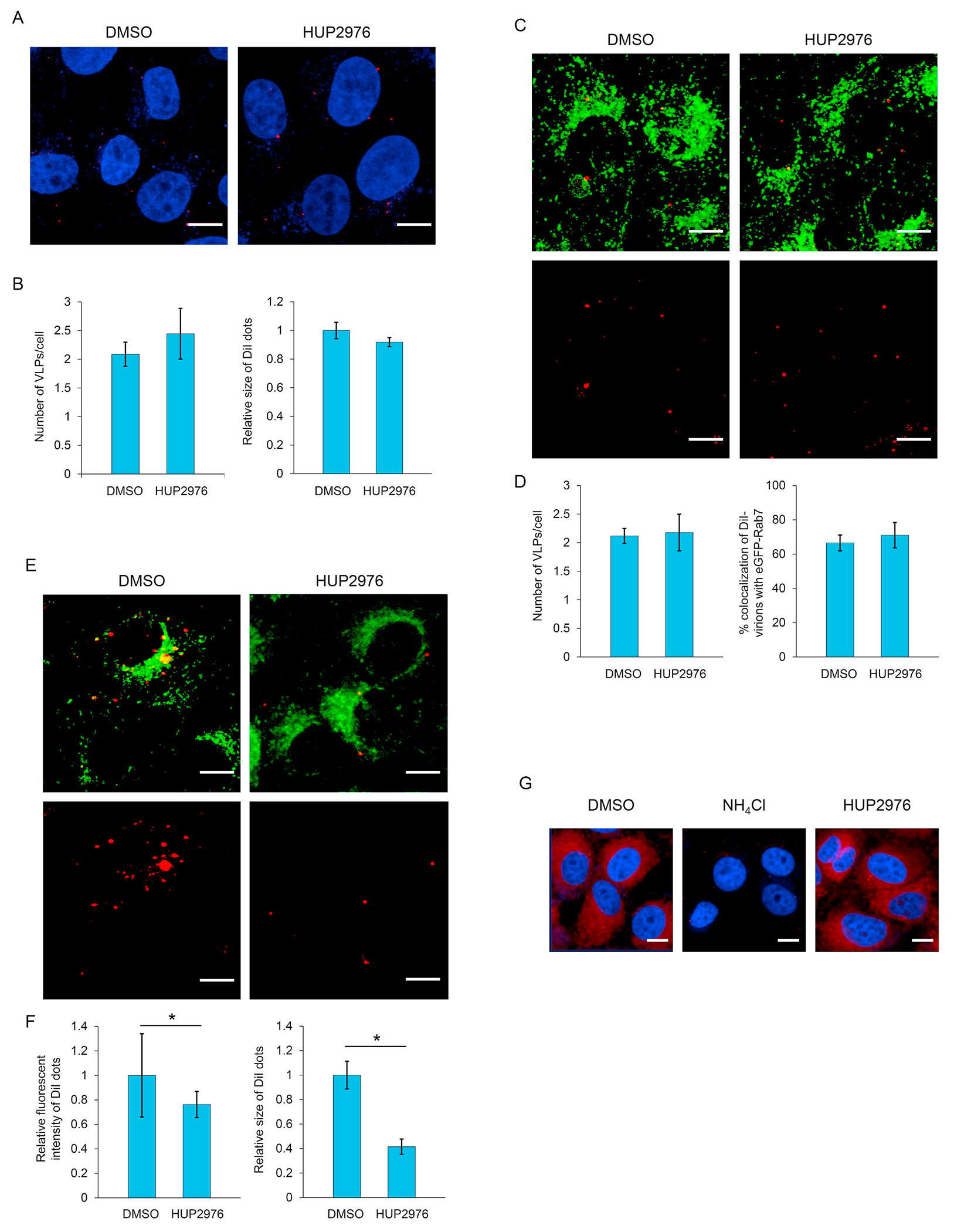
Ebolaviruses and marburgviruses, members of the family Filoviridae, are known to cause fatal diseases often associated with hemorrhagic fever. Recent outbreaks of Ebola virus disease in West African countries and the Democratic Republic of the Congo have made clear the urgent need for the development of therapeutics and vaccines against filoviruses. Using replication-incompetent vesicular stomatitis virus (VSV) pseudotyped with the Ebola virus (EBOV) envelope glycoprotein (GP), we screened a chemical compound library to obtain new drug candidates that inhibit filoviral entry into target cells. We discovered a biaryl sulfonamide derivative that suppressed in vitro infection mediated by GPs derived from all known human-pathogenic filoviruses. To determine the inhibitory mechanism of the compound, we monitored each entry step (attachment, internalization, and membrane fusion) using lipophilic tracer-labeled ebolavirus-like particles and found that the compound efficiently blocked fusion between the viral envelope and the endosomal membrane during cellular entry. However, the compound did not block the interaction of GP with the Niemann-Pick C1 protein, which is believed to be the receptor of filoviruses. Using replication-competent VSVs pseudotyped with EBOV GP, we selected escape mutants and identified two EBOV GP amino acid residues (positions 47 and 66) important for the interaction with this compound. Interestingly, these amino acid residues were located at the base region of the GP trimer, suggesting that the compound might interfere with the GP conformational change required for membrane fusion. These results suggest that this biaryl sulfonamide derivative is a novel fusion inhibitor and a possible drug candidate for the development of a pan-filovirus therapeutic.

Keywords: Compound; Ebolavirus; Entry inhibitor; Glycoprotein; Marburgvirus; Membrane fusion.


Figures

Fig. 1.
Fig. 1.
HUP2976 and its inhibitory activity against VSVs pseudotyped with filovirus GPs. (A) Chemical structure of HUP2976 (MW: 470.6). (B) Pseudotyped VSVs (1000 IU/ml) were mixed with equal volumes of HUP2976 and inoculated onto Vero E6 and Huh7 cells in 96-well plates. The cells were incubated at 37 °C for 18 h and the numbers of GFP-expressing cells were counted using IN Cell Analyzer. One experiment performed in triplicate is shown; averages and standard deviations are presented. (C) Vero E6 and Huh7 cells were incubated with the indicated concentrations of HUP2976 or 0.8% DMSO. Cell viabilities were measured after 24 h-incubation.
Fig. 2.
Fig. 2.
Inhibitory activity of HUP2976 against EBOV-GFP. EBOV-GFP was diluted to infect Vero E6 cells at high (0.5–1.0) and low (0.05–0.1) multiplicities of infection. Following infection, the cells were incubated with the indicated concentrations of HUP2976 for 2 days and fluorescent images were captured (A). EBOV-GFP was diluted (200 focus forming units/well) and inoculated to Vero E6 cells on 96-well plates. Then, the cells were incubated as described in Materials and Methods. The numbers of GFP-expressing foci were counted from triplicate wells (low moi samples) and averages and standard deviations are shown (B). Statistical analysis was performed using Student’s t-test (*p < 0.05).
Fig. 3.
Fig. 3.
Membrane fusion inhibition by HUP2976. DiI-labeled VLPs (red) were inoculated into confluent Vero E6 cells expressing eGFP-Rab7 (green) and incubated for 30 min on ice. After adsorption, the cells were incubated for 0 (A,B), 2 (C,D), or 6 h (E,F) at 37 °C in the presence of HUP2976 (25 μM) or 0.25% DMSO. The cells were fixed with 4% paraformaldehyde and nuclei were stained with DAPI (blue). DiI signals on the cell surface and in the cytoplasm were monitored by confocal laser scanning microscopy (A,C,E). Scale bars represent 10 μm. (B,D,F) Three microscopic fields were acquired randomly, and the number of DiI-labeled virions was measured in approximately 50 individual cells (B,D). Percentage of colocalization (D) and size (B,F) and fluorescence intensity (F) of DiI dots were measured in approximately 50 individual cells and quantified using Image J software. Averages and standard deviations of three independent experiments are shown (B,D,F). Statistical analysis was performed using Student’s t-test (*p < 0.05). (G) Vero E6/eGFP-Rab7 cells were incubated with HUP2976 (25 μM), NH4Cl (25 mM), or DMSO (0.25%) and stained with LysoTracker Red DND-99. Acidic endosomes are visualized in red.
Fig. 4.
Fig. 4.
Effect of HUP2976 on the GP-NPC1 interaction. ELISA plates were coated with thermolysin-treated VLPs, followed by incubation with HUP2976 or mAb114, HA-tagged NPC1 or mock cell lysates, a rat anti-HA antibody, and HRP-conjugated anti-rat IgG (H + L). The reaction was visualized with the TMB substrate. The OD values of mock cell lysates were subtracted from those of HUP2976- or mAb114-treated lysates at each concentration. The experiment was performed in triplicate and averages and standard deviations are shown.
Fig. 5.
Fig. 5.
Identification of amino acid substitutions allowing escape in EBOV GP. (A) The primary structure of GP and amino acid sequences at positions 45–70 are shown. The primary GP structure contains the base, glycan cap, mucin-like domain (MLD), internal fusion loop (IFL), and transmembrane region (TM) and cytoplasmic tail (CT). Amino acid substitutions found in the EBOV GP escape mutants selected under HUP2976 pressure are shown in red. (B) The trimeric structure of EBOV GP (PDB code: 5JQ3) was constructed using PyMOL 1.2r3pre (Schrödinger) and the colored corresponding sequence map above. (C) VSVs pseudotyped with wildtype and mutant EBOV GPs were diluted to 1000 IU/ml, mixed with equal volumes of HUP2976, and inoculated onto Vero E6 cells. The cells were incubated at 37 °C for 18 h, and GFP-expressing cells were counted with IN Cell Analyzer. Averages and standard deviations from 3 independent experiments are shown. (D) Amino acid sequences at positions 45–70 (EBOV numbering) of ebolavirus and marburgvirus GPs. Amino acid residues conserved among all of the filoviruses and those conserved among all of the ebolaviruses but not marburgvirus are highlighted in light blue and pink, respectively.
Fig. 6.
Fig. 6.
Substituted amino acid residues mapped on the GP trimeric structure. The amino acid residues at positions 47 ad 66 are mapped on a ribbon model (A) and surface model (B) of the EBOV GP trimeric structure constructed using PyMOL 1.2r3pre (Schrödinger) based on the crystal structure (PDB code: 6G95). A close-up of the EBOV GP inhibitor-binding pocket in a surface representation in the solid black square. GP1 and GP2 are shown in black and orange in a GP monomer and in gray and yellow in another monomer, respectively. Green and red spheres represent D47 and V66 residues, respectively.

Similar articles

Cited by

References

    1. Alvarez CP, Lasala F, Carrillo J, Muñiz O, Corbí AL, Delgado R, 2002. C-type lectins DC-SIGN and L-SIGN mediate cellular entry by ebola virus in cis and in trans. J. Virol 76, 6841–6844. 10.1128/jvi.76.13.6841-6844.2002. - DOI - PMC - PubMed
    1. Bai CQ, Mu JS, Kargbo D, Song Y. Bin, Niu WK, Nie WM, Kanu A, Liu WW, Wang YP, Dafae F, Yan T, Hu Y, Deng YQ, Lu HJ, Yang F, Zhang XG, Sun Yang, Cao YX, Su HX, Sun Yu, Liu W. Sen, Wang CY, Qian J, Liu L, Wang H, Tong YG, Liu ZY, Chen YS, Wang HQ, Kargbo B, Gao GF, Jiang JF, 2016. Clinical and virological characteristics of ebola virus disease patients treated with favipiravir (T-705) - Sierra Leone, 2014. Clin. Infect. Dis 63, 1288–1294. 10.1093/cid/ciw571. - DOI - PubMed
    1. Baseler L, Chertow DS, Johnson KM, Feldmann H, Morens DM, 2017. The pathogenesis of ebola virus disease. Annu. Rev. Pathol 12, 387–418. 10.1146/annurev-pathol-052016-100506. - DOI - PubMed
    1. Basler CF, Wang X, Muhlberger E, Volchkov V, Paragas J, Klenk H-D, Garcia-Sastre A, Palese P, 2000. The Ebola virus VP35 protein functions as a type IIFN antagonist. Proc. Natl. Acad. Sci. Unit. States Am 97, 12289–12294. 10.1073/pnas.220398297. - DOI - PMC - PubMed
    1. Cagigi A, Misasi J, Ploquin A, Stanley DA, Ambrozak D, Tsybovsky Y, Mason RD, Roederer M, Sullivan NJ, 2018. Vaccine generation of protective ebola antibodies and identification of conserved B-cell signatures. J. Infect. Dis 218, S528–S536. 10.1093/infdis/jiy333. - DOI - PMC - PubMed

Publication types