. 2020 Oct:172:106135.
doi: 10.1016/j.rmed.2020.106135. Epub 2020 Sep 11.

Pulmonary artery thrombi are co-located with opacifications in SARS-CoV2 induced ARDS

Affiliations

Pulmonary artery thrombi are co-located with opacifications in SARS-CoV2 induced ARDS

Katharina Mueller-Peltzer et al. Respir Med. 2020 Oct.

Abstract

Purpose: Patients hospitalized for infection with SARS-CoV-2 typically present with pneumonia. The respiratory failure is frequently complicated by pulmonary embolism in segmental pulmonary arteries. The distribution of pulmonary embolism in regard to lung parenchymal opacifications has not been investigated yet.

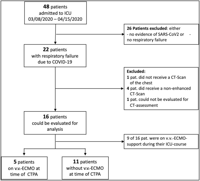
Methods: All patients with COVID-19 treated at a medical intensive care unit between March 8th and April 15th, 2020 undergoing computed tomography pulmonary angiography (CTPA) were included. All CTPA were assessed by two radiologists independently in respect to parenchymal changes and pulmonary embolism on a lung segment basis.

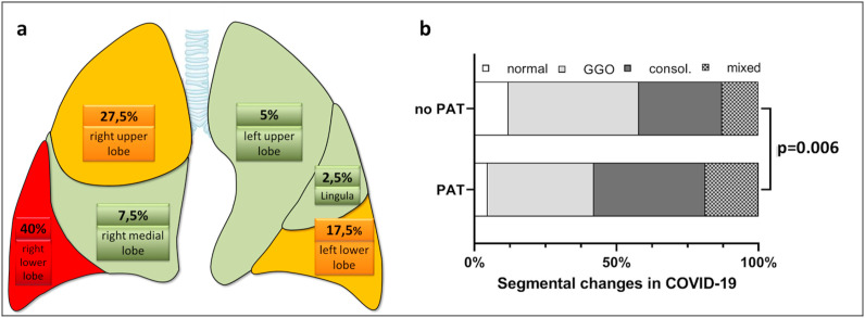
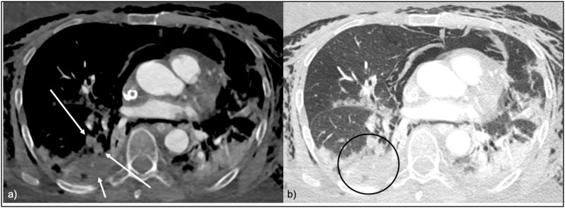
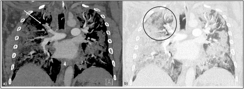
Results: Out of 22 patients with severe COVID-19 treated within the observed time period, 16 (age 60.4 ± 10.2 years, 6 female SAPS2 score 49.2 ± 13.9) underwent CT. A total of 288 lung segment were analyzed. Thrombi were detectable in 9/16 (56.3%) patients, with 4.4 ± 2.9 segments occluded per patient and 40/288 (13.9%) segments affected in the whole cohort. Patients with thrombi had significantly worse segmental opacifications in CT (p < 0.05) and all thrombi were located in opacitated segments. There was no correlation between d-dimer level and number of occluded segmental arteries.

Conclusions: Thrombi in segmental pulmonary arteries are common in COVID-19 and are located in opacitated lung segments. This might suggest local clot formation.

Keywords: COVID19; CT-Scan; CTPA; Pulmonary artery thrombosis; Pulmonary embolism; SARS-CoV2.


Conflict of interest statement

The authors declare that they have no conflicts of interests.

Figures

Fig. 1
Fig. 1
Flowchart: patient selection. CTPA computed tomography pulmonary angiography, COVID-19 Corona-Virus Disease 2019, ECMO extracorporeal membrane oxygenation.
Fig. 2
Fig. 2
a Distribution of pulmonary thrombosis on lung lobe level: most affected was the right lower lobe, followed by the right upper lobe. Colors: Red = high rate of pulmonary thrombosis, Yellow = median rate of pulmonary thrombosis, Green = low rate of pulmonary thrombosis. b Segmental analysis of patients with COVID-19. There is a significant difference in Patients with PAT compared to those without with more.
Fig. 3
Fig. 3
a CTPA in soft tissue window, axial view. PAT in the postero-basal lung segment of the right lower lobe with complete occlusion of the widened vessels (long red arrows). The affected lung segment shows areas of hypoenhancement consistent with embolic infarction (short red arrow). Fig. 3b CTPA in lung window, axial view. The affected postero-basal lung segment of the right lower lobe is consolidated (red circle). Abbreviation PAT pulmonary artery thrombosis.
Fig. 4
Fig. 4
a CTPA in soft tissue window, coronal view. PAT in the apical segment of the right upper lobe with subtotal occlusion of the vessel (red arrow). Fig. 4b CTPA in lung window, coronal view. The affected apical lung segment of the right upper lobe is opacified with a mixture of GGO and consolidations (red circle). Abbreviations: PAT pulmonary artery thrombosis; GGO ground-glass opacities.

Comment in

  • Covid-19 and in situ pulmonary artery thrombosis.
    Mandal AKJ, Kho J, Ioannou A, Van den Abbeele K, Missouris CG. Mandal AKJ, et al. Respir Med. 2021 Jan;176:106176. doi: 10.1016/j.rmed.2020.106176. Epub 2020 Oct 2. Respir Med. 2021. PMID: 33092932 Free PMC article. No abstract available.

References

    1. Wang D., Hu B., Hu C., et al. Clinical characteristics of 138 hospitalized patients with 2019 novel coronavirus–infected pneumonia in wuhan, China. J. Am. Med. Assoc. 2020;323:1061. doi: 10.1001/jama.2020.1585. - DOI - PMC - PubMed
    1. Chung M., Bernheim A., Mei X., et al. CT imaging features of 2019 novel coronavirus (2019-nCoV) Radiology. 2020;295:202–207. doi: 10.1148/radiol.2020200230. - DOI - PMC - PubMed
    1. Zhu J., Zhong Z., Li H., et al. CT imaging features of 4121 patients with COVID‐19: a meta‐analysis. J Med Virol jmv. 2020;25910 doi: 10.1002/jmv.25910. - DOI - PMC - PubMed
    1. Ding X., Xu J., Zhou J., Long Q. Chest CT findings of COVID-19 pneumonia by duration of symptoms. Eur. J. Radiol. 2020;127:109009. doi: 10.1016/j.ejrad.2020.109009. - DOI - PMC - PubMed
    1. Shen C., Yu N., Cai S., et al. Quantitative computed tomography analysis for stratifying the severity of Coronavirus Disease 2019. J. Pharmaceut. Biomed. Anal. 2020;10:123–129. doi: 10.1016/j.jpha.2020.03.004. - DOI - PMC - PubMed

Publication types

MeSH terms