Review
. 2020 Sep 16;21(18):6779.
doi: 10.3390/ijms21186779.

Structure, Folding and Stability of Nucleoside Diphosphate Kinases

Affiliations
Review

Structure, Folding and Stability of Nucleoside Diphosphate Kinases

Florian Georgescauld et al. Int J Mol Sci. .

Abstract

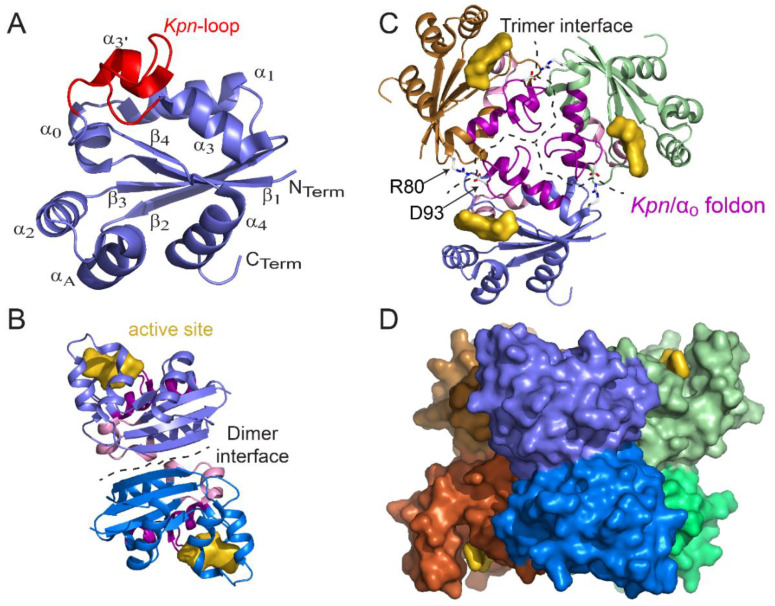
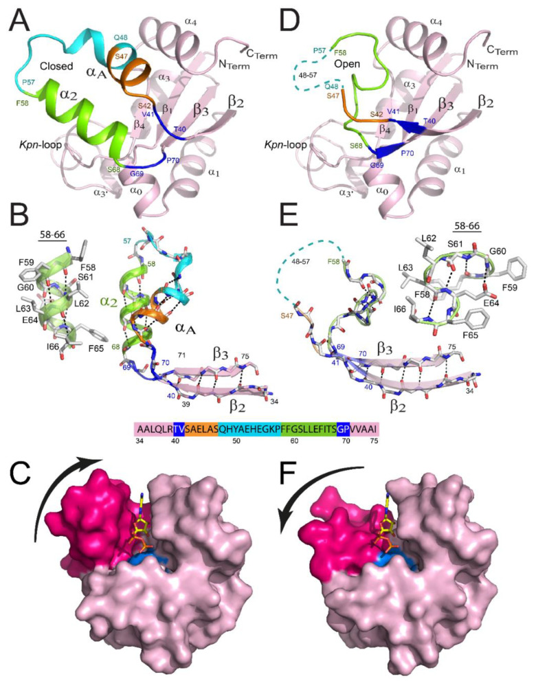
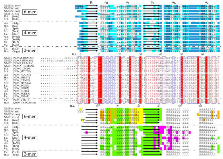
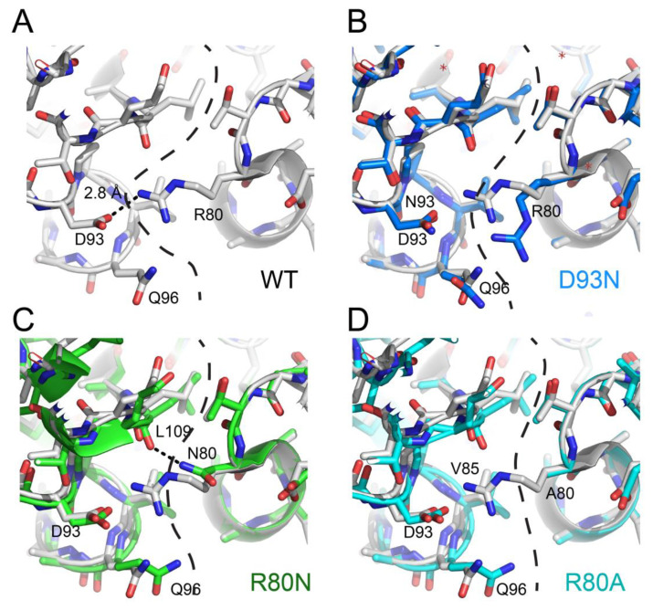
Nucleoside diphosphate kinases (NDPK) are oligomeric proteins involved in the synthesis of nucleoside triphosphates. Their tridimensional structure has been solved by X-ray crystallography and shows that individual subunits present a conserved ferredoxin fold of about 140 residues in prokaryotes, archaea, eukaryotes and viruses. Monomers are functionally independent from each other inside NDPK complexes and the nucleoside kinase catalytic mechanism involves transient phosphorylation of the conserved catalytic histidine. To be active, monomers must assemble into conserved head to tail dimers, which further assemble into hexamers or tetramers. The interfaces between these oligomeric states are very different but, surprisingly, the assembly structure barely affects the catalytic efficiency of the enzyme. While it has been shown that assembly into hexamers induces full formation of the catalytic site and stabilizes the complex, it is unclear why assembly into tetramers is required for function. Several additional activities have been revealed for NDPK, especially in metastasis spreading, cytoskeleton dynamics, DNA binding and membrane remodeling. However, we still lack the high resolution structural data of NDPK in complex with different partners, which is necessary for deciphering the mechanism of these diverse functions. In this review we discuss advances in the structure, folding and stability of NDPKs.

Keywords: histidine kinase; nucleoside diphosphate kinase structure; oligomeric state; protein folding; protein stability; quaternary structure.


Conflict of interest statement

The authors declare no conflict of interest.

Figures

Figure 2
Figure 2
Structure of monomer, dimer, trimer, and hexamer of NDPK from M. tuberculosis (Figure adapted with permission from [36]). (A) View of the monomer with labeled secondary structural elements. (B) Side view of a dimer showing the dimer interface (residues 17, 19–21, 23, 24, 27, 33–38, and 72) and the active site pocket (K10, Y50, R104, N114, H117, S119, and E128). (C) Top view of a trimer. At the trimer interface (residues 16, 25, 28–31, 79, 80, 83, 84, 87, 88, 93–96, 98–102, and 105–110), the Kpn0 foldon (the Kpn-loop and the α0 helix are colored magenta and pink, respectively) and the R80–D93 salt bridge (sticks) are involved in hexamer assembly. The trimer stacks in a “head-to-head” manner and not in a “head-to-tail” manner such that the Kpn0 foldon is exposed on either side of the hexamer. (D) Side view of the surface of the six-color hexamer Mt-NDPK. The active site is colored yellow. In panels B and C, the chains are colored as in panel D. All structure figures were drawn using PyMOL molecular graphic system [38].
Figure 3
Figure 3
Remodeling of the binding site of NDPK from M. tuberculosis. View of a monomer presenting a closed structure (A) versus the remodeled one (D). The β2αAα2β3 region is remodeled in the latter monomer, and details are provided in panels (B,E), respectively. Residues 40–41, 42–47, 48–57, 58–68, and 69–70 are colored blue, orange, cyan, green, and blue, respectively. The region covering residues 48–57 is not visible in the X-ray structure for monomers and is shown as a cyan dashed line in panels D and E. The α2 helix (B) and the successive β-turn structures (E) are shown as cartoons and ball-and-stick structures. The surface views of the monomer are displayed in the closed (C) and open (F) conformations. αA and α2 helices are colored magenta and the catalytic His117 is colored cyan. ADP is bound in the active site by homology with NDPK-A. (Figure adapted with permission from [41]).
Figure 1
Figure 1
Secondary structure, residue accessibility, sequence and interface alignments of hexamer, tetramer and dimer NDPKs (nucleoside diphosphate kinases) with known 3D structure. (i) The relative accessibility and the secondary structure of each residue were calculated using DSSP (Database of Secondary Structure of Proteins) [31]. The accessibility is rendered as colored boxes (marine, cyan and white for accessible, intermediate and buried, respectively). α-helices, β-strands and strict β-turns are displayed as squiggles, arrows and TT letters, respectively. The conventional names of the NDPK secondary structure elements and the Kpn-loop are indicated above. (ii) On sequence alignment, strictly conserved residues are in white on a red background while similar residues are in red on a white background with blue frames (with numbered residues of M.t. and M.x. marked above and below the alignment, respectively). The active site residues and the catalytic histidine are indicated by a green star, and a blue arrow, respectively. (iii) The various interfaces are highlighted in different colors: green for the common-dimer interface, maroon and yellow for the trimer interfaces, red for the CTerm trimer interface, magenta for tetramer interface. The interfaces were identified using PISA (Proteins, Interfaces, Structures and Assemblies) [32]. The figure was drawn with ESPript3 [33]. (NME 1 to 4, Non Metastatic human isoforms 1 to 4, D.d., D. discoideum; M.t., M. tuberculosis; A.a., A. aeolicus; H.p., H. pylori; C.j., C. jejuni; M.x., M. xanthus; E.c., E. coli; N.g., N. gonorrhoeae; A.b., A. baumannii; B.t., B. thailandensis; P.a., P. aeruginosa; V.c., V. cholerae; H. sp, Halomonas sp. 593). The PDB and Uniprot Ids are in parentheses.
Figure 1
Figure 1
Secondary structure, residue accessibility, sequence and interface alignments of hexamer, tetramer and dimer NDPKs (nucleoside diphosphate kinases) with known 3D structure. (i) The relative accessibility and the secondary structure of each residue were calculated using DSSP (Database of Secondary Structure of Proteins) [31]. The accessibility is rendered as colored boxes (marine, cyan and white for accessible, intermediate and buried, respectively). α-helices, β-strands and strict β-turns are displayed as squiggles, arrows and TT letters, respectively. The conventional names of the NDPK secondary structure elements and the Kpn-loop are indicated above. (ii) On sequence alignment, strictly conserved residues are in white on a red background while similar residues are in red on a white background with blue frames (with numbered residues of M.t. and M.x. marked above and below the alignment, respectively). The active site residues and the catalytic histidine are indicated by a green star, and a blue arrow, respectively. (iii) The various interfaces are highlighted in different colors: green for the common-dimer interface, maroon and yellow for the trimer interfaces, red for the CTerm trimer interface, magenta for tetramer interface. The interfaces were identified using PISA (Proteins, Interfaces, Structures and Assemblies) [32]. The figure was drawn with ESPript3 [33]. (NME 1 to 4, Non Metastatic human isoforms 1 to 4, D.d., D. discoideum; M.t., M. tuberculosis; A.a., A. aeolicus; H.p., H. pylori; C.j., C. jejuni; M.x., M. xanthus; E.c., E. coli; N.g., N. gonorrhoeae; A.b., A. baumannii; B.t., B. thailandensis; P.a., P. aeruginosa; V.c., V. cholerae; H. sp, Halomonas sp. 593). The PDB and Uniprot Ids are in parentheses.
Figure 4
Figure 4
Stabilizing mechanisms induced by mutations at subunit interfaces of M. tuberculosis NDPK. (A) WT (wild type; white), (B) D93N (marine) vs. WT (white), (C) R80N (green) vs. WT (white), and (D) R80A (cyan) vs. WT (white).

Similar articles

Cited by

References

    1. Boissan M., Schlattner U., Lacombe M.L. The NDPK/NME superfamily: State of the art. Lab. Investig. 2018;98:164–174. doi: 10.1038/labinvest.2017.137. - DOI - PubMed
    1. Desvignes T., Pontarotti P., Fauvel C., Bobe J. Nme protein family evolutionary history, a vertebrate perspective. BMC Evol. Biol. 2009;9:256. doi: 10.1186/1471-2148-9-256. - DOI - PMC - PubMed
    1. Lacombe M.L., Milon L., Munier A., Mehus J.G., Lambeth D.O. The human Nm23/nucleoside diphosphate kinases. J. Bioenerg. Biomembr. 2000;32:247–258. doi: 10.1023/A:1005584929050. - DOI - PubMed
    1. Lacombe M.L., Tokarska-Schlattner M., Boissan M., Schlattner U. The mitochondrial nucleoside diphosphate kinase (NDPK-D/NME4), a moonlighting protein for cell homeostasis. Lab. Investig. 2018;98:582–588. doi: 10.1038/s41374-017-0004-5. - DOI - PubMed
    1. Lascu I., Gonin P. The catalytic mechanism of nucleoside diphosphate kinases. J. Bioenerg. Biomembr. 2000;32:237–246. doi: 10.1023/A:1005532912212. - DOI - PubMed

Substances

LinkOut - more resources