. 2020 Nov 10;86(23):e02080-20.
doi: 10.1128/AEM.02080-20. Print 2020 Nov 10.

Competence-Stimulating-Peptide-Dependent Localized Cell Death and Extracellular DNA Production in Streptococcus mutans Biofilms

Affiliations

Competence-Stimulating-Peptide-Dependent Localized Cell Death and Extracellular DNA Production in Streptococcus mutans Biofilms

Ryo Nagasawa et al. Appl Environ Microbiol. .

Abstract

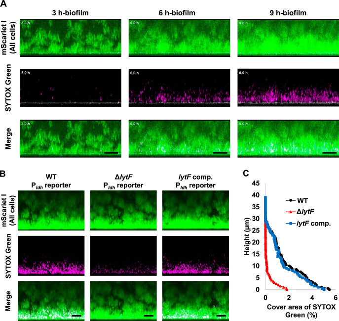
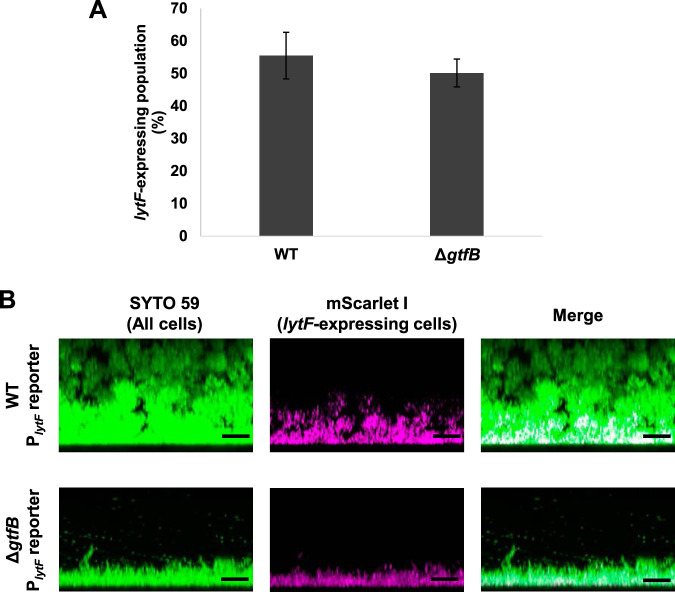
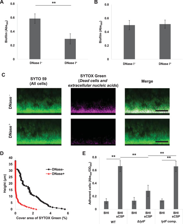
Extracellular DNA (eDNA) is a biofilm component that contributes to the formation and structural stability of biofilms. Streptococcus mutans, a major cariogenic bacterium, induces eDNA-dependent biofilm formation under specific conditions. Since cell death can result in the release and accumulation of DNA, the dead cells in biofilms are a source of eDNA. However, it remains unknown how eDNA is released from dead cells and is localized within S. mutans biofilms. We focused on cell death induced by the extracellular signaling peptide called competence-stimulating peptide (CSP). We demonstrate that nucleic acid release into the extracellular environment occurs in a subpopulation of dead cells. eDNA production induced by CSP was highly dependent on the lytF gene, which encodes an autolysin. Although lytF expression was induced bimodally by CSP, lytF-expressing cells further divided into surviving cells and eDNA-producing dead cells. Moreover, we found that lytF-expressing cells were abundant near the bottom of the biofilm, even when all cells in the biofilm received the CSP signal. Dead cells and eDNA were also abundantly present near the bottom of the biofilm. The number of lytF-expressing cells in biofilms was significantly higher than that in planktonic cultures, which suggests that adhesion to the substratum surface is important for the induction of lytF expression. The deletion of lytF resulted in reduced adherence to a polystyrene surface. These results suggest that lytF expression and eDNA production induced near the bottom of the biofilm contribute to a firmly attached and structurally stable biofilm.IMPORTANCE Bacterial communities encased by self-produced extracellular polymeric substances (EPSs), known as biofilms, have a wide influence on human health and environmental problems. The importance of biofilm research has increased, as biofilms are the preferred bacterial lifestyle in nature. Furthermore, in recent years it has been noted that the contribution of phenotypic heterogeneity within biofilms requires analysis at the single-cell or subpopulation level to understand bacterial life strategies. In Streptococcus mutans, a cariogenic bacterium, extracellular DNA (eDNA) contributes to biofilm formation. However, it remains unclear how and where the cells produce eDNA within the biofilm. We focused on LytF, an autolysin that is induced by extracellular peptide signals. We used single-cell level imaging techniques to analyze lytF expression in the biofilm population. Here, we show that S. mutans generates eDNA by inducing lytF expression near the bottom of the biofilm, thereby enhancing biofilm adhesion and structural stability.

Keywords: Streptococcus mutans; biofilms; cell death; cell-to-cell communication; extracellular DNA.


Figures

FIG 1
FIG 1
A subpopulation of cells releases nucleic acids into the extracellular environment. We inoculated WT cells at the exponential phase into a microfluidic device, which allowed the cells to grow two-dimensionally at 37°C in BHI with sCSP under flow conditions. The medium flowed from left to right in these images at a constant rate (150 μl/h). SYTOX Green dye (1.25 μM) was added to the medium to visualize dead cells and extracellular nucleic acids. Images before and after cell death are displayed. Green indicates dead cells and extracellular nucleic acids.
FIG 2
FIG 2
Cell death and eDNA production induced by sCSP depend on lytF expression via the Com system. Cells were grown in BHI with or without sCSP in an aerobic atmosphere containing 5% CO2 at 37°C for 6 h. (A) Dead cells were stained with 500 nM SYTOX Green for 15 min and quantified by flow cytometry. (B) eDNA was extracted from the 6-h culture supernatants and quantified using a Quant-iT PicoGreen dsDNA assay kit. The results were standardized by the OD600 value at 6 h of culture, since sCSP affects growth. These data indicate the mean ± standard deviations of the results from three independent experiments. The asterisks indicate a significant difference; **, adjusted P < 0.01 (post hoc Bonferroni test).
FIG 3
FIG 3
Cell death occurs in a subpopulation of lytF-expressing cells. The PlytF reporter strain was grown in BHI with sCSP in an aerobic atmosphere containing 5% CO2 at 37°C for 6 h. Dead cells were stained with 500 nM SYTOX Green for 15 min. (A) Cells observed with differential interference contrast (DIC) and fluorescence microscopy. mScarlet-I (lytF-expressing cells) and SYTOX Green (dead cells) are shown in red and green, respectively. The scale bars indicate 20 μm. (B) lytF-expressing cells and dead cells were quantified by flow cytometry. The x axis and y axis show the fluorescence intensities of mScarlet-I and SYTOX Green, respectively. Gating was determined based on unstained and single fluorescent samples. Representative data from independent experiments are presented.
FIG 4
FIG 4
The distribution of lytF-expressing cells in the biofilm is maintained near the bottom even when the culture time is extended. The Pldh and PlytF dual-reporter strain was grown in an aerobic atmosphere containing 5% CO2 at 37°C in BHIs with sCSP for 3, 6, or 9 h in glass-bottom dishes. Nonadherent cells were removed by washing twice with PBS. Areas of approximately 40 μm from the surface were observed using an inverted CLSM. Z-stacks were acquired at 0.5 μm intervals. (A) Images showing side views of the biofilms. Since the samples for each culture time were individually prepared, these images are not in the same field of view. mNeonGreen (all cells) and mScarlet-I (lytF-expressing cells) are shown in green and magenta, respectively. The scale bars indicate 10 μm. Representative images from independent experiments are presented. (B) Image analysis of the biofilms in panel A. In each two-dimensional image, the cover areas showing the fluorescence of mScarlet-I (lytF-expressing cells) and mNeonGreen (all cells) were calculated by ImageJ and Fiji.
FIG 5
FIG 5
Dead cells and eDNA are also abundant near the bottom of the biofilm. (A) The Pldh reporter was grown in a glass-bottom dish in BHIs with sCSP at 37°C. The medium was supplemented with 1.25 μM SYTOX Green to stain dead cells and extracellular nucleic acids. The same field of view within a range of approximately 40 μm from the glass surface was observed while culturing at 37°C. Z-stacks were acquired at 0.55 μm intervals. These images show side views of the biofilms, and the white line below each image shows the glass surface obtained by the reflection. (B) The WT, lytF deficient mutant (ΔlytF), and lytF-complemented (lytF comp) strains with a Pldh reporter background were grown in an aerobic atmosphere containing 5% CO2 at 37°C in BHIs with sCSP for 6 h in glass-bottom dishes. Planktonic cells were removed by washing twice with PBS. These images show side views of the biofilms. Z-stacks were acquired at 0.5 μm intervals. mScarlet-I (all cells) and SYTOX Green (dead cells and extracellular nucleic acids) are shown in green and magenta, respectively. The scale bars indicate 10 μm. Representative images from independent experiments are presented. (C) Image analysis of the 3D images shown in panel B. In each two-dimensional image, the cover areas showing the fluorescence of SYTOX Green were calculated by ImageJ and Fiji.
FIG 6
FIG 6
lytF-expressing cells are abundant near the bottom of the biofilm even in a thin biofilm formed by ΔgtfB cells. (A) PlytF reporter and ΔgtfB PlytF reporter strains were grown in an aerobic atmosphere containing 5% CO2 at 37°C in BHIs with sCSP for 6 h in a 6-well plate. Sonication was performed to suspend aggregated cells. lytF-expressing cells were quantified by flow cytometry. These data indicate the mean ± standard deviations of the results from three independent experiments. (B) Images showing side views of the biofilms for strains grown in an aerobic atmosphere containing 5% CO2 at 37°C in BHIs with sCSP for 6 h in a glass-bottom dish. Nonadherent cells were removed by washing twice with PBS. The biofilm cells were stained with 5 μM SYTO 59 and observed with inverted CLSM. Z-stacks were acquired at 0.5 μm intervals. SYTO 59 (all cells) and mScarlet-I (lytF-expressing cells) are shown in green and magenta, respectively. The scale bars indicate 10 μm. Representative data from independent experiments are presented.
FIG 7
FIG 7
CSP-induced lytF expression and eDNA production contribute to biofilm adhesion to surfaces. (A) DNase I inhibits biofilm formation. The overnight culture was diluted to an OD600 of 0.05 with BHIs with 1 μM CSP and cultured in an aerobic atmosphere containing 5% CO2 at 37°C for 6 h. DNase I was added to the medium at a concentration of 50 U/ml (DNase+) before the incubation. The biofilms were stained with CV. The absorbance at 595 nm (Abs595) was measured on a plate reader. (B to D) eDNA within the preformed biofilm was degraded by DNase I, but the biofilm structure was maintained. Biofilms were formed in BHIs with sCSP medium in an aerobic atmosphere containing 5% CO2 at 37°C for 6 h. The culture solutions were removed and washed twice with PBS. (B) The biofilms were treated with incubation buffer with or without DNase I (50 U/ml) at 37°C for 6 h. They were then stained with CV and the absorbance at 595 nm (Abs595) was measured on a plate reader. (C) Side views of biofilm cells stained with 5 μM SYTO 59 and 500 μM SYTOX Green for 30 min and observed with inverted CLSM. Z-stacks were acquired at 0.5 μm intervals. SYTO 59 (all cells) and SYTOX Green (eDNA and extracellular nucleic acids) are shown in green and magenta, respectively. The scale bars indicate 20 μm. (D) In each two-dimensional image in panel C, the cover areas showing the fluorescence of SYTOX Green were calculated by ImageJ and Fiji. (E) Quantification of adhered cells to the surface in the WT, ΔlytF, and lytF-complemented (lytF comp.) strains. The cells grown in BHI with sCSP were allowed to adhere to the surface by standing for 6 h at 4°C. Adherent cells were stained with CV for 15 min. The absorbance at 595 nm (Abs595) was measured with a plate reader. The results were standardized by the OD600 value at 6 h of culture. These data indicate the mean ± standard deviations of the results from three independent experiments. The asterisks indicate a significant difference; **, adjusted P < 0.01 (post hoc Bonferroni test).

Similar articles

Cited by

References

    1. Okshevsky M, Regina VR, Meyer RL. 2015. Extracellular DNA as a target for biofilm control. Curr Opin Biotechnol 33:73–80. doi:10.1016/j.copbio.2014.12.002. - DOI - PubMed
    1. Petersen FC, Tao L, Scheie AA. 2005. DNA binding-uptake system: a link between cell-to-cell communication and biofilm formation. J Bacteriol 187:4392–4400. doi:10.1128/JB.187.13.4392-4400.2005. - DOI - PMC - PubMed
    1. Thomas VC, Thurlow LR, Boyle D, Hancock LE. 2008. Regulation of autolysis-dependent extracellular DNA release by Enterococcus faecalis extracellular proteases influences biofilm development. J Bacteriol 190:5690–5698. doi:10.1128/JB.00314-08. - DOI - PMC - PubMed
    1. Qin Z, Ou Y, Yang L, Zhu Y, Tolker-Nielsen T, Molin S, Qu D. 2007. Role of autolysin-mediated DNA release in biofilm formation of Staphylococcus epidermidis. Microbiology (Reading) 153:2083–2092. doi:10.1099/mic.0.2007/006031-0. - DOI - PubMed
    1. Regev-Yochay G, Trzcinski K, Thompson CM, Malley R, Lipsitch M. 2006. Interference between Streptococcus pneumoniae and Staphylococcus aureus: in vitro hydrogen peroxide-mediated killing by Streptococcus pneumoniae. J Bacteriol 188:4996–5001. doi:10.1128/JB.00317-06. - DOI - PMC - PubMed

Publication types

MeSH terms

Substances

LinkOut - more resources