. 2020 Sep 18;6(38):eabb4920.
doi: 10.1126/sciadv.abb4920. Print 2020 Sep.

Artificial cells drive neural differentiation

Affiliations

Artificial cells drive neural differentiation

Ö Duhan Toparlak et al. Sci Adv. .

Abstract

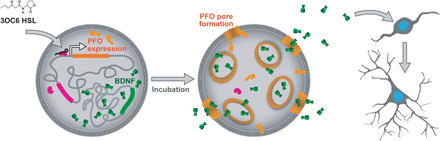
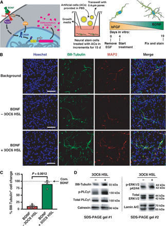
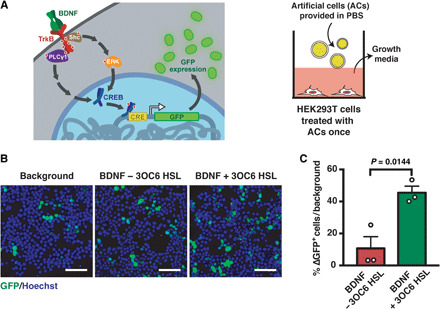
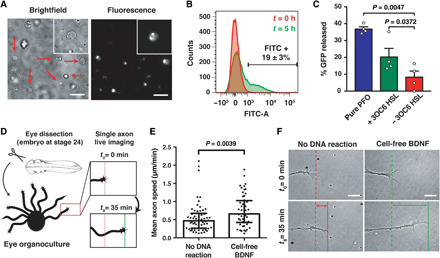
We report the construction of artificial cells that chemically communicate with mammalian cells under physiological conditions. The artificial cells respond to the presence of a small molecule in the environment by synthesizing and releasing a potent protein signal, brain-derived neurotrophic factor. Genetically controlled artificial cells communicate with engineered human embryonic kidney cells and murine neural stem cells. The data suggest that artificial cells are a versatile chassis for the in situ synthesis and on-demand release of chemical signals that elicit desired phenotypic changes of eukaryotic cells, including neuronal differentiation. In the future, artificial cells could be engineered to go beyond the capabilities of typical smart drug delivery vehicles by synthesizing and delivering specific therapeutic molecules tailored to distinct physiological conditions.


Figures

Fig. 1
Fig. 1. Communication between artificial and neural stem cells.
The synthesis of PFO monomers (light orange) is controlled by a genetic AND-gate that requires both LuxR and 3OC6 HSL for gene expression. Monomers of PFO assemble into pores in the presence of cholesterol-containing membranes, thereby releasing BDNF. Homodimers of mature BDNF (green) act on the cognate receptor TrkB, activating signaling pathways leading to neural stem cell differentiation and maturation. The figure was not drawn to scale.
Fig. 2
Fig. 2. Artificial cells drive neuronal differentiation.
(A) Overview of artificial cell treatment and mNS cell differentiation strategy. Left: Overview of signaling between artificial cells and mNS cells. Middle: Artificial cells were incubated with mNS cells for 15 days. Artificial cells were washed away, and fresh artificial cells in fresh medium were added every 24 hours. The cartoon represents a cross-section of the well. Right: Over the course of artificial cell treatment (days 4 to 19), BDNF-secreting artificial cells were gradually increased. (B) Representative immunostaining microscopy of the differentiation of mNS cells into mature neurons at 19 days. Biologically inactive artificial cells (sfGFP secreting, “Background”) were used for normalization. (C) Statistical analysis of βIII-tubulin–overexpressing mNS cells. Cultures treated with commercial BDNF (Com. BDNF) were taken as a reference (100% active, dashed line). The data from (B) were used to generate the plot. Raw data are in fig. S4 (F and G). (D) Western blot of predifferentiated mNS cells in response to treatment with artificial cells at 19 days. Scale bars, 50 μm. Data show mean ± SEM for n = 3 biological replicates, independent experiments. Statistical test was Student’s t test (unpaired, two-tailed). See the Supplementary Materials for detailed figure legend.
Fig. 3
Fig. 3. Artificial cells influence the behavior of HEK293T cells.
(A) Overview of artificial cell treatment of HEK293T cells. Left: Overview of signaling between artificial cells and neural stem cells. Artificial cells activate the expression of genes behind a CRE-regulated promoter, which was GFP. Right: Cartoon representation of the treatment and a cross-section of a cell culture well. (B) Representative microscopy images of GFP expression in genetically engineered HEK293T cells. As in Fig. 2, biologically inactive artificial cells were considered as a mock treatment and used for normalization to assess the signal change (indicated as “Background”). Scale bars, 50 μm. (C) Statistical analysis of GFP-expressing HEK293T cells treated with artificial cells. Change in number of GFP-expressing cells was calculated using background levels as 0%. The percent difference was calculated as a per capita change in GFP+ cells over the entire population with respect to the background. Data show mean ± SEM for n = 3 biological replicates, independent experiments. Statistical test was Student’s t test (unpaired, two-tailed).
Fig. 4
Fig. 4. Characterization of artificial cells and cell-free synthesized BDNF under physiological conditions.
(A) Representative microscopy images of artificial cells that synthesize a BDNF-sfGFP fusion protein at t = 5 hours. Arrows indicate artificial cells. Scale bars, 10 μm. (B) Representative flow cytometry of BDNF-sfGFP–producing artificial cells. (C) GFP release from artificial cells. Recombinant PFO, denoted as “pure PFO,” was provided at a final concentration of 4 μM (0.2 μg/μl). Data show mean ± SEM for independent experiments, n = 4 biological replicates. Statistical test was Student’s t test (paired, two-tailed). (D) Experimental overview of the single axon live-imaging strategy of Xenopus laevis ex vivo eye organocultures. (E) An increase of axon speed was observed when cell-free expressed BDNF was supplied. (F) Representative images of RGC axons from real-time imaging. Scale bars, 10 μm. Data show median with interquartile for n = 4 independent biological replicates. Statistical test was two-tailed Mann-Whitney test, where the data were not normally distributed (Shapiro-Wilk test). At least 55 growth cones were counted for each group.

Similar articles

Cited by

References

    1. Buddingh B. C., van Hest J. C. M., Artificial cells: Synthetic compartments with life-like functionality and adaptivity. Acc. Chem. Res. 50, 769–777 (2017). - PMC - PubMed
    1. Schwille P., Spatz J., Landfester K., Bodenschatz E., Herminghaus S., Sourjik V., Erb T. J., Bastiaens P., Lipowsky R., Hyman A., Dabrock P., Baret J.-C., Vidakovic-Koch T., Bieling P., Dimova R., Mutschler H., Robinson T., Tang T.-Y. D., Wegner S., Sundmacher K., MaxSynBio: Avenues towards creating cells from the bottom up. Angew. Chem. Int. Ed. Engl. 57, 13382–13392 (2018). - PubMed
    1. Deng N.-N., Yelleswarapu M., Zheng L., Huck W. T. S., Microfluidic assembly of monodisperse vesosomes as artificial cell models. J. Am. Chem. Soc. 139, 587–590 (2017). - PubMed
    1. Booth M. J., Schild V. R., Graham A. D., Olof S. N., Bayley H., Light-activated communication in synthetic tissues. Sci. Adv. 2, e1600056 (2016). - PMC - PubMed
    1. Elani Y., Law R. V., Ces O., Vesicle-based artificial cells as chemical microreactors with spatially segregated reaction pathways. Nat. Commun. 5, 5305 (2014). - PubMed

Publication types

LinkOut - more resources