Review
. 2020 Nov;61(11):1365-1376.
doi: 10.1194/jlr.R120001116. Epub 2020 Sep 18.

Chylomicronemia from GPIHBP1 autoantibodies

Affiliations
Review

Chylomicronemia from GPIHBP1 autoantibodies

Kazuya Miyashita et al. J Lipid Res. 2020 Nov.

Abstract

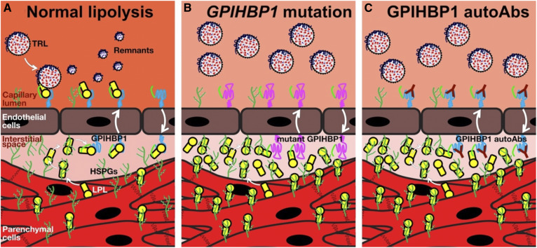
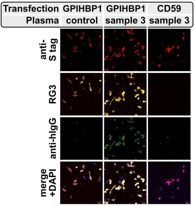
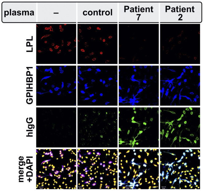
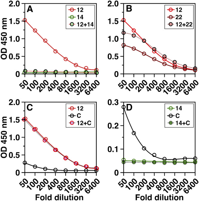
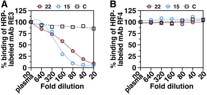
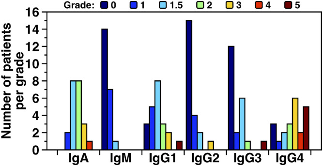
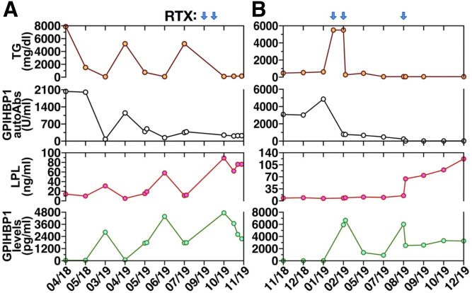
Some cases of chylomicronemia are caused by autoantibodies against glycosylphosphatidylinositol-anchored HDL binding protein 1 (GPIHBP1), an endothelial cell protein that shuttles LPL to the capillary lumen. GPIHBP1 autoantibodies prevent binding and transport of LPL by GPIHBP1, thereby disrupting the lipolytic processing of triglyceride-rich lipoproteins. Here, we review the "GPIHBP1 autoantibody syndrome" and summarize clinical and laboratory findings in 22 patients. All patients had GPIHBP1 autoantibodies and chylomicronemia, but we did not find a correlation between triglyceride levels and autoantibody levels. Many of the patients had a history of pancreatitis, and most had clinical and/or serological evidence of autoimmune disease. IgA autoantibodies were present in all patients, and IgG4 autoantibodies were present in 19 of 22 patients. Patients with GPIHBP1 autoantibodies had low plasma LPL levels, consistent with impaired delivery of LPL into capillaries. Plasma levels of GPIHBP1, measured with a monoclonal antibody-based ELISA, were very low in 17 patients, reflecting the inability of the ELISA to detect GPIHBP1 in the presence of autoantibodies (immunoassay interference). However, GPIHBP1 levels were very high in five patients, indicating little capacity of their autoantibodies to interfere with the ELISA. Recently, several GPIHBP1 autoantibody syndrome patients were treated successfully with rituximab, resulting in the disappearance of GPIHBP1 autoantibodies and normalization of both plasma triglyceride and LPL levels. The GPIHBP1 autoantibody syndrome should be considered in any patient with newly acquired and unexplained chylomicronemia.

Keywords: autoimmune disease; glycosylphosphatidylinositol-anchored high density lipoprotein binding protein 1; immunoglobulin A; immunoglobulin G4; lipoprotein lipase; triglycerides.


Conflict of interest statement

Conflict of interest—K,N. holds stock in Immuno-Biological Laboratories (IBL) and serves as a consultant for Skylight and Sysmex. All other authors declare that they have no conflicts of interest with the contents of this article.

Figures

Fig. 1.
Fig. 1.
Normal intravascular lipolysis (A), lipolysis in the setting of a GPIHBP1 missense mutation in the LU domain (B), or in the presence of GPIHBP1 autoantibodies (autoAbs) (C). Absent GPIHBP1 function, caused either by a GPIHBP1 missense mutation or GPIHBP1 autoAbs, eliminates transport of LPL to the capillary lumen, causing a dramatic reduction in intravascular triglyceride hydrolysis and severe hypertriglyceridemia (chylomicronemia).
Fig. 2.
Fig. 2.
Immunoassay interference—the first clue to the existence of GPIHBP1 autoantibodies. As part of an effort to validate a monoclonal antibody-based sandwich ELISA designed to measure the levels of GPIHBP1 in human plasma, we tested whether the GPIHBP1 ELISA would faithfully detect higher levels of GPIHBP1 in plasma samples that had been “spiked” with recombinant human GPIHBP1 (35). A total of 62.5 pg of human GPIHBP1 was spiked into plasma samples from 40 human patients (31 from patients with hypertriglyceridemia and 9 from normolipidemic controls). This graph shows the ability of the ELISA to recover the 62.5 pg of GPIHBP1 that was spiked into each plasma sample. In 38 out of the 40 samples, the recovery (±SD) of the spiked recombinant human GPIHBP1 was excellent (98.8 ± 3.8%). However, in plasma samples from two patients (both of whom had severe hypertriglyceridemia), the recovery of the spiked GPIHBP1 was negligible (6.8% and 4.4%). The inability of the ELISA to detect GPIHBP1 in these two patients indicated immunoassay interference, which can be caused by autoantibodies. Subsequent studies showed that the plasma from the two patients contained autoantibodies against GPIHBP1. This figure is reproduced with permission from Beigneux et al. (35).
Fig. 3.
Fig. 3.
Detection, by confocal immunofluorescence microscopy, of GPIHBP1 autoantibodies in the plasma of a patient with chylomicronemia. CHO cells were transfected with expression vectors encoding S-protein tagged versions of human GPIHBP1 or a related LU family member (CD59). After 24 h, the cells were incubated with the plasma from a normolipidemic subject (control) or a patient with chylomicronemia (sample 3 in Table 1). GPIHBP1 and CD59 were both detected with an antibody against the S-protein epitope tag (red); GPIHBP1 was also detected with the human GPIHBP1–specific mAb RG3 (yellow). The presence of GPIHBP1 autoantibodies on the GPIHBP1-transfected cells was detected with an antibody against human IgG (green). DNA was stained with DAPI (blue). This figure is reproduced with permission from Beigneux et al. (35).
Fig. 4.
Fig. 4.
GPIHBP1 autoantibodies block the binding of LPL to GPIHBP1 on the surface of GPIHBP1-transfected cells, as judged by confocal fluorescence microscopy. GPIHBP1-transfected cells were preincubated with a plasma sample from a normolipidemic subject (control) or from patients with chylomicronemia from GPIHBP1 autoantibodies (patients 2 and 7 in Table 1) and subsequently incubated with the conditioned media from human LPL–transfected cells. GPIHBP1 autoantibodies (green) in plasma samples from patients 2 and 7 bound avidly to the GPIHBP1 (blue) on the surface of GPIHBP1-transfected cell. The binding of autoantibodies prevented the binding of LPL (red) to GPIHBP1. DNA was stained with DAPI (yellow). This figure is reproduced with permission from Beigneux et al. (35).
Fig. 5.
Fig. 5.
GPIHBP1 autoantibodies from different patients differ in their capacity to interfere with the detection of GPIHBP1 by a monoclonal antibody–based ELISA. Here, we show the ability of the ELISA to detect GPIHBP1 in plasma samples from three different patients with the GPIHBP1 autoantibody syndrome (patients 12, 14, and 22 in Table 1) and a normolipidemic control “C”. The plasma of patient 14 had undetectable levels of GPIHBP1, reflecting the presence of GPIHBP1 autoantibodies that interfered with the ELISA. Patients 12 and 22 had high levels of GPIHBP1, indicating that their autoantibodies against GPIHBP1 had little capacity to interfere with the ELISA. A: The high GPIHBP1 levels in the plasma of patient 12 were no longer detectable after mixing the plasma of patient 12 with the plasma from patient 14. B: Mixing plasma samples from patients 12 and 22 did not interfere with detection of GPIHBP1. C: Mixing the plasma from patient 12 with a normolipidemic control (sample C) did not alter the ability of the ELISA to detect GPIHBP1. D: Mixing plasma from patient 14 with the plasma from the control subject (subject C) abolished detection of GPIHBP1 by the ELISA. Results show the optical density (OD) in the GPIHBP1 ELISA at each sample dilution (1:50 to 1:6,400).
Fig. 6.
Fig. 6.
ELISAs testing the ability of dilutions of plasma from a control subject “C” and two GPIHBP1 autoantibody patients (patients 15 and 22 in Table 1) to compete with two different HRP-labeled GPIHBP1-specific monoclonal antibodies (mAb RE3, mAb RF4) for binding to recombinant human GPIHBP1. RE3 binds to GPIHBP1’s LU domain and abolishes GPIHBP1’s ability to bind LPL (38). RF4 binds to an epitope located between GPIHBP1’s acidic domain and LU domain (8). A: Plasma samples from the two GPIHBP1 autoantibody patients, but not the control plasma, inhibited the binding of mAb RE3 to GPIHBP1. B: Neither the control plasma nor the plasma samples from the two GPIHBP1 autoantibody patients inhibited the binding of mAb RF4 to recombinant GPIHBP1. Plasma samples were tested over a range of dilutions (1:20 to 1:640).
Fig. 7.
Fig. 7.
IgA and IgG4 autoantibodies are predominant in the GPIHBP1 autoantibody syndrome. Shown here are numbers of GPIHBP1 autoantibody syndrome patients having IgA, IgM, IgG1, IgG2, IgG3, and IgG4 autoantibodies, organized according to “titer grade.” The titer grade for each immunoglobulin class and subclass was assessed for 22 patients with the GPIHBP1 autoantibody syndrome. The titer was assessed by ELISAs in which 1:1,000 dilutions of plasma samples were loaded onto wells coated with recombinant GPIHBP1. After washing the plates, HRP-labeled immunoglobulin class- and subclass-specific antibodies were added to the plates. The binding of those antibodies was graded according to the optical density (OD) in the ELISA. Grades ranged from below detection (grade 0, dark blue bars) to very high levels (grade 5, brown bars). Grade 0, OD < 0.1; grade 1, OD 0.1–0.25; grade 1.5, OD 0. 25–0.5; grade 2, OD 0.5–1.0; grade 3, OD 1.0–2.0; grade 4, OD 2.0–3.0; grade 5, OD > 3.0.
Fig. 8.
Fig. 8.
Plasma triglyceride levels (TG, brown circles), GPIHBP1 autoantibodies (autoAbs, black circles), LPL mass (red circles), and GPIHBP1 mass levels (green circles) in two patients with the GPIHBP1 autoantibody syndrome. Values are shown both before and after treatment with rituximab (RTX); blue arrows indicate timing of RTX infusions. A: TG, GPIHBP1 autoantibodies, LPL mass, and GPIHBP1 mass measurements in a GPIHBP1 autoantibody patient (patient 13 in Table 1) described by Ashrafi et al. (75). Panel A modified, with permission, from the publication by Ashraf et al. (75). B: TG, GPIHBP1 autoantibodies, LPL mass, and GPIHBP1 mass measurements in a GPIHBP1 autoantibody patient (patient 11 in Table 1) described by Lutz et al. (76).

References

    1. Fong L. G., Young S. G., Beigneux A. P., Bensadoun A., Oberer M., Jiang H., and Ploug M.. 2016. GPIHBP1 and plasma triglyceride metabolism. Trends Endocrinol. Metab. 27: 455–469. - PMC - PubMed
    1. Young S. G., Fong L. G., Beigneux A. P., Allan C. M., He C., Jiang H., Nakajima K., Meiyappan M., Birrane G., and Ploug M.. 2019. GPIHBP1 and lipoprotein lipase, partners in plasma triglyceride metabolism. Cell Metab. 30: 51–65. - PMC - PubMed
    1. Davies B. S. J., Beigneux A. P., Barnes R. H. II, Tu Y., Gin P., Weinstein M. M., Nobumori C., Nyrén R., Goldberg I. J., Olivecrona G., et al. . 2010. GPIHBP1 is responsible for the entry of lipoprotein lipase into capillaries. Cell Metab. 12: 42–52. - PMC - PubMed
    1. Allan C. M., Larsson M., Jung R. S., Ploug M., Bensadoun A., Beigneux A. P., Fong L. G., and Young S. G.. 2017. Mobility of “HSPG-bound” LPL explains how LPL is able to reach GPIHBP1 on capillaries. J. Lipid Res. 58: 216–225. - PMC - PubMed
    1. Goulbourne C. N., Gin P., Tatar A., Nobumori C., Hoenger A., Jiang H., Grovenor C. R., Adeyo O., Esko J. D., Goldberg I. J., et al. . 2014. The GPIHBP1-LPL complex is responsible for the margination of triglyceride-rich lipoproteins in capillaries. Cell Metab. 19: 849–860. - PMC - PubMed

Publication types

MeSH terms