

Sphingolipids as critical players in retinal physiology and pathology
- PMID: 32948663
- PMCID: PMC7933806
- DOI: 10.1194/jlr.TR120000972
Sphingolipids as critical players in retinal physiology and pathology
Abstract
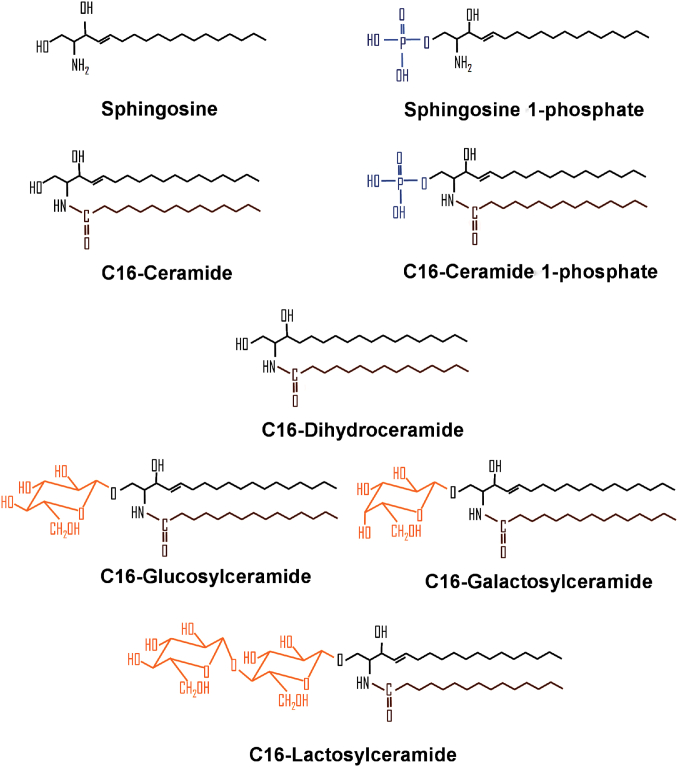
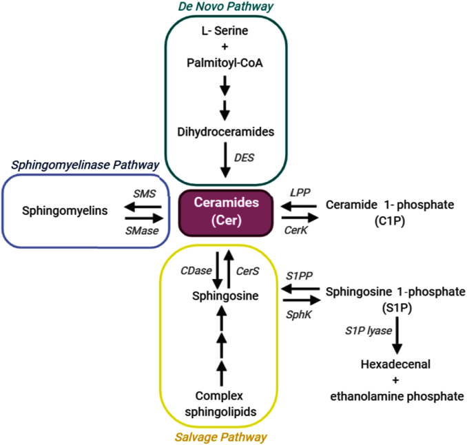
Sphingolipids have emerged as bioactive lipids involved in the regulation of many physiological and pathological processes. In the retina, they have been established to participate in numerous processes, such as neuronal survival and death, proliferation and migration of neuronal and vascular cells, inflammation, and neovascularization. Dysregulation of sphingolipids is therefore crucial in the onset and progression of retinal diseases. This review examines the involvement of sphingolipids in retinal physiology and diseases. Ceramide (Cer) has emerged as a common mediator of inflammation and death of neuronal and retinal pigment epithelium cells in animal models of retinopathies such as glaucoma, age-related macular degeneration (AMD), and retinitis pigmentosa. Sphingosine-1-phosphate (S1P) has opposite roles, preventing photoreceptor and ganglion cell degeneration but also promoting inflammation, fibrosis, and neovascularization in AMD, glaucoma, and pro-fibrotic disorders. Alterations in Cer, S1P, and ceramide 1-phosphate may also contribute to uveitis. Notably, use of inhibitors that either prevent Cer increase or modulate S1P signaling, such as Myriocin, desipramine, and Fingolimod (FTY720), preserves neuronal viability and retinal function. These findings underscore the relevance of alterations in the sphingolipid metabolic network in the etiology of multiple retinopathies and highlight the potential of modulating their metabolism for the design of novel therapeutic approaches.
Keywords: age-related macular degeneration; ceramide; ceramide-1-phosphate; photoreceptor degeneration; retinitis pigmentosa; sphingosine-1-phosphate.
Copyright © 2021 The Authors. Published by Elsevier Inc. All rights reserved.
Conflict of interest statement
Conflict of interest The authors declare that they have no conflicts of interest with the contents of this article.
Figures
Similar articles
-
Updates on sphingolipids: Spotlight on retinopathy.Biomed Pharmacother. 2021 Nov;143:112197. doi: 10.1016/j.biopha.2021.112197. Epub 2021 Sep 21. Biomed Pharmacother. 2021. PMID: 34560541 Review.
-
Sphingolipids as Emerging Mediators in Retina Degeneration.Front Cell Neurosci. 2019 Jun 11;13:246. doi: 10.3389/fncel.2019.00246. eCollection 2019. Front Cell Neurosci. 2019. PMID: 31244608 Free PMC article. Review.
-
Sphingosine-1-phosphate and ceramide-1-phosphate promote migration, pro-inflammatory and pro-fibrotic responses in retinal pigment epithelium cells.Exp Eye Res. 2022 Nov;224:109222. doi: 10.1016/j.exer.2022.109222. Epub 2022 Aug 27. Exp Eye Res. 2022. PMID: 36041511
-
Metabolism and biological functions of two phosphorylated sphingolipids, sphingosine 1-phosphate and ceramide 1-phosphate.Prog Lipid Res. 2007 Mar;46(2):126-44. doi: 10.1016/j.plipres.2007.03.001. Epub 2007 Mar 14. Prog Lipid Res. 2007. PMID: 17449104 Review.
-
Regulating survival and development in the retina: key roles for simple sphingolipids.J Lipid Res. 2010 Jun;51(6):1247-62. doi: 10.1194/jlr.R003442. Epub 2010 Jan 25. J Lipid Res. 2010. PMID: 20100817 Free PMC article. Review.
Cited by
-
Optimized Lipidomics Extraction of Sphingosine and Sphinganine from Optic Nerve for Signaling Studies.Methods Mol Biol. 2024;2816:25-33. doi: 10.1007/978-1-0716-3902-3_3. Methods Mol Biol. 2024. PMID: 38977585
-
Phenome-wide Mendelian randomisation analysis identifies causal factors for age-related macular degeneration.Elife. 2023 Jan 27;12:e82546. doi: 10.7554/eLife.82546. Elife. 2023. PMID: 36705323 Free PMC article.
-
Dynamic lipid turnover in photoreceptors and retinal pigment epithelium throughout life.Prog Retin Eye Res. 2022 Jul;89:101037. doi: 10.1016/j.preteyeres.2021.101037. Epub 2021 Dec 29. Prog Retin Eye Res. 2022. PMID: 34971765 Free PMC article. Review.
-
Complement activation, lipid metabolism, and mitochondrial injury: Converging pathways in age-related macular degeneration.Redox Biol. 2020 Oct;37:101781. doi: 10.1016/j.redox.2020.101781. Epub 2020 Nov 2. Redox Biol. 2020. PMID: 33162377 Free PMC article. Review.
-
Dysregulated lipid metabolism in a retinal pigment epithelial cell model and serum of patients with age-related macular degeneration.BMC Biol. 2025 Apr 12;23(1):96. doi: 10.1186/s12915-025-02198-8. BMC Biol. 2025. PMID: 40221802 Free PMC article.
References
-
- Dressler K.A., Mathias S., Kolesnick R.N. Tumor necrosis factor-alpha activates the sphingomyelin signal transduction pathway in a cell-free system. Science. 1992;255:1715–1718. - PubMed
-
- Hannun Y.A., Loomis C.R., Merrill A.H., Bell R.M. Sphingosine inhibition of protein kinase C activity and of phorbol dibutyrate binding in vitro and in human platelets. J. Biol. Chem. 1986;261:12604–12609. - PubMed
-
- Kolesnick R.N. 1,2-Diacylglycerols but not phorbol esters stimulate sphingomyelin hydrolysis in GH3 pituitary cells. J. Biol. Chem. 1987;262:16759–16762. - PubMed
-
- Obeid L.M., Linardic C.M., Karolak L.A., Hannun Y.A. Programmed cell death induced by ceramide. Science. 1993;259:1769–1771. - PubMed
-
- Cuvillier O., Pirianov G., Kleuser B., Vanek P.G., Cosot O.A., Gutkind J.S., Spiegel S. Suppression of ceramide-mediated programmed cell death by sphingosine-1-phosphate. Nature. 1996;381:800–803. - PubMed
Publication types
MeSH terms
Substances
Grants and funding
LinkOut - more resources
Full Text Sources
Other Literature Sources
Medical
