. 2020 Sep 18;11(9):778.
doi: 10.1038/s41419-020-02972-2.

Preclinical evaluation of a regimen combining chidamide and ABT-199 in acute myeloid leukemia

Affiliations

Preclinical evaluation of a regimen combining chidamide and ABT-199 in acute myeloid leukemia

Kai Chen et al. Cell Death Dis. .

Abstract

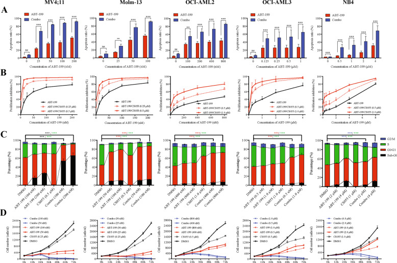
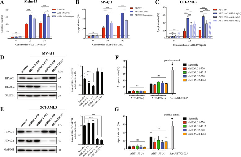
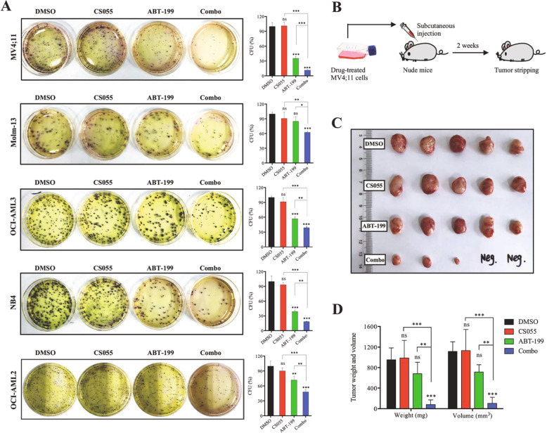
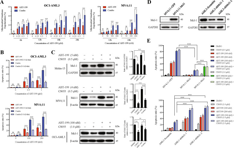
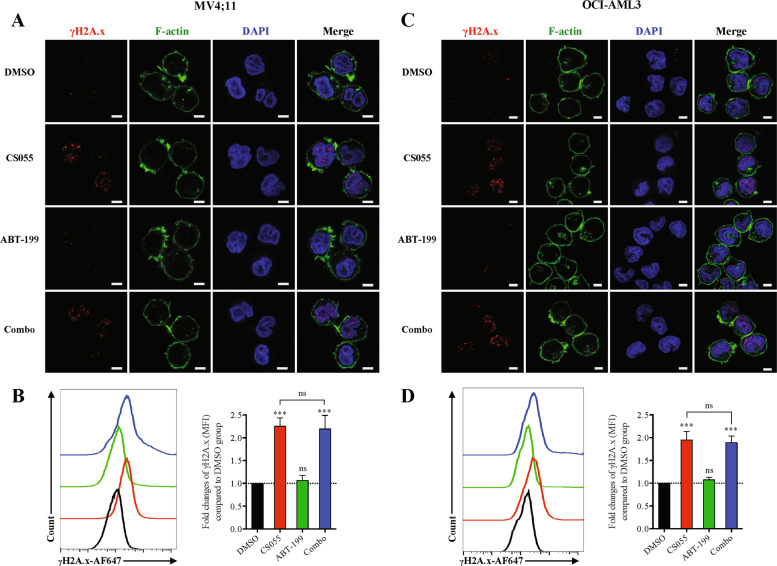
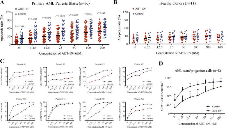
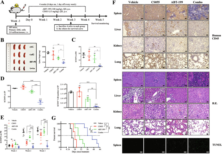
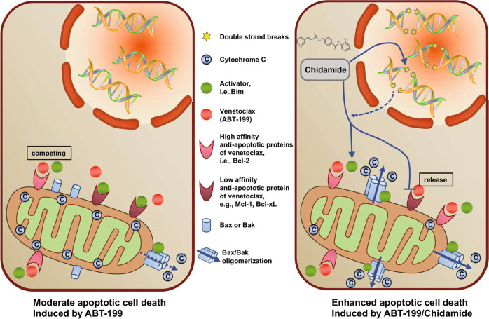
Acute myeloid leukemia (AML) is a heterogeneous myeloid neoplasm with poor clinical outcome, despite the great progress in treatment in recent years. The selective Bcl-2 inhibitor venetoclax (ABT-199) in combination therapy has been approved for the treatment of newly diagnosed AML patients who are ineligible for intensive chemotherapy, but resistance can be acquired through the upregulation of alternative antiapoptotic proteins. Here, we reported that a newly emerged histone deacetylase inhibitor, chidamide (CS055), at low-cytotoxicity dose enhanced the anti-AML activity of ABT-199, while sparing normal hematopoietic progenitor cells. Moreover, we also found that chidamide showed a superior resensitization effect than romidepsin in potentiation of ABT-199 lethality. Inhibition of multiple HDACs rather than some single component might be required. The combination therapy was also effective in primary AML blasts and stem/progenitor cells regardless of disease status and genetic aberrance, as well as in a patient-derived xenograft model carrying FLT3-ITD mutation. Mechanistically, CS055 promoted leukemia suppression through DNA double-strand break and altered unbalance of anti- and pro-apoptotic proteins (e.g., Mcl-1 and Bcl-xL downregulation, and Bim upregulation). Taken together, these results show the high therapeutic potential of ABT-199/CS055 combination in AML treatment, representing a potent and alternative salvage therapy for the treatment of relapsed and refractory patients with AML.


Conflict of interest statement

The authors declare that they have no conflict of interest.

Figures

Fig. 1
Fig. 1. Low and a sublethal dose of CS055 potentiates the anti-leukemic activity of ABT-199 toward various acute myeloid leukemia (AML) cell lines.
Five human AML cell lines were exposed to indicated concentrations of ABT-199 ± CS055 (0.5 μM for Molm-13 and MV4;11; 1.0 μM for OCI-AML2, OCI-AML3, and NB4) for 24 or 48 h, after which cells were subjected to the following analyses: a the percentage of apoptotic cells were determined at 48 h using Annexin V/PI double staining by flow cytometry. b The inhibition rate of cell viability was measured at 48 h using the CCK-8 kit. c Cell cycle distribution was assessed at 24 h by flow cytometry (black asterisk, red asterisk, green asterisk, and blue asterisk present the statistical significance of Sub-G0, G0/G1, S, G2/M between DMSO and Combo group, respectively). d Cell growth was monitored every 12 h for consecutive 3 days. Values indicate mean ± SD for at least three independent experiments performed in triplicate (*P < 0.05, **P < 0.01, and ***P < 0.001, ns not significant).
Fig. 2
Fig. 2. CS055 shows a superior resensitization effect than romidepsin in potentiation of ABT-199 lethality in acute myeloid leukemia (AML) cells.
a Molm-13, b MV4;11, and c OCI-AML3 cells were exposed to the indicated concentrations of ABT-199 ± CS055 (0.25 µM for Molm-13 and MV4;11, and 1.0 µM for OCI-AML3) or romidepsin (1.5 nM for Molm-13 and MV4;11, and 2.5–3 nM for OCI-AML3) for 48 h, after which the percentage of apoptotic cells was determined using Annexin V/PI double staining by flow cytometry. d, e Western blot analysis was performed to validate downregulation efficiency of HDAC1 and HDAC2 in MV4;11 and OCI-AML3 cells. f, g Cells were exposed to ABT-199 (50 nM for MV4;11, 1.0 µM for OCI-AML3), and then the percentage of apoptotic cells was determined by flow cytometry. Scramble shRNA-transfected cells exposed for 48 h to combined treatment with ABT-199 (50 nM for MV4;11, 1.0 µM for OCI-AML3) and CS055 (0.5 µM for MV4;11, 1.0 µM for OCI-AML3) serve as a positive control. Values indicate mean ± SD for at least three independent experiments performed in triplicate (*P < 0.05, **P < 0.01, ***P < 0.001, ns not significant).
Fig. 3
Fig. 3. ABT-199 in combination with CS055 impairs colony- and tumor-forming ability of acute myeloid leukemia (AML) cells in vitro and in vivo.
a AML cells were treated with ABT-199 (200 nM for MV4;11, 100 nM for Molm-13, 800 nM for OCI-AML2, 1 μM for OCI-AML3, and 4 μM for NB4) ± CS055 (0.5 μM for MV4;11/Molm-13, and 1.0 μM for the other lines) for 12 h, after which the clonogenicity assay was performed to determine the percentage of CFU (left, representative images; right, bar graphs). Values indicate mean ± SD for at least three independent experiments performed in triplicate (*P < 0.05, **P < 0.01, and ***P < 0.001; ns not significant for CS055 vs vehicle). b The scheme for the procedure of the in vivo experiment. MV4;11 cells were pre-incubated for 12 h with ABT-199 ± CS055, followed by subcutaneous injection on the left flank of nude mice. c, d Two weeks after cell inoculation, tumors were removed (c) and measured for weight and size (d).
Fig. 4
Fig. 4. CS055 enhances mitochondrion-dependent apoptosis induced by ABT-199 by downregulation of Mcl-1.
a OCI-AML3 (left) and MV4;11 cells (right) were exposed to the indicated concentrations of ABT-199 ± CS055 (1.0 μM for OCI-AML3; 0.5 μM for MV4;11) for 6–24 h, after which mitochondrial membrane potential (MMP) was measured using the JC-1 kit by flow cytometry. b OCI-AML3 (upper) and MV4;11 cells (lower) were pre-incubated with 20 μM Z-VAD-fmk for 2 h, followed by treatment with indicated concentrations of ABT-199 ± CS055 (1.0 μM for OCI-AML3; 0.5 μM for MV4;11) for additional 24 h. The percentage of apoptosis was then assessed by flow cytometry. c Western blot analysis was performed to monitor the expression of Mcl-1 in Molm-13, MV4;11, and OCI-AML3 cells treated with the indicated concentration of ABT-199 ± CS055. Blots were probed for β-actin or GAPDH as loading controls. d Mcl-1 was ectopically expressed in MV4;11 cells (left) or knocked down by shRNA in OCI-AML3 cells (right). e MV4;11 cells overexpressing Mcl-1 (upper) and OCI-AML3 cells with Mcl-1 knockdown (lower) were treated with indicated concentrations of ABT-199 ± CS055 for 48 h, after which the percentage of apoptosis was determined by flow cytometry. For panels a, b and e, values indicate mean ± SD for at least three independent experiments performed in triplicate (*P < 0.05, **P < 0.01, and ***P < 0.001, ns not significant).
Fig. 5
Fig. 5. CS055 induces expression and foci formation of γH2AX, an event not enhanced by ABT-199.
MV4;11 (a, b) and OCI-AML3 cells (c, d) were treated with ABT-199 (5 nM for MV4;11, 100 nM for OCI-AML3) ± CS055 (0.5 μM for MV4;11, 1.0 μM for OCI-AML3) for 18 h, after which cells were subjected to immunofluorescent staining for Ser139 phosphorylation of histone H2A.X (H2A.X, red) and confocal microscopy (a, c; phalloidin—green, DAPI—blue; scale bars: 5 μm.) or flow cytometry for monitoring H2A.X expression (b, d). Values indicate mean ± SD for at least three independent experiments performed in triplicate (***P < 0.001, ns not significant).
Fig. 6
Fig. 6. ABT-199/CS055 combination regimen is effective toward primary acute myeloid leukemia (AML) blasts and leukemic stem/progenitor cells while sparing normal hematopoietic progenitors.
a, b Mononuclear cells isolated from bone marrow samples of patients with AML (a; n = 36) and peripheral blood of healthy donors (b; n = 11) were exposed to the indicated concentrations of ABT-199 ± 1.0 μM CS055 for 48 h, after which flow-cytometric analysis was performed to determine the percentage of Annexin V+ apoptotic cells. Each dot represents one individual. c, d After treatment with the indicated concentrations of ABT-199 ± 1.0 μM CS055 for 48 h, the percentage of apoptotic cells in primary CD34+CD38 AML stem/progenitor cells was determined by flow cytometry in eight patients (#1, #6, #11, #13, #15, #16, #21, and #22 as shown in Supplementary Table S2) who had enough number of CD34+CD38 cells available for this analysis (c). The results obtained from these eight patients were then analyzed together (d).
Fig. 7
Fig. 7. The ABT-199/CS055 combination regimen is highly active in vivo in a PDX mouse model generated from an acute myeloid leukemia (AML) patient carrying FLT3-ITD mutation.
a The scheme for the process of the experiments using a PDX mouse model generated by tail vein injection of NOD-Prkdc−/−IL2rg−/− mice with cells obtained from a patient carrying FLT3-ITD (#13 as shown in Supplementary Table S2). Three weeks after cell inoculation, mice were randomly assigned into four groups and received vehicle, CS055 (15 mg/kg), ABT-199 (100 mg/kg), or the combination by oral gavage for four consecutive weeks by following a weekly schedule of 6 days on and 1 day off. b Images of spleens removed from three representative mice were shown (left), and the weight of the spleens was measured (right). ce Flow-cytometric analysis was performed to determine tumor burden of human CD45+ leukemic cells in femur bone marrow (BM; c), and spleen (SP; d, left— percentage and right—absolute number of human CD45+ leukemic cells). e Flow-cytometric analysis was performed to monitor the percentage of human CD45+ leukemic cells in peripheral blood (PB) every week. f Immunohistochemical staining for human CD45 (upper) and H&E staining (middle) were performed to examine infiltration of tumor cells in the spleen, liver, kidney, and lung (scale bar: 25 µm). Alternatively, TUNEL staining was performed to visualize apoptotic cells in the spleen (bottom; scale bar: 100 µm. g Kaplan–Meier analysis was performed to assess animal survival (**P < 0.01).
Fig. 8
Fig. 8. Mechanism of action of the combined treatment.
CS055 induces DNA double-strand break accumulation and alters the balance of pro-apoptotic vs. antiapoptotic Bcl-2 proteins, by which CS055 interacts with ABT-199 to overcome the acquired resistance to ABT-199 in acute myeloid leukemia (AML).

References

    1. Papaemmanuil E, et al. Genomic classification and prognosis in acute myeloid leukemia. N. Engl. J. Med. 2016;374:2209–2221. - PMC - PubMed
    1. Dohner H, Weisdorf DJ, Bloomfield CD. Acute myeloid leukemia. N. Engl. J. Med. 2015;373:1136–1152. - PubMed
    1. Perini GF, Ribeiro GN, Neto JVP, Campos LT, Hamerschlak N. BCL-2 as therapeutic target for hematological malignancies. J. Hematol. Oncol. 2018;11:65. - PMC - PubMed
    1. Delbridge ARD, Strasser A. The BCL-2 protein family, BH3-mimetics and cancer therapy. Cell Death Differ. 2015;22:1071. - PMC - PubMed
    1. Hanahan D, Weinberg RA. Hallmarks of cancer: the next generation. Cell. 2011;144:646–674. - PubMed

Publication types

MeSH terms