. 2020 Sep 18;11(1):4467.
doi: 10.1038/s41467-020-18304-z.

DsbA-L mediated renal tubulointerstitial fibrosis in UUO mice

Affiliations

DsbA-L mediated renal tubulointerstitial fibrosis in UUO mice

Xiaozhou Li et al. Nat Commun. .

Abstract

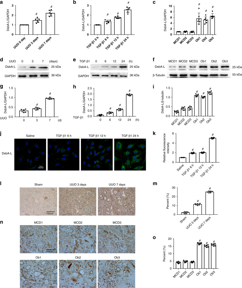
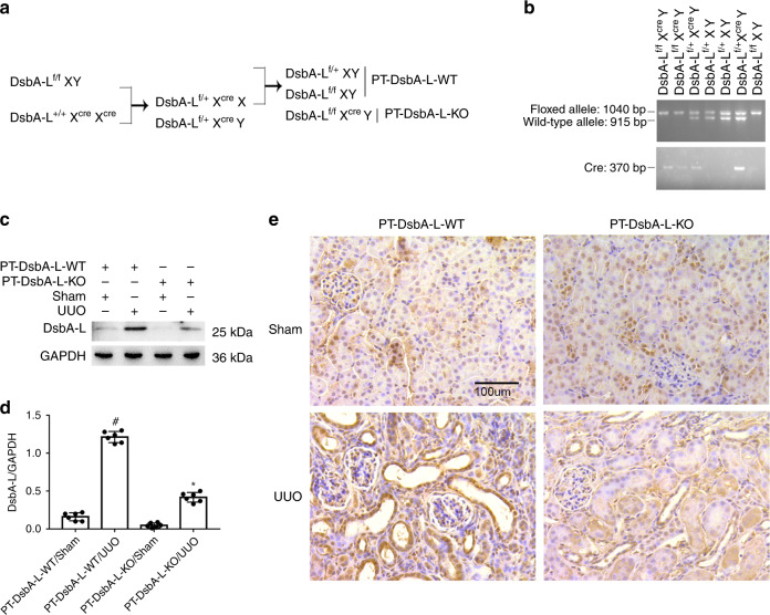
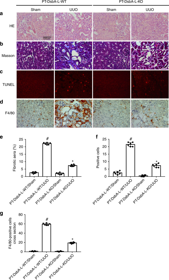
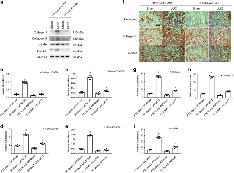
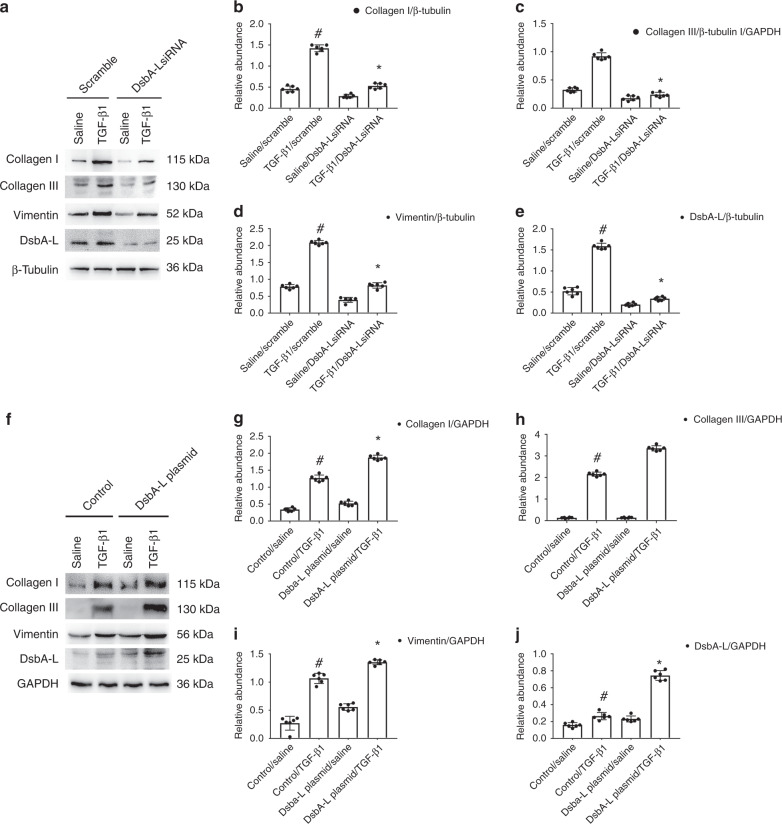
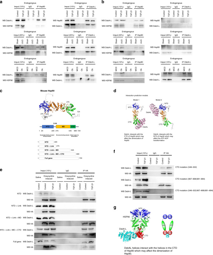
Recent studies have reported that upregulation of disulfide-bond A oxidoreductase-like protein (DsbA-L) prevented lipid-induced renal injury in diabetic nephropathy (DN). However, the role and regulation of proximal tubular DsbA-L for renal tubulointerstitial fibrosis (TIF) remains unclear. In current study, we found that a proximal tubules-specific DsbA-L knockout mouse (PT-DsbA-L-KO) attenuated UUO-induced TIF, renal cell apoptosis and inflammation. Mechanistically, the DsbA-L interacted with Hsp90 in mitochondria of BUMPT cells which activated the signaling of Smad3 and p53 to produce connective tissue growth factor (CTGF) and then resulted in accumulation of ECM of BUMPT cells and mouse kidney fibroblasts. In addition, the progression of TIF caused by UUO, ischemic/reperfusion (I/R), aristolochic acid, and repeated acute low-dose cisplatin was also alleviated in PT-DsbA-L-KO mice via the activation of Hsp90 /Smad3 and p53/CTGF axis. Finally, the above molecular changes were verified in the kidney biopsies from patients with obstructive nephropathy (Ob). Together, these results suggest that DsbA-L in proximal tubular cells promotes TIF via activation of the Hsp90 /Smad3 and p53/CTGF axis.


Conflict of interest statement

The authors declare no competing interests.

Figures

Fig. 1
Fig. 1. Induction of DsbA-L in BUMPT cells treated by TGF-β1 and the kidneys of UUO mice and Ob patients.
af RT-qPCR and immunoblot analysis of DsbA-L and GAPDH in BUMPT cells treated with or without TGF-β1 and the kidneys of UUO mice and Ob patients. g–i Analysis of the grayscale image between them. j, l, n Immunofluorescence or immunohistochemical staining of DsbA-L in BUMPT cells treated with or without TGF-β1 as well as in the kidneys of both UUO mice and Ob patients. k, m, o Quantification of DsbA-L staining. Original magnification ×400. Scale bar: 100 µM. Data are expressed as means ± s.d. (n = 6). #P < 0.05: versus saline group, sham group, or MCD group. Each experiment (j, l, n) was repeated six times independently with similar results. ac, gi, k, m, o indicate the statistical Student’s t test used (means ± s.d., n = 6, P < 0.05).
Fig. 2
Fig. 2. Generation and features of the PT-DsbA-L-KO mouse model.
a Breeding procedure for the creation of PT-DsbA-L-KO mice. b PCR-based genotyping of wild-type and floxed alleles of DsbA-L and PEPCK-Cre allele. c Cortices and outer medulla of kidneys from PT-DsbA-L-KO and PT-DsbA-L-WT littermate mice following UUO injury were collected for immunoblot analysis of DsbA-L and GAPDH. d Analysis of the grayscale image between them. e Immunohistochemical staining of DsbA-L in the kidney cortical tissues of wild-type and DsbA-L-KO mice following UUO injury. Original magnification ×400. Scale bar: 100 µM. Data are expressed as means ± s.d. (n = 6). #P < 0.05 versus sham group. *P < 0.05 versus PT-DsbA-L-WT with UUO group. Each experiment (c, e) was repeated six times independently with similar results. d indicate the statistical Student’s t test used (means ± s.d., n = 6, P < 0.05).
Fig. 3
Fig. 3. Amelioration of UUO-induced renal fibrosis and tubular cell apoptosis in PT-DsbA-L-KO mice.
The left ureter of PT-DsbA-L-KO and PT-DsbA-L-WT littermate mice was ligated for 7 days to establish a UUO model. a Hematoxylin and eosin staining. b Representative Masson’s trichrome staining. c Representative sections of TUNEL-positive cells. d Immunohistochemistry of F4/80 staining. e Quantification of tubulointerstitial fibrosis in the kidney cortex. f The number of TUNEL-positive cells. g Quantification of the F4/80-positive cells in the kidney cortex. Original magnification ×400. Scale bar: 100 µM. Data are expressed as means ± s.d. (n = 6). #P < 0.05 versus sham group. *P < 0.05 versus PT-DsbA-L-WT with the UUO group. Each experiment (ad) was repeated six times independently with similar results. eg indicate the statistical Student’s t test used (means ± s.d., n = 6, P < 0.05).
Fig. 4
Fig. 4. Attenuation of UUO-induced expression of Col I&III and ɑ-SMA in PT-DsbA-L-KO mice.
The left ureter of PT-DsbA-L-KO and PT-DsbA-L-WT littermate mice was ligated for 7 days to establish a UUO model. a The immunoblot analysis of Col I&III, ɑ-SMA, and DsbA-L. be Analysis of the grayscale image between them. f Immunohistochemical staining of Col I&III and ɑ-SMA. gi Quantification of immunohistochemical staining. Original magnification ×400. Scale bar:100 µM. Data are expressed as means ± s.d. (n = 6). #P < 0.05 versus sham group. *P < 0.05 versus PT-DsbA-L-WT with the UUO group. Each experiment (a, f) was repeated six times independently with similar results. be, gi indicate the statistical Student’s t test used (means ± s.d., n = 6, P < 0.05).
Fig. 5
Fig. 5. DsbA-L-mediated the TGF-β1-induced expression of Col I&III and vimentin in BUMPT cells.
The plasmid and siRNA of DabA-L were transfected into BUMPT cells and treated with or without 5 ng/ml TGF-β1 for 24 h. a, f Immunoblot analysis of Col I&III, vimentin, and DsbA-L. b–e, gj Analysis of the gray scale image between them. Data are expressed as means ± s.d. (n = 6). #P < 0.05 versus scramble with the saline group. *P < 0.05 versus scramble with the TGF-β1 group. Each experiment (a, f) was repeated six times independently with similar results. be, gj indicate the statistical Student’s t test used (means ± s.d., n = 6, P < 0.05).
Fig. 6
Fig. 6. Interaction between DsbA-L and Hsp90 in BUMPT cells treated with TGF-β1 and the kidneys of UUO mice and Ob patients.
a, b The whole or mitochondrial lysate was extracted for reciprocal co-immunoprecipitation of DsbA-L and Hsp90 in BUMPT cells treated with TGF-β1 and the kidneys of UUO mice and Ob patients. c The structure and five sections of Hsp90. d Interaction of a predictive models. e, f Anti-HA immunoprecipitates were analyzed for HA and DsbA-L by immunoblotting. g The model of DsbA-L interaction with CTD of Hsp90β. Each experiment (a, b, e, f) was repeated six times independently with similar results.

References

    1. Qi R, Yang C. Renal tubular epithelial cells: the neglected mediator of tubulointerstitial fibrosis after injury. Cell Death Dis. 2018;9:1126. - PMC - PubMed
    1. Gewin LS. Renal fibrosis: primacy of the proximal tubule. Matrix Biol. 2018;68–69:248–262. - PMC - PubMed
    1. Forbes MS, et al. Fight-or-flight: murine unilateral ureteral obstruction causes extensive proximal tubular degeneration, collecting duct dilatation, and minimal fibrosis. Am. J. Physiol. Ren. Physiol. 2012;303:F120–F129. - PMC - PubMed
    1. Forbes MS, Thornhill BA, Chevalier RL. Proximal tubular injury and rapid formation of atubular glomeruli in mice with unilateral ureteral obstruction: a new look at an old model. Am. J. Physiol. Ren. Physiol. 2011;301:F110–F117. - PMC - PubMed
    1. Li H, et al. Atg5-mediated autophagy deficiency in proximal tubules promotes cell cycle G2/M arrest and renal fibrosis. Autophagy. 2016;12:1472–1486. - PMC - PubMed

Publication types

MeSH terms

Substances