. 2020 Oct 29;103(5):951-965.
doi: 10.1093/biolre/ioaa143.

Increased FOXL2 expression alters uterine structures and functions†

Affiliations

Increased FOXL2 expression alters uterine structures and functions†

Rong Li et al. Biol Reprod. .

Abstract

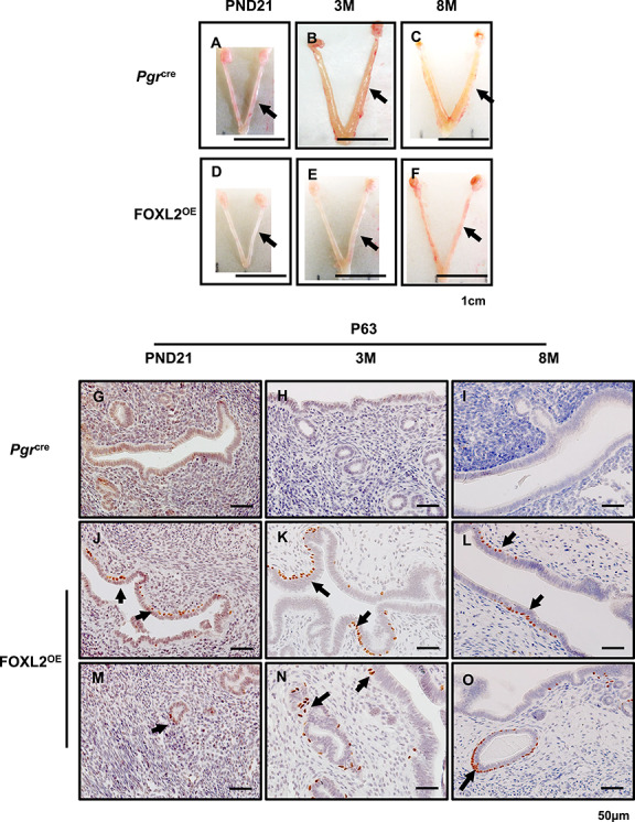
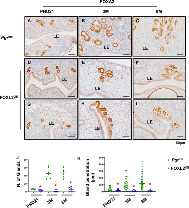
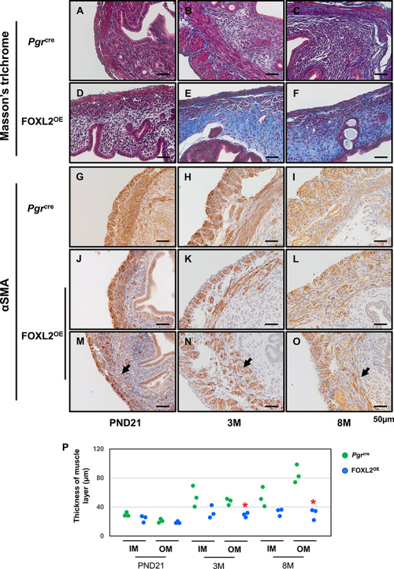
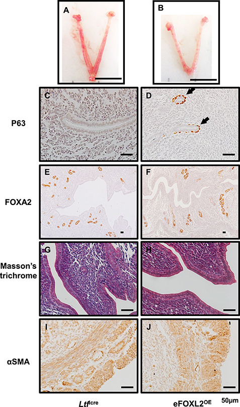
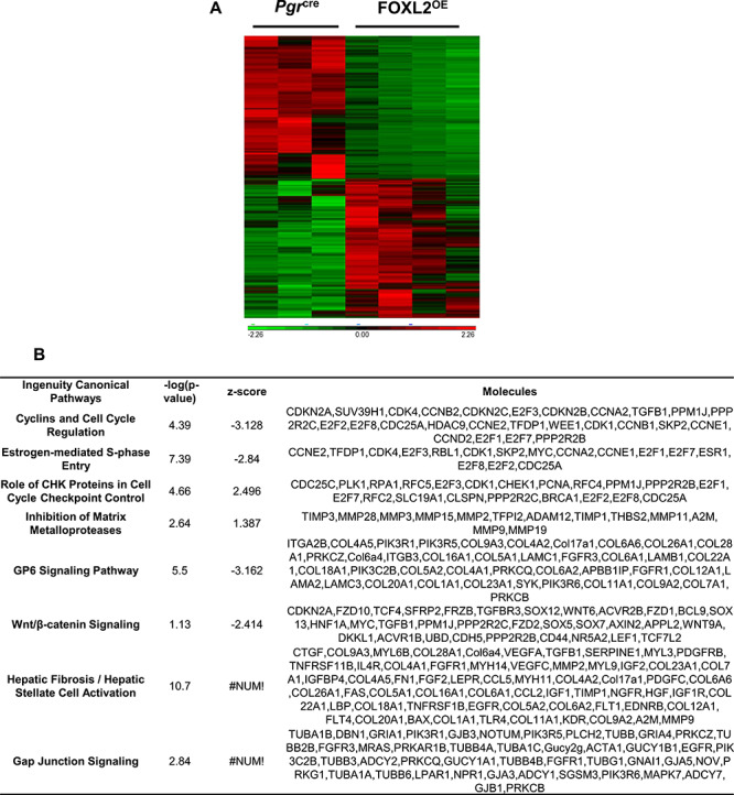
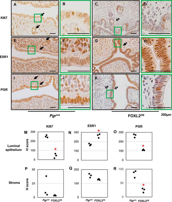
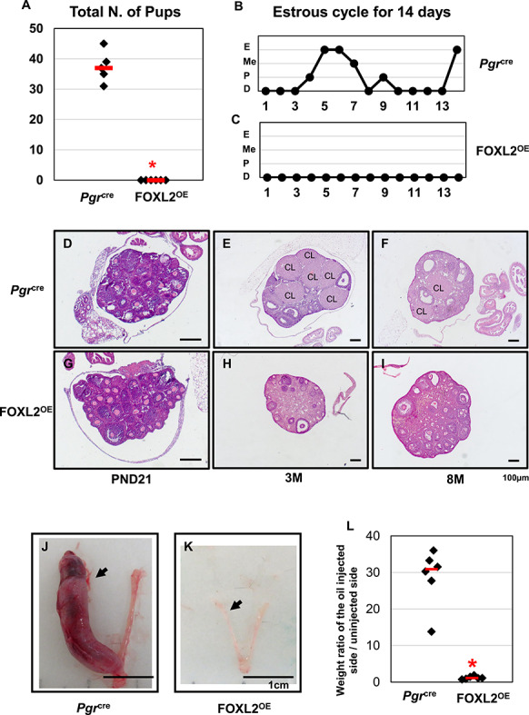
The transcription factor forkhead box L2 (FOXL2) regulates sex differentiation and reproductive function. Elevated levels of this transcription factor have been observed in the diseases of the uterus, such as endometriosis. However, the impact of elevated FOXL2 expression on uterine physiology remains unknown. In order to determine the consequences of altered FOXL2 in the female reproductive axis, we generated mice with over-expression of FOXL2 (FOXL2OE) by crossing Foxl2LsL/+ with the Progesterone receptor Pgrcre model. FOXL2OE uterus showed severe morphological abnormality including abnormal epithelial stratification, blunted adenogenesis, increased endometrial fibrosis, and disrupted myometrial morphology. In contrast, increasing FOXL2 levels specifically in uterine epithelium by crossing the Foxl2LsL/+ with the lactoferrin Ltficre mice resulted in the eFOXL2OE mice with uterine epithelial stratification but without defects in endometrial fibrosis and adenogenesis, demonstrating a role of the endometrial stroma in the uterine abnormalities of the FOXL2OE mice. Transcriptomic analysis of 12 weeks old Pgrcre and FOXL2OE uterus at diestrus stage showed multiple signaling pathways related with cellular matrix, wnt/β-catenin, and altered cell cycle. Furthermore, we found FOXL2OE mice were sterile. The infertility was caused in part by a disruption of the hypophyseal ovarian axis resulting in an anovulatory phenotype. The FOXL2OE mice failed to show decidual responses during artificial decidualization in ovariectomized mice demonstrating the uterine contribution to the infertility phenotype. These data support that aberrantly increased FOXL2 expressions in the female reproductive tract can disrupt ovarian and uterine functions.

Keywords: FOXL2; adenogenesis; fibrosis; myometrial; stratification.


Figures

Figure 1
Figure 1
FOXL2OE mice displayed thin uterus and stratified uterine epithelium. The representative uterine images of Pgrcre (A–C) and FOXL2OE mice (D–F) at PND21 (A and D), 3 M (B and E), and 8 M (C and F). The uteri were much thinner in FOXL2OE mice compared with Pgrcre mice at 3 M and 6 M. Basal cell marker, P63, staining in the uterus of Pgrcre (G–I) and FOXL2OE mice (J–O) at PND21 (G, J, and M), 3 M (H, K, and N), and 6 M (I, L, and O). P63 positive basal cells in the uterus indicate epithelium stratification. They were found at the basal side of some luminal epithelium (J–L) and some glandular cells (M–O) in the FOXL2oe uterus. PND: postnatal day; M: months old. Arrow indicates stratified epithelium. N = 3 for each genotype and age group. Arrow in A–F refers one uterine horn. Arrow in J–O indicates stratified epithelium.
Figure 2
Figure 2
Disrupted adenogenesis in FOXL2OE mice. FOXA2 labeled glands in Pgrcre (A–C) and FOXL2OE (D–I) uterus at PND21 (A, D, and G), 3 M (B, E, and H), and 8 M (C, F, and I). The number of glands was calculated in two cross sections per mouse, in total six mice (J). The penetration of glands was defined as the closest distance of the glands to the adjacent luminal epithelium and calculated in one longitudinal section per mouse, in total six mice (K). PND: postnatal day; M: months old. *P < 0.05. FOXL2OE compared with Pgrcre mice at the same age.
Figure 3
Figure 3
Stroma fibrosis and defective myometrium in FOXL2OE mice. Masson’s trichrome staining in Pgrcre (A–C) and FOXL2oe (D–F) uterus at PND21 (A and D), 3 M (B and E), 8 M (C and F). Increased blue staining in the stroma of FOXL2OE uterus suggested increased collagen deposition. αSMA staining in Pgrcre (G–I) and FOXL2OE (J–O) uterus at PND21 (G, J, and M), 3 M (H, K, and N), 8 M (I, L, and O). The thickness of the inner (circular) and outer (longitudinal) muscle layer was calculated in one cross-section of each mouse (P). The thickness of outer layer was decreased at 3 M and 8 M and the inner muscle layer was discontinued in the FOXL2OE uterus. PND: postnatal day; M: months old; IM: Inner muscle layer; OM: Outer muscle layer. The arrow indicates the discontinued muscle layer. N = 3 for each genotype and age group. *P < 0.05, FOXL2OE compared with Pgrcre mice at the same age.
Figure 4
Figure 4
eFOXL2OE exhibited epithelium stratification but no adenogenesis, stroma fibrosis, and myometrial defects. The representative uterine images of Ltficre (A) and eFOXL2OE mice (B) at Pregnancy D3.5. P63 (C and D), FOXA2 (E and F), Masson’s trichrome (G and H), and αSMA (I and J) staining in Ltficre (C, E, G, and I) and eFOXL2OE (D, E, H, and J) uterus. Basal cells with P63 positive staining was detected in the luminal and glandular epithelium of eFOXL2OE uterus. No changes were observed in FOXA2 labeled uterine glands, Masson’s trichrome staining, and αSMA labeled muscle layers. Arrow indicates stratified epithelium. N = 3.
Figure 5
Figure 5
Transcriptomic changes of Pgrcre and FOXL2OE at 3 M diestrus stage (P < 0.05, fold change > 1.5, <−1.5). Heating map showed cluster of differentially expressed genes in Pgrcre and FOXL2OE (A). Ingenuity pathway analysis identified top altered signaling pathways (B). N = 3–4.
Figure 6
Figure 6
Protein expressions of KI67, ESR1, and PGR in FOXL2OE uterus at 3 M diestrus stage. Immunohistochemistry of KI67 (A–D), ESR1 (E–H), PGR (I–L) in Pgrcre (A, B, E, F, I, and J), and FOXL2OE (C, D, G, H, K, and L) uterus. H-score of the immunohistochemistry of Ki67 (M and P), ESR1 (N and Q), and PGR (O and R) in the uterine luminal epithelium and stroma showed ESR1 was increased in the epithelium, PGR and KI67 were decreased in the epithelium of the FOXL2OE uterus. Solid arrow indicates cells with strong staining. Open arrow indicates cells with weak staining. N = 3.
Figure 7
Figure 7
FOXL2OE mice were infertile with abolished decidual responses. All the Pgrcre mice but none of the FOXL2OE mice delivered any pups during 6-month breeding trial (A). Representative pictures of estrous cycle showed continuous diestrus in FOXL2OE mice (C) in contrast to the estrous cycle showed in Pgrcre mice (B) during 14-day period. Representative ovarian histology pictures of Pgrcre (D–F) and FOXL2OE (G–I) virgin mice at diestrus stage at the age of PND21 (D and G), 3 M (E and H), and 8 M (F and I). FOXL2OE ovaries were depleted of corpus luteum. The representative pictures of the hormone primed uterus after 5-day oil injections in the Pgrcre (J) and FOXL2OE (K) mice. The weight ratio of the oil-injected side over the un-injected side uterine horn (L) confirmed that the decidualization occurred in all the Pgrcre but none of the FOXL2OE mice. Arrow indicates the oil-injected side. D: diestrus; P: proestrus; Me: metestrus; E: estrus; PND: postnatal day; M: months old; CL: corpus luteum. The arrow indicates oil injected uterine horn. N = 6 for each genotype and age group.

Similar articles

Cited by

References

    1. Ellsworth BS, Egashira N, Haller JL, Butts DL, Cocquet J, Clay CM, Osamura RY, Camper SA. FOXL2 in the pituitary: Molecular, genetic, and developmental analysis. Mol Endocrinol 2006; 20:2796–2805. - PubMed
    1. Egashira N, Takekoshi S, Takei M, Teramoto A, Osamura RY. Expression of FOXL2 in human normal pituitaries and pituitary adenomas. Mod Pathol 2011; 24:765–773. - PubMed
    1. Governini L, Carrarelli P, Rocha AL, Leo VD, Luddi A, Arcuri F, Piomboni P, Chapron C, Bilezikjian LM, Petraglia F. FOXL2 in human endometrium: Hyperexpressed in endometriosis. Reprod Sci 2014; 21:1249–1255. - PubMed
    1. Brody JR, Histologic CGR. Morphometric, and Immunocytochemical analysis of Myometrial development in rats and mice. 1. Normal Development. American Journal of Anatomy 1989; 186:1–20. - PubMed
    1. Schmidt D, Ovitt CE, Anlag K, Fehsenfeld S, Gredsted L, Treier AC, Treier M. The murine winged-helix transcription factor Foxl2 is required for granulosa cell differentiation and ovary maintenance. Development 2004; 131:933–942. - PubMed

Publication types

Substances

Supplementary concepts