. 2021 Feb;20(1):281-291.
doi: 10.1007/s10237-020-01383-8. Epub 2020 Sep 19.

Evaluation of nucleus pulposus fluid velocity and pressure alteration induced by cartilage endplate sclerosis using a poro-elastic finite element analysis

Affiliations

Evaluation of nucleus pulposus fluid velocity and pressure alteration induced by cartilage endplate sclerosis using a poro-elastic finite element analysis

Chaudhry Raza Hassan et al. Biomech Model Mechanobiol. 2021 Feb.

Abstract

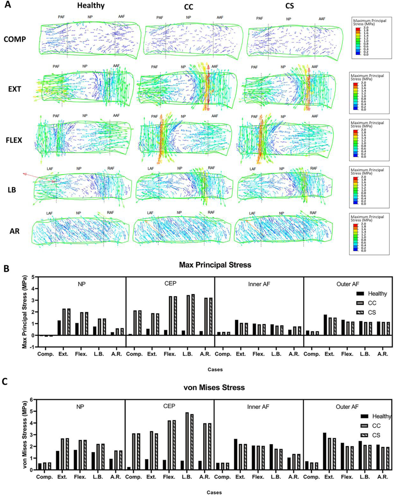
The nucleus pulposus (NP) in the intervertebral disk (IVD) depends on diffusive fluid transport for nutrients through the cartilage endplate (CEP). Disruption in fluid exchange of the NP is considered a cause of IVD degeneration. Furthermore, CEP calcification and sclerosis are hypothesized to restrict fluid flow between the NP and CEP by decreasing permeability and porosity of the CEP matrix. We performed a finite element analysis of an L3-L4 lumbar functional spine unit with poro-elastic constitutive equations. The aim of the study was to predict changes in the solid and fluid parameters of the IVD and CEP under structural changes in CEP. A compressive load of 500 N was applied followed by a 10 Nm moment in extension, flexion, lateral bending, and axial rotation to the L3-L4 model with fully saturated IVD, CEP, and cancellous bone. A healthy case of L3-L4 physiology was then compared to two cases of CEP sclerosis: a calcified cartilage endplate and a fluid constricted sclerotic cartilage endplate. Predicted NP fluid velocity increased for the calcified CEP and decreased for the calcified + less permeable CEP. Decreased NP fluid velocity was prominent in the axial direction through the CEP due to a less permeable path available for fluid flux. Fluid pressure and maximum principal stress in the NP were predicted to increase in both cases of CEP sclerosis compared to the healthy case. The porous medium predictions of this analysis agree with the hypothesis that CEP sclerosis decreases fluid flow out of the NP, builds up fluid pressure in the NP, and increases the stress concentrations in the NP solid matrix.

Keywords: Cartilage endplate sclerosis; Disk degeneration; Finite element analysis; Fluid pressure; Fluid velocity.


Figures

Fig. 1:
Fig. 1:
(A) L3-4 lumbar spine 3D surface geometry was extracted from a healthy human male CT scans and meshed to obtain hexahedral mesh. Ligaments were added using truss elements. (B) Sagittal cross-section of L3-4 spine shows an internal structure including cartilage endplate and nucleus pulposus.
Fig. 2:
Fig. 2:
(A) Intradiscal pressure (IDP) and (B) loss in height of L3-4 lumbar spine under 500 N of compression. The load was applied in 1 s and held for 15 mins for creep response. Maximum predicted pressure was 0.56 MPa, which decreased to 0.30 MPa after creep. Loss in IVD height was 0.15 mm at the end of 15 mins creep.
Fig. 3:
Fig. 3:
(A) Stress tensor flow maps of the L3-4 disc under compression (COMP), extension (EXT), flexion (FLEX), lateral bending (LB), and axial rotation (AR) for healthy, calcified cartilage (CC), and constricted sclerotic (CS) cases. COMP, EXT, and FLEX are shown in the sagittal view while LB and AR are shown in a coronal view. Predicted (B) maximum principal and (C) von Mises stress in the nucleus pulposus (NP), inner and outer annulus fibrosus (AF), and cartilage endplate (CEP). Both stresses increased in calcified sclerotic cases. (PAF: posterior annulus fibrosus; AAF: anterior annulus fibrosus; LAF: left annulus fibrosus; RAF: right annulus fibrosus; NP: nucleus pulposus).
Fig. 4:
Fig. 4:
(A) Flow maps of the L3-4 disc under compression (COMP), extension (EXT), flexion (FLEX), lateral bending (LB), and axial rotation (AR) for healthy, calcified cartilage (CC), and constricted sclerotic (CS) cases. COMP, EXT, and FLEX are shown in the sagittal view while LB and AR are shown in a coronal view. (B) Maximum predicted fluid velocity in the nucleus pulposus (NP), inner and outer annulus fibrosus (AF), and cartilage endplate (CEP). (PAF: posterior annulus fibrosus; AAF: anterior annulus fibrosus; LAF: left annulus fibrosus; RAF: right annulus fibrosus; NP: nucleus pulposus).
Fig. 5:
Fig. 5:
(A) Contour plots the L3-4 disc under compression (COMP), extension (EXT), flexion (FLEX), lateral bending (LB), and axial rotation (AR) for healthy, calcified cartilage (CC), and constricted sclerotic (CS) cases. COMP, EXT, and FLEX are shown in the sagittal view while LB and AR are shown in a coronal view. (B) Maximum predicted pore fluid pressure in the nucleus pulposus (NP), inner and outer annulus fibrosus (AF), and cartilage endplate (CEP). Maximum fluid pressure increased in NP and inner AF but decreased in outer AF for all diseased cases. (PAF: posterior annulus fibrosus; AAF: anterior annulus fibrosus; LAF: left annulus fibrosus; RAF: right annulus fibrosus; NP: nucleus pulposus).
Fig. 6:
Fig. 6:
Range of motion (ROM) of L3-4 lumbar spine under extension (EXT), flexion (FLEX), lateral bending (LB), and axial rotation (AR) for healthy, calcified cartilage (CC), and constricted sclerotic (CS) cases. ROM decreased for calcified cases compared to healthy.

References

    1. Adams M, McNally D, Dolan P (1996) ‘Stress’ distribution inside intervertebral discs J Bone Joint Surg Br 78:965–972 - PubMed
    1. Ayotte DC, Ito K, Perren SM, Tepic S (2000) Direction-dependent constriction flow in a poroelastic solid: the intervertebral disc valve J Biomech Eng 122:587–593 doi:10.1115/1.1319658 - DOI - PubMed
    1. Bian Q et al. (2016) Excessive Activation of TGFβ by Spinal Instability Causes Vertebral Endplate Sclerosis Scientific reports 6:27093–27093 doi:10.1038/srep27093 - DOI - PMC - PubMed
    1. Brinckmann P, Grootenboer H (1991) Change of Disc Height, Radial Disc Bulge, and Intradiscal Pressure From Discectomy An in Vitro Investigation on Human Lumbar Discs Spine 16:641–646 - PubMed
    1. Dreischarf M et al. (2014) Comparison of eight published static finite element models of the intact lumbar spine: predictive power of models improves when combined together Journal of biomechanics 47:1757–1766 - PubMed

MeSH terms

LinkOut - more resources