. 2020 Nov:183:104933.
doi: 10.1016/j.antiviral.2020.104933. Epub 2020 Sep 17.

A synthetic STING agonist inhibits the replication of human parainfluenza virus 3 and rhinovirus 16 through distinct mechanisms

Affiliations

A synthetic STING agonist inhibits the replication of human parainfluenza virus 3 and rhinovirus 16 through distinct mechanisms

Qingyuan Zhu et al. Antiviral Res. 2020 Nov.

Abstract

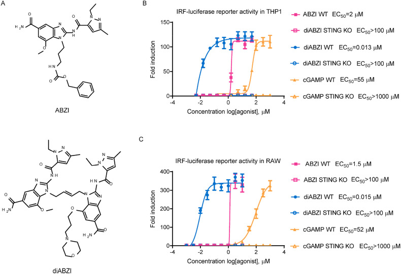
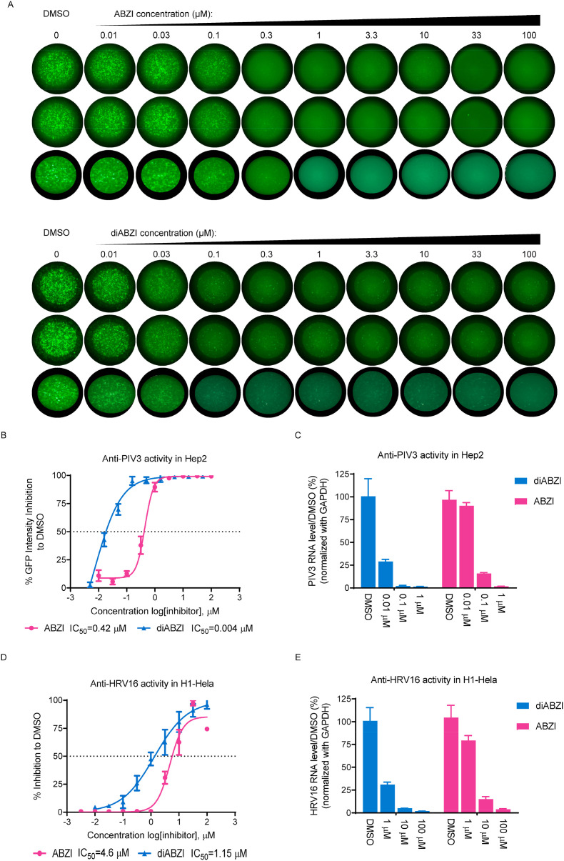
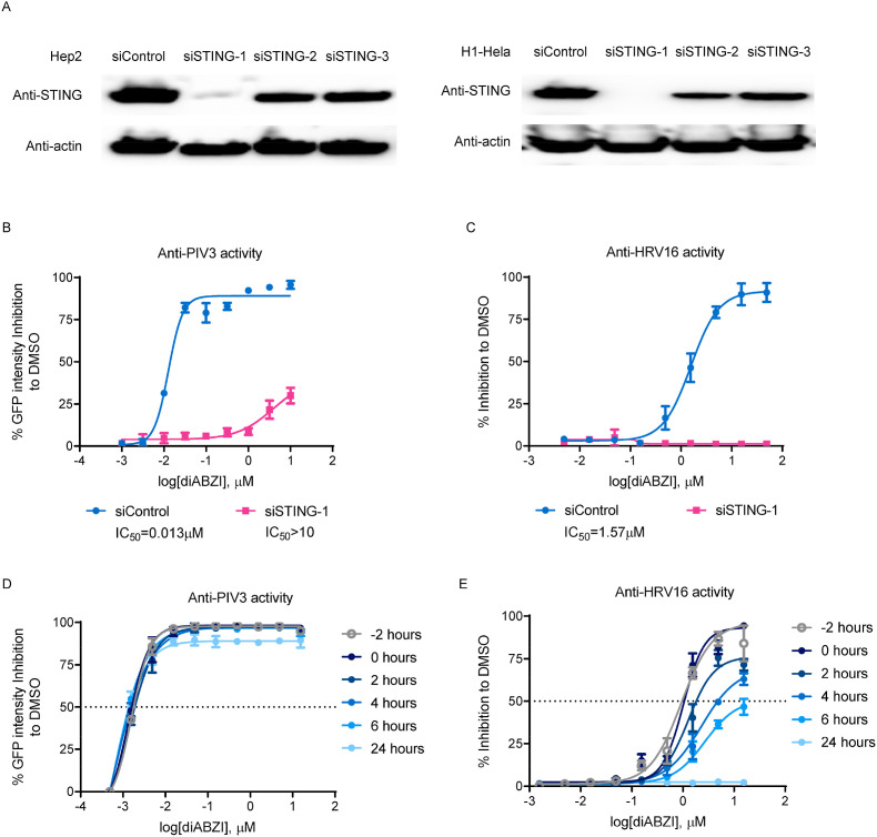
Stimulator of interferon genes (STING), as a signaling hub in innate immunity, plays a central role for the effective initiation of host defense mechanisms against microbial infections. Upon binding of its ligand cyclic dinucleotides (CDNs) produced by the cyclic GMP-AMP synthase (cGAS) or invading bacteria, STING is activated, leading to the induction of both type I interferon responses and autophagy, which are critical for the control of certain microbial infections. RNA viruses, such as Parainfluenza virus (PIV) and Rhinovirus (HRV), are among the leading causes of respiratory infections that affect human health without effective treatments. Activation of STING pathway may provide a new therapeutic approach fighting against these viruses. However, the role of STING in the control of RNA virus infection remains largely unexplored. In this study, using dimeric amidobenzimidazole (diABZI), a newly discovered synthetic small molecule STING receptor agonist with much higher potency than CDNs, we found that activation of STING elicits potent antiviral effects against parainfluenza virus type 3 (PIV3) and human rhinovirus 16 (HRV16), two representative respiratory viral pathogens. Notably, while anti-PIV3 activity was depend on the induction of type I interferon responses through TANK-binding kinase 1 (TBK1), anti-HRV16 activity required the induction of autophagy-related gene 5 (ATG5)-dependent autophagy, indicating that two distinct antiviral mechanisms are engaged upon STING activation. Antiviral activity and individual specific pathway was further confirmed in infected primary bronchial epithelial cells. Our findings thus demonstrate the distinct antiviral mechanisms triggered by STING agonist and uncover the potential of therapeutic effect against different viruses.

Keywords: Autophagy; Parainfluenza virus; Rhinovirus; STING agonist; diABZI.


Conflict of interest statement

The authors are employees of F. Hoffman-La Roche Ltd.

Figures

Fig. 1
Fig. 1
STING agonist stimulates the IRF pathway via STING in both human and mouse cells. (A) The chemical structures of ABZI-based STING receptor agonists. (B) Wild-type or STING knockout human THP-1 or (C) mouse RAW264.7 IRF-inducible luciferase reporter cells were treated with indicated compounds at various concentrations for 24 h and the culture supernatants were collected for luciferase activity determination. The fold induction relative to DMSO treatment was plotted. The EC50 values were calculated using Prism software. The data are presented as the means ± SDs of triplicate samples from one experiment and are representative of at least three independent experiments.
Fig. 2
Fig. 2
STING agonist inhibits PIV and HRV replication. (A) Hep2 cells were seeded in triplicate wells of 96-well plates. The cells were infected with PIV3-GFP at MOI of 0.01 and treated with different doses of the indicated compounds for 48 h. Green fluorescence images of each well were acquired with a fluorescence ELISpot reader. (B) The GFP signal intensity was quantified in each well using AID ELISpot Software. The percent inhibition relative to DMSO treatment was plotted. (C) 24 h after PIV3-GFP infection (MOI = 0.01), total cellular RNA was isolated and reverse transcribed to cDNA. Viral RNA and GAPDH mRNA levels were then quantified via RT-PCR. The PIV3 RNA levels were normalized to the GAPDH mRNA leveland the percentage inhibition relative to DMSO treatment were plotted. (D) H1–HeLa cells were infected with HRV16 at MOI of 0.01 and treated with indicated concentrations of ABZI or diABZI for 72 h. The CPE was determined by a CCK-8 assay and the percent inhibition relative to DMSO treatment was plotted. (E) 24 h after HRV16 infection (MOI = 0.01), total cellular RNA was isolated and reverse transcribed to cDNA. Viral RNA and GAPDH mRNA levels were then quantified via RT-PCR. The HRV16 RNA levels were normalized to the GAPDH mRNA leveland the percentage inhibition relative to DMSO treatment were plotted. The IC50 values were calculated using Prism software. The data are presented as the means ± SDs of triplicate samples from one experiment and are representative of at least three independent experiments.
Fig. 3
Fig. 3
The Antiviral activity of the STING receptor agonist depends on STING expression. (A) Hep2 (left panel) or H1–HeLa (right panel) cells were transfected with 20 pM STING-targeted siRNA (siSTING-1, 2, or 3) or control siRNA (siControl) and the knockdown efficiency was determined by western blotting after 48 h. (B) Hep2 or (C) H1–HeLa cells were transfected with 20 pM STING-targeted siSTING-1 for 24 h and were infected with PIV3-GFP at MOI of 0.01 (Hep2) or with HRV16 at an MOI of 0.01 (H1–HeLa) and treated with diABZI at the indicated concentrations for 48 h (Hep2) or 72 h (H1–HeLa). To determine the anti-PIV activity, the GFP signal intensity was quantified (left panel). To determine the anti-HRV activity, the CPE was assessed by a CCK-8 assay (right panel). The percent inhibition relative to DMSO treatment was plotted. (D) Hep2 cells were infected with PIV3-GFP at an MOI of 0.01 and treated with different doses of diABZI at the indicated times. After 48 h, the GFP signal intensity was quantified and the percent inhibition relative to DMSO treatment was plotted. (E) H1–HeLa cells were infected with HRV16 at an MOI of 0.01 and treated with different doses of diABZI at the indicated times. After 72 h, the CPE was determined by a CCK-8 assay and the percent inhibition relative to DMSO treatment was plotted. The data are presented as the means ± SDs of triplicate samples from one experiment and are representative of at least three independent experiments.
Fig. 4
Fig. 4
The STING receptor agonist inhibits PIV and HRV through distinct mechanisms. (A) H1–HeLa (upper panels) or Hep2 (lower panels) cells were treated with the indicated concentrations of diABZI or diABZI derivative for 24 h. Cells were lysed and lysates were subjected to Western blot analysis of LC3, p-IRF3 and actin with specific antibodies. (B) Hep2 or (C) H1–HeLa cells were infected with PIV3-GFP at an MOI of 0.01 and treated with the indicated concentrations of diABZI in the presence or absence of 1 μM (Hep2)/5 μM (H1–HeLa) of TBK1 inhibitor (TBKi, BX795) or 50 μM (Hep2)/25 μM (H1–HeLa) CQ for 48 h. The GFP signal intensity was quantified and the percent inhibition relative to DMSO treatment was plotted. (D) H1–HeLa cells were infected with HRV16 at an MOI of 0.01 and treated with the indicated concentrations of diABZI in the presence or absence of 5 μM TBKi (BX795) or 25 μM CQ for 72 h. The CPE was determined by a CCK-8 assay and the percent inhibition relative to DMSO treatment was plotted. The data are presented as the means ± SDs of triplicate samples from one experiment and are representative of at least three independent experiments. *P < 0.05, **P < 0.01.
Fig. 5
Fig. 5
The STING agonist exhibits anti-PIV3 and anti-HRV16 activity in PBECs. (A) PBECs were seeded in triplicate wells of collagen-coated 96-well plates, infected with PIV3-GFP at an MOI 0.001 and treated with the indicated concentrations of diABZI for 48 h. Green fluorescence images of each well were acquired with a fluorescence ELISpot reader. (B) The GFP signal intensity was quantified and the percent inhibition relative to DMSO treatment was plotted. The IC50 values were calculated using Prism software. (C) PBECs were infected with HRV16 at an MOI of 10 and treated with the indicated concentrations of diABZI for 6 h or 24 h. Total cellular RNA was isolated and reverse transcribed to cDNA. Viral RNA and GAPDH mRNA were then quantified via RT-PCR. The HRV RNA level was normalized to the GAPDH mRNA level and the percent inhibition relative to DMSO treatment was plotted. (D) PBECs were seeded on collagen-coated 96-well plates, then infected with PIV3-GFP at an MOI 0.001 and treated with the indicated concentrations of diABZI in the presence or absence of CQ (50 μM)/IFNR antibody (anti-IFNR mAb 5 μg/ml)/isotype control antibody (control mAb, 5 μg/ml) for 48 h. The GFP signal intensity was quantified and the percent inhibition relative to DMSO treatment was plotted. (E) PBECs were infected with HRV16 at an MOI of 10 and treated with 30 μM of diABZI for 24 h in the presence or absence of CQ (50 μM)/IFNR antibody (anti-IFNR mAb 5 μg/ml)/isotype control antibody (control mAb, 5 μg/ml). Total cellular RNA was isolated and reverse transcribed to cDNA. Viral RNA and GAPDH mRNA were then quantified via RT-PCR. The HRV RNA level was normalized to the GAPDH mRNA level and the percent inhibition relative to DMSO treatment was plotted. The data are presented as the means ± SD of triplicate samples from one experiment and are representative of at least three independent experiments. *P < 0.05, **P < 0.01.

References

    1. Aguirre S., Maestre A.M., Pagni S., Patel J.R., Savage T., Gutman D., Maringer K., Bernal-Rubio D., Shabman R.S., Simon V., Rodriguez-Madoz J.R., Mulder L.C., Barber G.N., Fernandez-Sesma A. DENV inhibits type I IFN production in infected cells by cleaving human STING. PLoS Pathog. 2012;8 - PMC - PubMed
    1. Atreya P.L., Kulkarni S. Respiratory syncytial virus strain A2 is resistant to the antiviral effects of type I interferons and human MxA. Virology. 1999;261:227–241. - PubMed
    1. Barber G.N. STING: infection, inflammation and cancer. Nat. Rev. Immunol. 2015;15:760–770. - PMC - PubMed
    1. Bloom B., Cohen R.A., Freeman G. Summary health statistics for U.S. Children: national health interview survey, 2008. Vital Health Stat. 2009;10:1–81. - PubMed
    1. Bossios A., Psarras S., Gourgiotis D., Skevaki C.L., Constantopoulos A.G., Saxoni-Papageorgiou P., Papadopoulos N.G. Rhinovirus infection induces cytotoxicity and delays wound healing in bronchial epithelial cells. Respir. Res. 2005;6:114. - PMC - PubMed

Publication types

MeSH terms