. 2020 Oct:60:102991.
doi: 10.1016/j.ebiom.2020.102991. Epub 2020 Sep 16.

Dengue and Zika virus infections are enhanced by live attenuated dengue vaccine but not by recombinant DSV4 vaccine candidate in mouse models

Affiliations

Dengue and Zika virus infections are enhanced by live attenuated dengue vaccine but not by recombinant DSV4 vaccine candidate in mouse models

Rahul Shukla et al. EBioMedicine. 2020 Oct.

Abstract

Background: A tetravalent live attenuated dengue vaccine, Dengvaxia, sensitised naïve recipients to severe dengue illness upon a subsequent natural dengue infection and is suspected to be due to antibody-dependent enhancement (ADE). ADE has also been implicated in the severe neurological outcomes of Zika virus (ZIKV) infection. It has become evident that cross-reactive antibodies targeting the viral pre-membrane protein and fusion-loop epitope are ADE-competent. A pre-clinical tetravalent dengue sub-unit vaccine candidate, DSV4, eliminates these ADE-competent epitopes.

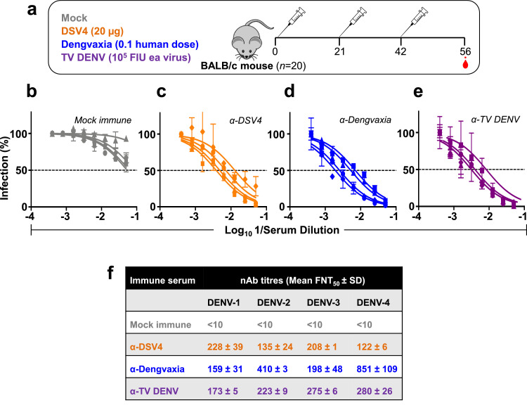
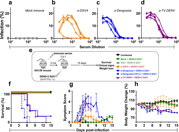
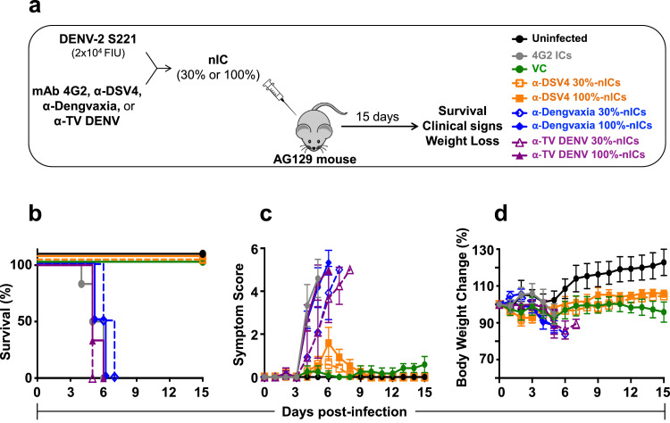
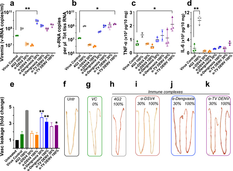
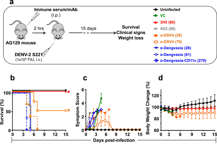
Methods: We compared protective efficacy and ADE-competence of murine polyclonal antibodies induced by DSV4, Dengvaxia and an 'in house' tetravalent mixture of all four laboratory DENV strains, TV DENV, using established mouse models.

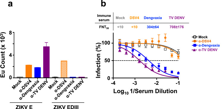
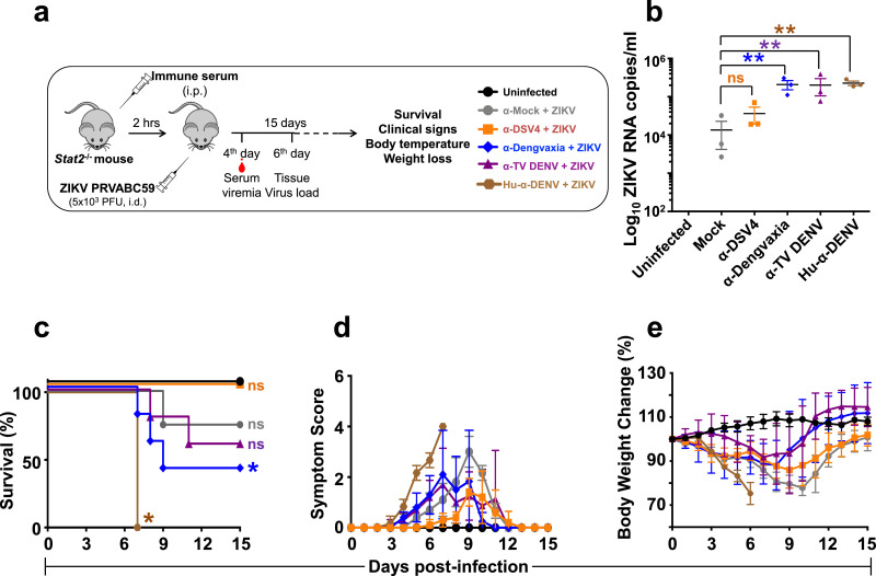
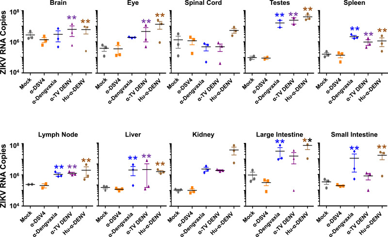
Findings: DSV4-induced antibodies, known to be predominantly type-specific, provided significant protection against lethal DENV challenge, but did not promote ADE of either DENV or ZIKV infection in vivo. Antibodies elicited by Dengvaxia and TV DENV, which are predominantly cross-reactive, not only failed to offer protection against lethal DENV challenge, but also promoted ADE of both DENV and ZIKV infection in vivo.

Interpretation: Protective efficacy against DENV infection may be linked to the induction of neutralising antibodies which are type-specific rather than cross-reactive. Whole virus-based dengue vaccines may be associated with ADE risk, despite their potent virus-neutralising capacity. Vaccines designed to eliminate ADE-competent epitopes may help eliminate/minimise ADE risk.

Funding: This study was supported partly by ICGEB, India, the National Biopharma Mission, DBT, Government of India, Sun Pharmaceutical Industries Limited, India, and NIAID, NIH, USA.

Keywords: AG129; Antibody-dependent enhancement; C57BL/6 Stat2(−/−); DSV4; Dengue VLP vaccine; Dengue virus; Dengvaxia; LAV; Zika virus.


Figures

Fig. 1:
Fig. 1
Generation of immune sera in BALB/c mice. (a) Schematic representation of the vaccination schedule. Groups of BALB/c mice (n=20) were immunised (i.m.) with three doses of (i) DSV4 (20 μg/dose, formulated in alum); (ii) Dengvaxia (1/10th the human dose); or (iii) TV DENV (a tetravalent mixture containing WHO reference strains of DENV-1, DENV-2, DENV-3 and DENV-4, 105 FIU each). Mice were bled 2 weeks after the last dose for seroanalysis. A fourth group of mice (Mock, n=20) did not receive any vaccine (these received PBS plus alum). (b-e) The graphs depict DENV nAb titration curves determined using mock-immune serum (grey curves, panel ‘b’), α-DSV4 antiserum (orange curves, panel ‘c’), α-Dengvaxia antiserum (blue curves, panel ‘d’) and α-TV DENV antiserum (purple curves, panel ‘e’), against DENV-1 (squares), DENV-2 (circles), DENV-3 (triangles) and DENV-4 (diamonds). (f) The mean nAb titres (± SD, standard deviation) against the four DENV serotypes (DENV-1, -2, -3 and -4), for each immune serum, based on the DENV neutralisation curves shown in panels ‘b-e’. The nAb titres in the mock immune sera, depicted as <10, denote that there was no discernible DENV neutralisation, even at the lowest serum dilution tested, which was 1:10 (For interpretation of the references to colour in this figure legend, the reader is referred to the web version of this article).
Fig. 2:
Fig. 2
Analysis of the capacity of anti-DSV4, anti-Dengvaxia and anti-TV DENV immune sera to enhance DENV infection in vitro and in vivo. (a–d) The graphs depict infection profiles of K562 cells by DENV-1 (squares), DENV-2 (circles), DENV-3 (triangles) and DENV-4 (diamonds) as a function of dilution of mock- (grey curves, panel ‘a’), α-DSV4- (orange curves, panel ‘b’), α-Dengvaxia- (blue curves, panel ‘c’) and α-TV DENV (purple curves, panel ‘d’) immune sera (raised in BALB/c mice), determined by flow cytometry. (e-h) Groups of AG129 mice (n=5) were administered either mAb 4G2 (grey curves), mock- (green curves), DSV4- (orange curves), Dengvaxia- (blue curves) or TV DENV- (purple curves) immune sera by passive transfer (10 µg mAb or 20 µl immune serum in a final volume of 200 µl 1x PBS), then challenged with a sub-lethal dose of DENV-2 S221 (panel ‘e’) and monitored for survival (panel ‘f’), clinical signs (panel ‘g’) and body weight change (panel ‘h’) over the next 15 days. A group of mice which received no treatment (Uninfected, black curves) was monitored in parallel for comparison. Survival data (in panel ‘f’) were analysed by Log-Rank (Mantel-Cox) test for significant difference in survival rates. Survival of mice which received either mock immune serum or α-DSV4 before challenge was not significantly different from that of Uninfected mice (100% survival). In comparison, survival was significantly reduced (p<0.05) in mice which received α-Dengvaxia immune serum and very significantly reduced (p<0.002) in mice which received α-TV DENV immune serum or mAb 4G2. In panel ‘g’, clinical scores were based on a 5 point system, with the maximum score denoting death: 0.5, mild ruffled fur; 1.0, ruffled fur; 1.5, compromised eyes; 2, compromised eyes with hunched back; 2.5, loose stools; 3.0, limited movement; 3.5, no movement/hind leg paralysis; 4.0, euthanised if cumulative score was 5. In panel ‘h’, body weight was monitored twice a day in the morning and evening, and the mean taken for plotting. Note: in panels ‘e-h’ a second pool of anti-Dengvaxia immune serum (α-Dengvaxia-CD11c+DENV-2 S221, dashed blue curves), raised by immunising CD11c-Ifnar1−/− mice, was also tested (all other murine immune sera are from BALB/c mice) (For interpretation of the references to colour in this figure legend, the reader is referred to the web version of this article).
Fig. 3:
Fig. 3
Evaluation of in vivo ADE capacity of BALB/c immune sera by inoculation of nICs into AG129 mice. Groups of AG129 mice (n=6) were given intravenous (retro-orbital) injections of nICs, generated by pre-incubating 20,000 FIUs of DENV-2 S221 with α-DSV4 antiserum, α-Dengvaxia antiserum or α-TV DENV antiserum, on day 0 (panel ‘a’). The amounts of antisera used were such that the virus in the nIC was either fully (100%, using ~5 µl immune serum) or partially (30%, using 0.08–0.32 µl immune serum) neutralised (based on residual infectivity measured using the Vero cell-based FACS assay). In parallel, control groups of mice which received no treatment (Uninfected), only the virus in the absence of any antiserum (virus control, VC) or nICs (containing 100% neutralised virus) generated using the FLE-specific CR mAb 4G2 (10 µg), were also included. The mice were monitored for survival (panel ‘b’, Kaplan-Meir survival analysis), clinical symptoms (panel ‘c’) and body weight change (panel d’), for up to 15 days post nIC inoculation. In panels ‘b–d’, the data for the different groups are indicated as follows: the Uninfected (black curves), VC (green curves) and mAb4G2-IC (grey curves) groups are shown by solid curves in different colours as indicated. For groups receiving nICs made using α-DSV4 (orange curves), α-Dengvaxia (blue curves) or α-TV DENV (purple curves) antisera, those receiving the 100% and 30% neutralised ICs, are indicated by solid and dashed curves, respectively. Survival data (panel ‘b’) were analysed by Log-Rank (Mantel-Cox) test for significant difference in survival rates. Survival of mice in α-DSV4 30% and 100% nIC groups was not significantly different from that of uninfected mice. In comparison, survival of mice in all the other experimental groups was significantly compromised (p≤0.001). Clinical scoring in panel ‘c’ and body weight measurements in panel ‘d’, were as described in Fig. 2 legend (For interpretation of the references to colour in this figure legend, the reader is referred to the web version of this article).
Fig. 4:
Fig. 4
Analysis of serum viremia, virus load, cytokine production and vascular leakage in the small intestines of nIC-inoculated AG129 mice. Groups of mice (n=3) corresponding to each of the experimental groups described in Fig. 3 were set up in parallel and euthanised on day 4 post-nIC inoculation for serum viremia determination (panel ’a’), determination of virus load (panel ‘b’), TNF-α (panel ‘c’) and IL 6 (panel ‘d’) production and vascular leakage (quantitative analysis in panel ‘e’; qualitative analysis in panel ‘f-k’) in the small intestines. Vascular leakage was quantified by measuring the amount of Evan's blue extravasation into the small intestines by spectrophotometry. Vascular leakage is expressed as fold change in small intestinal dye content with reference to that in the ‘Untreated’ mice, taken as 1.0. The single and double asterisks (panels ‘a-e’; in all cases, the test groups are compared to the ‘Virus Control’ group) denote statistically significant and very significant differences, respectively, with reference to the virus control group (using the two-way ANOVA test with Bonferroni correction). In all other nIC groups, differences with reference to the cognate virus control group were not significant. The percent neutralisation of the challenge virus is indicated as 0 (panel ‘g’), 30 or 100 (panels ‘h-k’) (For interpretation of the references to colour in this figure legend, the reader is referred to the web version of this article).
Fig. 5:
Fig. 5
The capacity of α-DSV4 and α-Dengvaxia immune sera to confer protection against lethal DENV-2 S221 challenge. (a) Schematic representation of the experimental design. Groups of AG129 mice (n=3 or 4) were administered (i.p) BALB/c immune sera (in two dosage levels, 100 µl and 300 µl/mouse) or mAbs (100 µg/mouse). The immune sera used were anti-DSV4 (α-DSV4, orange) and anti-Dengvaxia (α-Dengvaxia, blue) and the mAbs used were a DENV-2 TS mAb (3H5, red) and a pan-DENV CR mAb (4G2, grey). Two hours after passive transfer the mice were bled (for the determination of circulating DENV-2 nAb titers, shown in parenthesis in the legends in panel ‘a’), and then challenged shortly thereafter with a lethal dose (105 FIU/mouse) of DENV-2 S221. All groups were monitored over the next 15 days for survival (Kaplan-Meir survival curves, panel ‘b’), clinical symptoms (panel ‘c’) and body weight change (panel ‘d’). In parallel, one group received just the challenge virus (VC, green), and another group received neither passive antibody transfer nor the challenge virus (Uninfected, black). In panel ‘b-d’, data for mice which received 100 µl and 300 µl immune sera are shown using dashed and solid curves, respectively, in the indicated colours. Survival data (panel ‘b’) were analysed by Log-Rank (Mantel-Cox) test for significant difference in survival rates [the asterisks denote that survival in the 3H5 and α-DSV4 (70) groups were significantly higher in comparison to the 4G2 group (p<0.05), for both]. Clinical scoring in panel ‘c’ and body weight measurements in panel ‘d’, were as described in Fig. 2 legend. Note: a second pool of anti-Dengvaxia immune serum (α-Dengvaxia-CD11c, dashed blue curves), raised by immunising CD11c-Ifnar1−/− mice, was also tested for its efficacy against lethal DENV-2 S221 challenge (other murine immune sera are from BALB/c mice) (For interpretation of the references to colour in this figure legend, the reader is referred to the web version of this article).
Fig. 6:
Fig. 6
The capacity of α-DSV4, α-Dengvaxia and α-TV DENV immune sera to bind to recombinant ZIKV E and EDIII proteins and neutralise ZIKV in vitro. (a) TRF-IA of pooled BALB/c mock-immune sera (mock, grey), anti-DSV4 (α-DSV4, orange), anti-Dengvaxia (α-Dengvaxia, blue) and anti-TV DENV (α-TV-DENV, purple) antisera using insect cell-expressed recombinant ZIKV E protein (ZIKV E) and yeast-expressed ZIKV EDIII (ZIKV EDIII) as the capture antigens. Bound total antibodies were detected using anti-mouse IgG-Eu3+ conjugate by TRF. (b) The graph depicts nAb titration curves determined using mock-immune serum (grey curve), α-DSV4 antiserum (orange curve), α-Dengvaxia antiserum (blue curve) and α-TV DENV antiserum (purple curve), against ZIKV PRVABC59. The mean ZIKV nAb titers (FNT50 ± SD) are shown above the graph (For interpretation of the references to colour in this figure legend, the reader is referred to the web version of this article).
Fig. 7:
Fig. 7
The capacity of α-DSV4, α-Dengvaxia and α-TV immune sera to cause ADE of ZIKV infection in C57BL/6 Stat2−/− mice. (a) Groups (n=6-8) of C57BL/6 Stat2−/− mice were administered mock- (grey), α-DSV4- (orange), α-Dengvaxia- (blue), α-TV DENV- (purple) immune sera or dengue patient plasma (‘Hu-α-DENV’, brown), by passive transfer (20 µl/mouse), two hours before infection with ZIKV strain PRVABC59. A subset (n=3) of challenged mice were bled on day 4 (see panel ‘b’) and euthanised on day 6 post ZIKV challenge (see Fig. 8). The rest of the challenged mice were monitored for up to 15 days (see panels ‘c-e’). (b) Day 4 post infection ZIKV viral RNA in serum, isolated from a subset (n=3) of mice of each of the experimental groups shown in panel ‘a’, was quantified by RT-qPCR as described in methods. ZIKV RNA levels were determined with reference to an in vitro-transcribed RNA standard. Each data point denotes viremia level in a single mouse. The horizontal lines represent the mean viremia levels. ‘ns’ (not significant) and the twin asterisk symbol (very significant), denote statistical significance between the groups indicated, determined by two-way ANOVA with Bonferroni's correction. The rest of the infected mice were monitored for survival (panel ‘c’, ‘ns’ and the single asterisk denote ‘not significant’ and ‘significant’ survival differences for each of the groups with reference to survival of ‘Uninfected’ mice which was 100%), clinical signs (panel ‘d’) and body weight change (panel ‘e’), over the next 15 days. A group of mice which received no treatment (Uninfected, black curves) was monitored in parallel for comparison. In panel ‘d’, clinical scores were based on a 4-point system to grade signs of illness: 0, no symptom; 0.5, mild ruffling of fur; 1.0, ruffled fur; 1.5, compromised eyes; 2.0, hunched back position; 2.5, very limited movement/one leg paralysis; 3.0, no movement and paralysis of both hind legs; 4.0, dead or euthanised. In panel ‘e’, body weight was monitored daily at the same time and expressed as a percent of initial body weight (For interpretation of the references to colour in this figure legend, the reader is referred to the web version of this article).
Fig. 8:
Fig. 8
Determination of systemic virus loads in several organs of C57BL/6 Stat2−/− mice infected with ZIKV PRVABC59 in the presence of α-DSV4, α-Dengvaxia and α-TV DENV immune sera. A subset of mice (n=3) from the different immune sera passive transfer groups in the experiment described in Fig. 7, were euthanised on day 6 following ZIKV infection. Equal amounts of total RNA, extracted from the indicated tissues of these mice, were reverse-transcribed to cDNA and subjected to qPCR. Viral RNA genomes were quantified with reference to an in vitro-transcribed RNA standard. Each data point denotes tissue RNA level in a single mouse. The horizontal lines represent the mean tissue virus load. The double asterisks indicate statistical significance (two-way ANOVA with Bonferroni correction) of the data for group above which they are placed compared to that of the ‘Mock’ group. All other groups did not differ significantly in comparison to the mock group.

References

    1. Pierson TC, Diamond MS. Flaviviruses. In: Knipe DM, Howley PM, editors. Fields Virology. 6th ed. Wolters Kluwer and Lippincott Williams & Wilkins; Philadelphia, PA: 2013. pp. 747–794.
    1. Bhatt S, Gething PW, Brady OJ. The global distribution and burden of dengue. Nature. 2013;496:504–507. - PMC - PubMed
    1. Halstead SB. Neutralization and antibody-dependent enhancement of dengue viruses. Adv Virus Res. 2003;60:421–467. - PubMed
    1. Guzman MG, Alvarez M, Halstead SB. Secondary infection as a risk factor for dengue hemorrhagic fever/dengue shock syndrome: an historical perspective and role of antibody-dependent enhancement of infection. Arch Virol. 2013;158:1445–1459. - PubMed
    1. Taylor A, Foo SS, Bruzzone R, Dinh LV, King NJC, Mahalingam S. Fc receptors in antibody-dependent enhancement of viral infections. Immunol Reviews. 2015;268:340–364. - PMC - PubMed

MeSH terms