Review
. 2021 May;39(5):519-538.
doi: 10.1016/j.tibtech.2020.08.007. Epub 2020 Sep 16.

Enhancing Biopolymer Hydrogel Functionality through Interpenetrating Networks

Affiliations
Review

Enhancing Biopolymer Hydrogel Functionality through Interpenetrating Networks

Abhishek P Dhand et al. Trends Biotechnol. 2021 May.

Abstract

Traditional hydrogels are strong candidates for biomedical applications; however, they may suffer from drawbacks such as weak mechanics, static properties, and an inability to fully replicate aspects of the cellular microenvironment. These challenges can be addressed through the incorporation of second networks to form interpenetrating polymer network (IPN) hydrogels. The objective of this review is to establish clear trends on the enhanced functionality achieved by incorporating secondary networks into traditional, biopolymer-based hydrogels. These include mechanical reinforcement, 'smart' systems that respond to external stimuli, and the ability to tune cell-material interactions. Through attention to network structure and chemistry, IPN hydrogels may advance to meet challenging criteria for a wide range of biomedical fields.

Keywords: biopolymers; cell–material interactions; double networks; interpenetrating network hydrogels; mechanical reinforcement; stimuli-responsive materials.


Figures

Figure 1. Key Figure
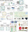
Figure 1. Key Figure. Overview of Interpenetrating Polymer Network (IPN) Hydrogels Employed for Applications in Tissue Engineering, Regenerative Medicine, Drug Delivery, and In Vitro Disease Models
In comparison with conventional single network hydrogels, the incorporation of secondary networks to form IPN hydrogels (two or more independent, yet entangled networks shown as green and blue) often results in advantageous properties such as enhanced mechanical strength, the ability to respond to stimuli, and mimicry of the native cellular microenvironment. The tuning of polymer components (concentration, charge, molecular weight, degradability, cell-adhesion ligand density) and control over network parameters (crosslinking density, network ratio, and sequence of formation) allow for the synthesis of functional IPN hydrogels with diverse properties. IPN hydrogels can be processed into various shapes and architectures as cell-laden or acellular structures using a variety of techniques (i.e., 3D printing, electrospinning, microfluidics, and lithography).
Figure 2.
Figure 2.
Mechanical Reinforcement of Hydrogels through an Interpenetrating Secondary Network. (A) Schematic representation of energy dissipation facilitated by rupture of a sacrificial primary network under tensile loading. (B) Investigation of the load-bearing capacity and crack resistance of a notched double network (DN) hydrogel via tensile stretching. The hydrogel was comprised of a chitosan/polyacrylamide DN soaked under alkaline conditions to yield a chitosan microcrystalline network (PAM-CS-A DN). Adapted, with permission, from [35] (C) Chitosan/gelatin/phytate (C-G-P) formed an interpenetrating polymer network (IPN) hydrogel (i.e., conjoined network) through noncovalent interactions. Graphical representation of tensile stress-strain curve for DN and conjoined network hydrogel. Adapted, with permission, from [36] (D) Design of an alginate/polyacrylamide (PAAm) DN tissue adhesive consisting of a dissipative matrix (made of ionically and covalently crosslinked networks) and adhesive surface through a bridging polymer with primary amine groups. In vivo hydrogel adhesion performance on a beating porcine heart with exposed blood. Adapted, with permission, from [41].
Figure 3.
Figure 3.
Building ‘Smart’ Hydrogels through an Interpenetrating Polymer Network (IPN) Approach. (A) Schematic illustration of the release and delivery of a therapeutic payload (bioactive drugs, proteins, growth factors, nucleotides, cells, spheroids) via a stimuli-responsive IPN hydrogel. The release can be facilitated either via: (i) changes in the hydrogel mesh size, or (ii) through selective network degradation. (B) (i) Temperature-induced collapse of Poly(N-isopropylacrylamide) (PNIPAAm) [at temperature higher than lower critical solution temperature (LCST)] results in tighter physical entanglement between an IPN of PNIPAAm and heparin and increased release of vascular endothelial growth factor (VEGF) when compared with single network hydrogels. Adapted, with permission, from [47]. (ii) Schematic illustration of IPN hydrogel comprised of poly(glycerol sebacate)-co-poly(ethylene glycol)-g-catechol (PEGSD) and ureido-pyrimidinone (UPy) modified gelatin. Gel–sol transition of the IPN hydrogel occurs when irradiated with near infrared (NIR) (808 nm) or increase in temperature (37°C). Adapted, with permission, from [76]. (C) Shear-thinning and self-healing behavior of IPN hydrogels that permit extrusion through a syringe. Whereas single networks [methacrylated hyaluronic acid (MeHA)] disperse rapidly when injected into an aqueous environment, the double network [supramolecular adamantane (Ad)-HA and β-cyclodextrin (CD)-HA guest–host (GH) network and covalently crosslinked MeHA network) is stable due to the presence of reversible physical bonds. Adapted, with permission, from [70]. (D) Schematic representation of components [alginate and poly(ethylene glycol) diacrylate (PEGDA)] for a 3D printable double network hydrogel ink. The 3D printed hydrogel constructs in the shape of ear, nose, hollow cube, porous mesh, and twisted bundle. Adapted, with permission, from [72].
Figure 4.
Figure 4.
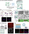
Interpenetrating Network Hydrogels Designed to Tune and Study Cell–Material Interactions. (A) Schematic illustration of enhanced cell behavior within an interpenetrating polymer network (IPN) hydrogel. (B) (i) Schematic of dynamic, viscoelastic IPN hydrogel formed by the combination of guest–host (GH) interactions [between adamantine- and cyclodextrin-modified hyaluronic acid (HA)] and covalent crosslinking [methacrylated hyaluronic acid (MeHA)]. Cytoskeletal staining (i.e., actin) indicates increased cell spreading as a function of the concentration of the viscous GH network. Adapted, with permission, from [98] (ii) Design of viscoplastic IPN hydrogels consisting of alginate and reconstituted basement membrane matrix. Enhanced matrix plasticity promoted increased spreading and motility of MDA-MB-231 breast cancer cells, independent of protease activity (arrows indicate differences in molecular weight). Adapted, with permission, from [95]. (C) Schematic representation of photopatterning or photostiffening IPN hydrogels. (i) Hydrogel discs formed from hydrazide-aldehyde-modified HA and covalently crosslinked norbornene-modified HA were photopatterned with thiolated rhodamine. Adapted, with permission, from [75]. (ii) Photo-micropatterning of IPN (bright) and semi-IPN (dark) regions within glycidyl methacrylate-modified HA/self-assembling peptide (Puramatrix®). Adapted, with permission, from [100]. (D) Microvasculature-on-a-chip model fabricated using agarose/gelatin IPN hydrogel. Human umbilical vein endothelial cells seeded on the microfluidic device attach to gelatin and appropriately remodel the microenvironment, as seen in vivo, illustrated with confocal images after 14 days for cadherin and the basement membrane proteins laminin and collagen IV. Adapted, with permission, from [102].
Figure I.
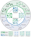
Figure I.. Synthesis of Interpenetrating Polymer Network (IPN) Hydrogels.
IPN synthesis techniques can be broadly classified into sequential (swelling of first network in a secondary monomer/macromer) or simultaneous (orthogonal crosslinking of both first and second networks). The selection of biopolymer and type of crosslinking chemistry utilized for each network influences the interactions between networks within IPN hydrogels. Representative modes of physical crosslinking (top, green) include ionic crosslinking (e.g., alginate, chitosan), temperature-induced coil–helix transition (e.g., agar, gelatin), hydrogen bonding (e.g., cellulose, dextran), and hydrophobicity (e.g., guest–host-modified biopolymers). Chemicals such as glutaraldehyde, genipin, or carbodiimide-based crosslinking can be used for the direct crosslinking of biopolymers via primary amines on biopolymers such as gelatin, chitosan, or collagen. Biopolymers can also be chemically modified with functional groups for covalent crosslinking (bottom, blue), such as acylhydrazone, diol-boronic acid, Schiff-base, Michael addition, Diels-Alder, disulfide linkage, azide-alkyne or thiol-ene/thiol-yne click chemistry, and free-radical polymerization (e.g., methacrylates, methacrylamides).

References

    1. Zhang YS and Khademhosseini A (2017) Advances in engineering hydrogels. Science 356, 3627 - PMC - PubMed
    1. Neves SC et al. (2020) Leveling up hydrogels: hybrid systems in tissue engineering. Trends Biotechnol. 38, 292–315 - PubMed
    1. Gong JP (2010) Why are double network hydrogels so tough? Soft Matter 6, 2583–2590
    1. Costa AMS and Mano JF (2015) Extremely strong and tough hydrogels as prospective candidates for tissue repair - a review. Eur. Polym. J 72, 344–364
    1. Higa K et al. (2016) Fundamental biomaterial properties of tough glycosaminoglycan-containing double network hydrogels newly developed using the molecular stent method. Acta Biomater. 43, 38–49 - PubMed

Publication types

MeSH terms