Review
. 2020 Oct:37:101696.
doi: 10.1016/j.redox.2020.101696. Epub 2020 Aug 27.

ROS systems are a new integrated network for sensing homeostasis and alarming stresses in organelle metabolic processes

Affiliations
Review

ROS systems are a new integrated network for sensing homeostasis and alarming stresses in organelle metabolic processes

Yu Sun et al. Redox Biol. 2020 Oct.

Abstract

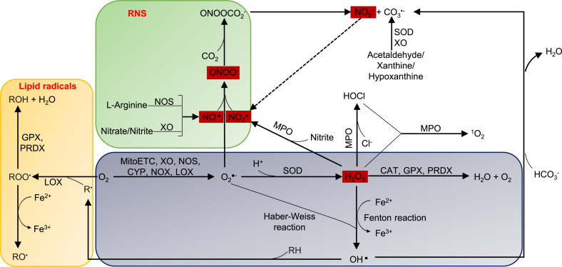
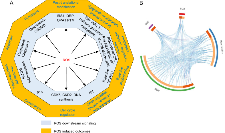
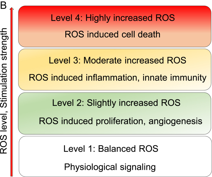
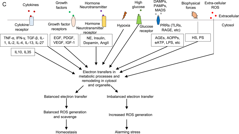
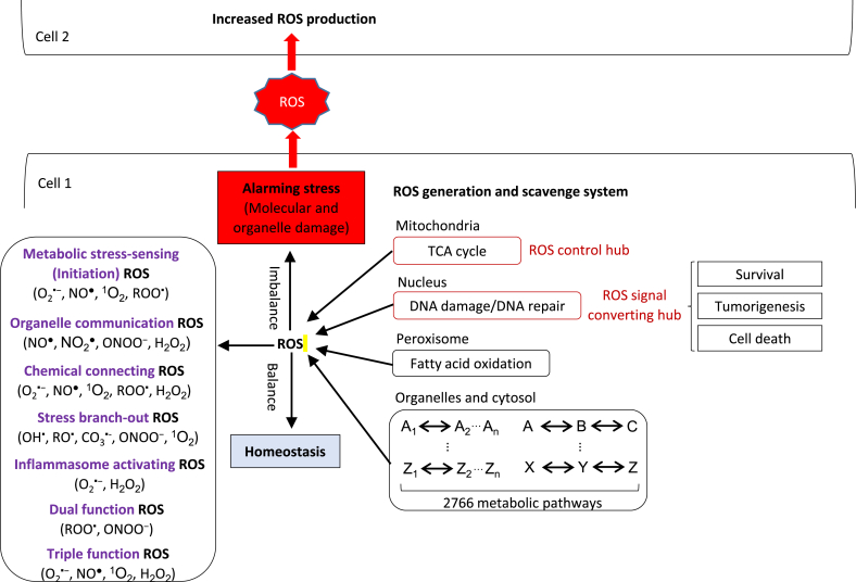
Reactive oxygen species (ROS) are critical for the progression of cardiovascular diseases, inflammations and tumors. However, the mechanisms of how ROS sense metabolic stress, regulate metabolic pathways and initiate proliferation, inflammation and cell death responses remain poorly characterized. In this analytic review, we concluded that: 1) Based on different features and functions, eleven types of ROS can be classified into seven functional groups: metabolic stress-sensing, chemical connecting, organelle communication, stress branch-out, inflammasome-activating, dual functions and triple functions ROS. 2) Among the ROS generation systems, mitochondria consume the most amount of oxygen; and nine types of ROS are generated; thus, mitochondrial ROS systems serve as the central hub for connecting ROS with inflammasome activation, trained immunity and immunometabolic pathways. 3) Increased nuclear ROS production significantly promotes cell death in comparison to that in other organelles. Nuclear ROS systems serve as a convergent hub and decision-makers to connect unbearable and alarming metabolic stresses to inflammation and cell death. 4) Balanced ROS levels indicate physiological homeostasis of various metabolic processes in subcellular organelles and cytosol, while imbalanced ROS levels present alarms for pathological organelle stresses in metabolic processes. Based on these analyses, we propose a working model that ROS systems are a new integrated network for sensing homeostasis and alarming stress in metabolic processes in various subcellular organelles. Our model provides novel insights on the roles of the ROS systems in bridging metabolic stress to inflammation, cell death and tumorigenesis; and provide novel therapeutic targets for treating those diseases. (Word count: 246).

Keywords: A sensing network for metabolic stress; Inflammation; Nuclear signaling; Reactive oxygen species (ROS); Trained immunity.


Conflict of interest statement

None.

Figures

Fig. 1
Fig. 1
Four types of ROS (red box) that are generated by nitrogen and oxygen are communication ROS, which are, connected with different types of ROS systems. NO▪, NO2 ▪, H2O2 and ONOO− have the ability to cross lipid membrane and are classified as communication ROS. Three types of nitrogen-containing ROS are communication ROS. Abbreviations: O2▪−—superoxide, NO▪—nitric oxide radical, NO2 ▪—nitrogen dioxide, OH▪—hydroxyl radical, ROO▪—peroxyl radical, RO▪— alkoxyl radical, CO3▪-—carbonate radical, H2O2—hydrogen peroxide, ONOO−—peroxynitrite, 1O2—singlet oxygen, HOCl—hypochlorous acid, ONOOCO2 −—nitrocarbonate, MitoETC: Mitochondrial electron transport chain, XO—xanthine oxidase, NOS—nitric oxide synthase, CYP— cytochrome P450, NOX—NADPH oxidase, LOX—lipoxygenase, SOD—superoxide dismutase, MPO—myeloperoxidase, MAO— monoaminoxidase. GPX—glutathione peroxidase, PRDX—peroxiredoxin, CAT—catalase. (For interpretation of the references to color in this figure legend, the reader is referred to the Web version of this article.)
Fig. 2
Fig. 2
A. Left panel (a): ROS induces ten types of cell functions (yellow) via various specific downstream pathways (grey). The ten cell functions include: 1) physiological signaling, 2) proliferation, 3) gene regulation, 4) cell cycle regulation, 5) epigenetic modification, 6) post-translational modification, 7) inflammation/innate immunity, 8) pyroptosis, 9) apoptosis, and 10) senescence. Right panel (b): The circus plot indicates that four major ROS generating and anti-ROS regulatory systems have both shared genes and unique genes. On the outside, each arc represents the identity of each gene list。On the inside, each arc represent a gene list, where each gene has a spot on the arc. Dark orange color represents the genes that appear in multiple lists and light orange color represents genes that are unique to that gene list. Purple lines link the same gene that are shared by multiple gene lists. Blue lines link the different genes where they fall into the same ontology them. (also see the detailed information in a supplemental table). Detailed information of Fig. 2A-b see supplemental Table 4. Abbreviations: IRS1—insulin receptor substrate 1, DRP--dynamin-related protein, OPA1--optic atrophy 1, PTM—post-translational modification, H3K14Ac—histone 3 lysine 14 acetylation, PI3K--phosphatidylinositol-3-kinase, Akt--protein kinase B, mTOR--mammalian target of rapamycin, Ras—GTPase, MEK--MAPK kinase, ERK--extracellular signals-regulated kinase, NF-kB--nuclear factor kappa-lightchain- enhancer of activated B cells, MAPK-- mitogen-activated protein kinases, JNK-- MAPK-Jun N-terminal kinase, Nrf-- Nuclear factor erythroid2-related factor, CDK5--cyclin dependent kinase 5, CKD2-- cyclin dependent kinase 2, NLRP3-- NOD-, LRR- and pyrin domain containing protein 3, GSDMD—gasdermin D, SOD-- superoxide dismutase, LOX--lipoxygenase, NOX--NADPH oxidase. B. The ladder indicates different levels of ROS contribute to different cellular functions. In redox homeostasis state, ROS contributes to physiological signaling. Low ROS increase contributes to proliferation and angiogenesis. Moderated ROS increase contributes to inflammatory response and innate immunity. High ROS increase contributes to various types of cell death. C. Seven types of stimulations promote ROS production, including cytokines, growth factors, hormone/neurotransmitters, hypoxia, high glucose, DAMPs and extracellular ROS. Abbreviations: TNF-α--tumor necrosis factor- α, IFN-γ-- interferon gamma, IL-1--interleukin 1, IL-2--interleukin 2, IL-4--interleukin 4, IL-13-- interleukin 13, IL-27--interleukin 27, IL-10--interleukin 10, IL-35--interleukin 35, EGF-- epidermal growth factor, PDGF--Platelet derived growth factor, VEGF-- vascular endothelial growth factor, IGF-1--Insulin-like growth factor 1, NE—Norepinephrine, AngII—angiotensin II, AGE-- AGEsadvanced glycation end products, AOPP-- advanced oxidation protein products, ATP-- adenosine triphosphate, LPS—lipopolysaccharide, HS-- Hemodynamic strain, PS-- Pulmonary stretch.
Fig. 2
Fig. 2
A. Left panel (a): ROS induces ten types of cell functions (yellow) via various specific downstream pathways (grey). The ten cell functions include: 1) physiological signaling, 2) proliferation, 3) gene regulation, 4) cell cycle regulation, 5) epigenetic modification, 6) post-translational modification, 7) inflammation/innate immunity, 8) pyroptosis, 9) apoptosis, and 10) senescence. Right panel (b): The circus plot indicates that four major ROS generating and anti-ROS regulatory systems have both shared genes and unique genes. On the outside, each arc represents the identity of each gene list。On the inside, each arc represent a gene list, where each gene has a spot on the arc. Dark orange color represents the genes that appear in multiple lists and light orange color represents genes that are unique to that gene list. Purple lines link the same gene that are shared by multiple gene lists. Blue lines link the different genes where they fall into the same ontology them. (also see the detailed information in a supplemental table). Detailed information of Fig. 2A-b see supplemental Table 4. Abbreviations: IRS1—insulin receptor substrate 1, DRP--dynamin-related protein, OPA1--optic atrophy 1, PTM—post-translational modification, H3K14Ac—histone 3 lysine 14 acetylation, PI3K--phosphatidylinositol-3-kinase, Akt--protein kinase B, mTOR--mammalian target of rapamycin, Ras—GTPase, MEK--MAPK kinase, ERK--extracellular signals-regulated kinase, NF-kB--nuclear factor kappa-lightchain- enhancer of activated B cells, MAPK-- mitogen-activated protein kinases, JNK-- MAPK-Jun N-terminal kinase, Nrf-- Nuclear factor erythroid2-related factor, CDK5--cyclin dependent kinase 5, CKD2-- cyclin dependent kinase 2, NLRP3-- NOD-, LRR- and pyrin domain containing protein 3, GSDMD—gasdermin D, SOD-- superoxide dismutase, LOX--lipoxygenase, NOX--NADPH oxidase. B. The ladder indicates different levels of ROS contribute to different cellular functions. In redox homeostasis state, ROS contributes to physiological signaling. Low ROS increase contributes to proliferation and angiogenesis. Moderated ROS increase contributes to inflammatory response and innate immunity. High ROS increase contributes to various types of cell death. C. Seven types of stimulations promote ROS production, including cytokines, growth factors, hormone/neurotransmitters, hypoxia, high glucose, DAMPs and extracellular ROS. Abbreviations: TNF-α--tumor necrosis factor- α, IFN-γ-- interferon gamma, IL-1--interleukin 1, IL-2--interleukin 2, IL-4--interleukin 4, IL-13-- interleukin 13, IL-27--interleukin 27, IL-10--interleukin 10, IL-35--interleukin 35, EGF-- epidermal growth factor, PDGF--Platelet derived growth factor, VEGF-- vascular endothelial growth factor, IGF-1--Insulin-like growth factor 1, NE—Norepinephrine, AngII—angiotensin II, AGE-- AGEsadvanced glycation end products, AOPP-- advanced oxidation protein products, ATP-- adenosine triphosphate, LPS—lipopolysaccharide, HS-- Hemodynamic strain, PS-- Pulmonary stretch.
Fig. 3
Fig. 3
A novel working model: 1) ROS are a novel integrated network for sensing homeostasis and alarming stress in organelle- or cytosolic-metabolic stresses; and 2) ROS also serve as cellular communication signaling to increase neighboring cell ROS production. Abbreviations: O2▪−—superoxide, NO▪—nitric oxide radical, NO2 ▪—nitrogen dioxide, OH▪—hydroxyl radical, ROO▪—peroxyl radical, RO▪— alkoxyl radical, CO3▪-—carbonate radical, H2O2—hydrogen peroxide, ONOO−—peroxynitrite, 1O2—singlet oxygen, HOCl—hypochlorous acid, TCA: tricarboxylic acid cycle.

References

    1. Panth N., Paudel K.R., Parajuli K. Reactive oxygen species: a key hallmark of cardiovascular disease. Advances in medicine. 2016;2016 - PMC - PubMed
    1. He F., Zuo L. Redox roles of reactive oxygen species in cardiovascular diseases. Int. J. Mol. Sci. 2015;16(11):27770–27780. - PMC - PubMed
    1. Senoner T., Dichtl W. Oxidative stress in cardiovascular diseases: still a therapeutic target? Nutrients. 2019;11(9):2090. - PMC - PubMed
    1. Yin Y. Early hyperlipidemia promotes endothelial activation via a caspase-1-sirtuin 1 pathway. Arterioscler. Thromb. Vasc. Biol. 2015;35(4):804–816. - PMC - PubMed
    1. Li Y.F. Caspase-1 mediates hyperlipidemia-weakened progenitor cell vessel repair. Front. Biosci. 2016;21:178–191. - PMC - PubMed

Substances

LinkOut - more resources