. 2020 Dec 1:180:108296.
doi: 10.1016/j.neuropharm.2020.108296. Epub 2020 Sep 17.

Sex differences in corticotropin releasing factor peptide regulation of inhibitory control and excitability in central amygdala corticotropin releasing factor receptor 1-neurons

Affiliations

Sex differences in corticotropin releasing factor peptide regulation of inhibitory control and excitability in central amygdala corticotropin releasing factor receptor 1-neurons

Abigail E Agoglia et al. Neuropharmacology. .

Abstract

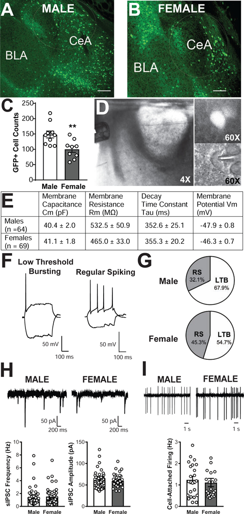
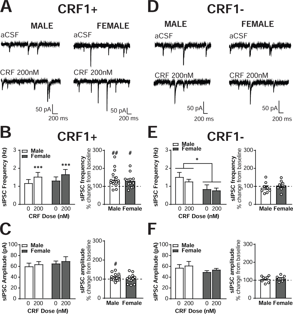
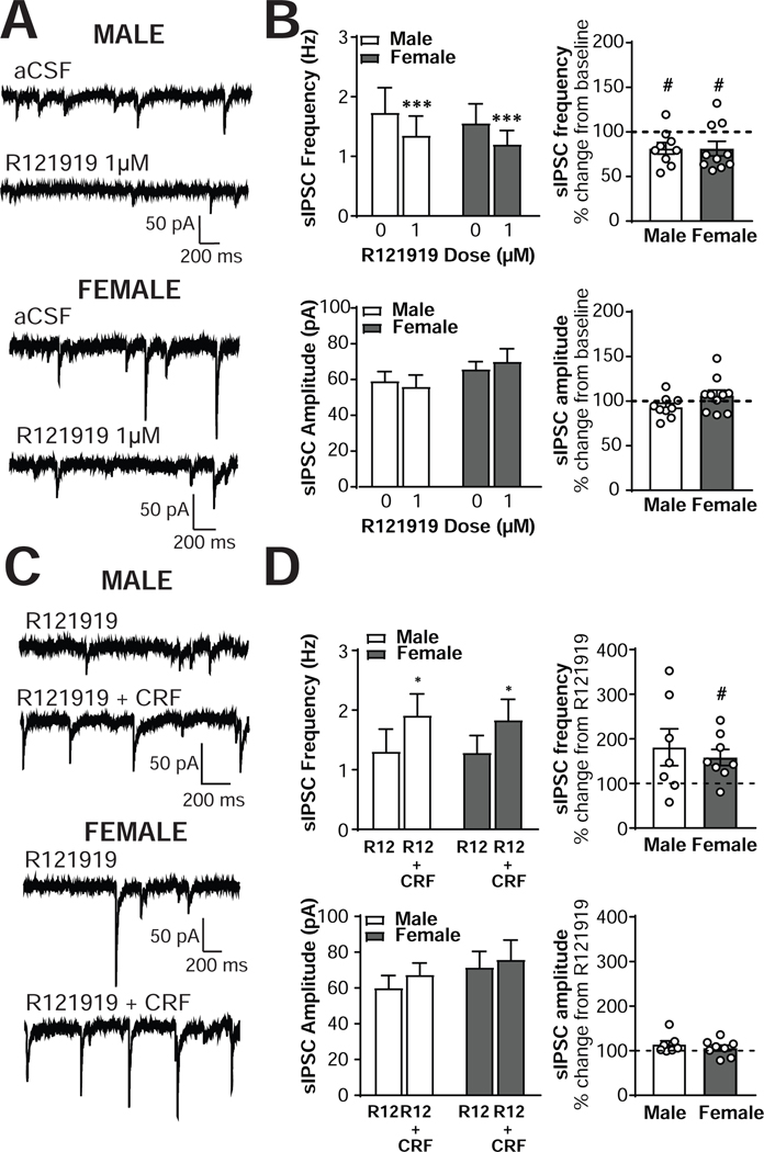
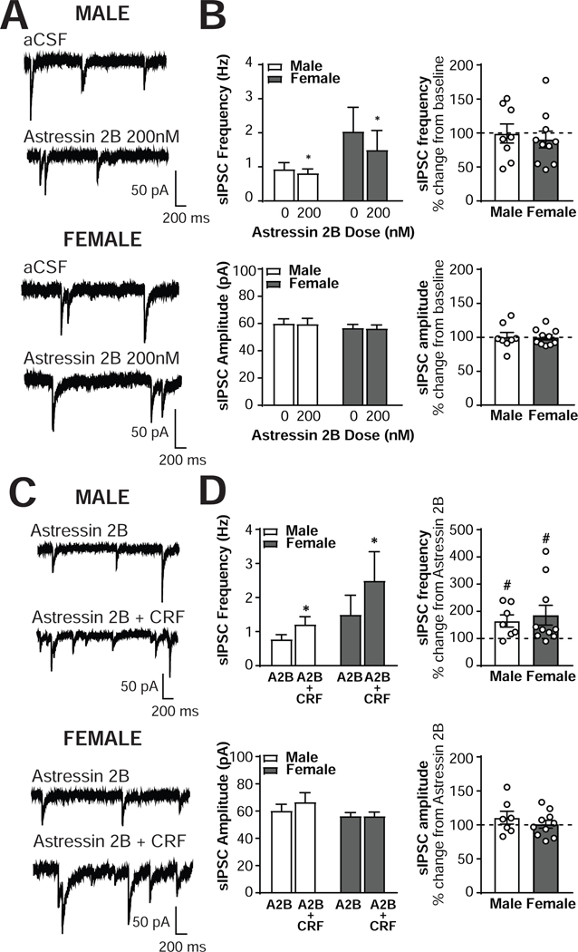
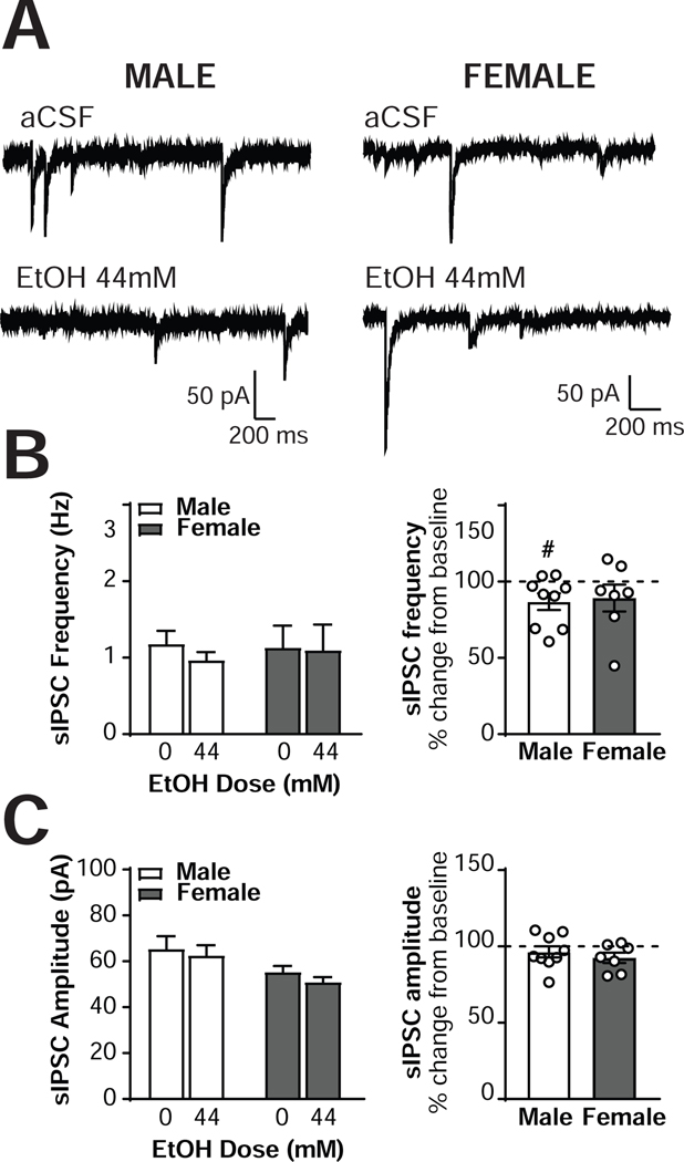
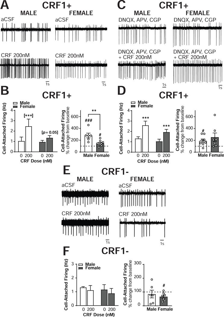
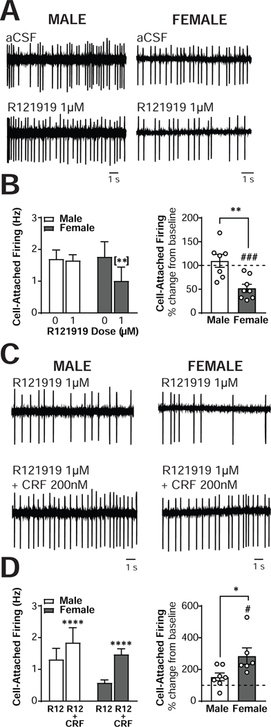
The central amygdala (CeA) is a critical regulator of emotional behavior that has been implicated in psychiatric illnesses, including anxiety disorders and addiction. The CeA corticotropin releasing factor receptor 1 (CRF1) system has been implicated in alcohol use disorder (AUD) and mood disorders, and has been shown to regulate anxiety-like behavior and alcohol consumption in rodents. However, the effects of CRF signaling within the CRF receptor 1-containing (CRF1+) population of the CeA remain unclear, and the effects of ethanol and CRF1 manipulations in female rodents have not been assessed. Here, we characterized inhibitory control and CRF1 signaling in male and female CRF1-GFP reporter mice. Male and female CRF1+ CeA neurons exhibited similar baseline GABAergic signaling and excitability and were comparably sensitive to CRF-induced increases in presynaptic GABA release. CRF1 antagonism reduced GABA release onto CRF1-containing neurons comparably in both males and females. Acute ethanol application reduced GABA release onto CRF1+ neurons from males, but female CRF1+ neurons were insensitive to ethanol. Exogenous CRF increased the firing rate of CRF1-containing neurons to a greater extent in male cells versus female cells, and CRF1 antagonism reduced firing in females but not males. Together, these findings indicate a critical sex-specific role for the CRF system in regulating inhibitory control and excitability of CRF1-containing neurons in the central amygdala. Sex differences in sensitivity of CRF/CRF1 signaling provide useful context for the sex differences in psychiatric illness reported in human patients, particularly AUD.

Keywords: Astressin 2B; CRF; CeA; Female; GABA; R121919.


Figures

Figure 1.
Figure 1.. Characterization of CRF1+ neurons in the male and female CeA.
Representative 10x photomicrographs of GFP expression in coronal CeA slices from male (A) and female (B) CRF1:GFP mice. Scale bar = 100μM. (C) Quantification of GFP+ cell counts in male and female CeA. ** = p < 0.01 via unpaired t-test. (D) 4x magnification photomicrograph depicting recording configuration of medial central amygdala (CeA) with y-tube and recording pipette (left), with insets depicting a GFP+ neuron in florescent optics (60x magnification, upper right) and in infrared differential interference contrast optics (60x magnification, lower right). (E) Summary of membrane characteristics of male and female CRF1+ CeA neurons. (F) Representative current clamp recordings illustrating low-threshold bursting (left) and regular spiking (right) firing types exhibited by CeA CRF1+ neurons. (G) Representation of each firing type in CeA neurons from male (upper) and female (lower) CeA. (H) Representative voltage-clamp recordings showing baseline sIPSCs from male (upper left) and female (upper right) CRF1+ CeA neurons and quantification of sIPSC frequency (lower left) and amplitude (lower right) of male and female CRF1+ CeA neurons.(I) Representative cell-attached recordings showing baseline firing in male (upper left) and female (upper right) CRF1+ CeA neurons and quantification (lower) of discharge rate.
Figure 2.
Figure 2.. CRF increases sIPSC frequency in male and female CRF1+, but not CRF1-, CeA neurons.
(A) Representative voltage-clamp recordings from male (left) and female (right) CRF1+ CeA neurons during focal application of aCSF (top) or CRF (200nM; bottom). (B) Quantification of sIPSC frequency in male and female CRF1+ CeA neurons following aCSF or CRF (200nM) focal application expressed in Hz (left) and percent of control (right). (C) Quantification of sIPSC amplitude in male and female CRF1+ CeA neurons following aCSF or CRF (200nM) focal application expressed in picoamps (left) and percent of control (right). (D) Representative voltage-clamp recordings from male (left) and female (right) CRF1- CeA neurons during focal application of aCSF (top) or CRF (200nM; bottom). (E) Quantification of sIPSC frequency in male and female CRF1- CeA neurons following aCSF or CRF (200nM) focal application expressed in Hz (left) and percent of control (right). (F) Quantification of sIPSC amplitude in male and female CRF1- CeA neurons following aCSF or CRF (200nM) focal application expressed in picoamps (left) and percent of control (right). * = p < 0.05 by two-way RM ANOVA, *** = p < 0.001 by two-way RM ANOVA, # = p < 0.05 by one-sample t-test, ## = p < 0.01 by one-sample t-test.
Figure 3.
Figure 3.. The CRF1 antagonist R121919 alters basal but not CRF-stimulated inhibitory control of male and female CRF1+ CeA neurons.
(A) Representative voltage-clamp recordings from male (top) and female (bottom) CRF1+ CeA neurons during focal application of aCSF (upper) or R121919 (1μM; lower). (B) Quantification of sIPSC frequency in male and female CRF1+ CeA neurons following aCSF or R121919 (1μM) focal application expressed in Hz (upper left) and percent of control (upper right). Quantification of sIPSC amplitude in male and female CRF1+ CeA neurons following aCSF or R121919 (1μM) focal application expressed in picoamps (lower left) and percent of control (lower right). (C) Representative voltage-clamp recordings from male (top) and female (bottom) CRF1+ CeA neurons during focal application of R121919 (1μM; upper) or R121919 (1μM) plus CRF (200nM) co-application (lower). (D) Quantification of sIPSC frequency in male and female CRF1+ CeA neurons following focal application of R121919 (1μM) and R121919 (1μM) plus CRF (200nM) co-application expressed in Hz (upper left) and percent of control (upper right). Quantification of sIPSC amplitude in male and female CRF1+ CeA neurons following focal application of R121919 (1μM) and R121919 (1μM) plus CRF (200nM) co-application expressed in picoamps (lower left) and percent of control (lower right). * = p < 0.05 by two-way RM ANOVA , *** = p < 0.001 by two-way RM ANOVA, # = p < 0.05 by one-sample t-test.
Figure 4.
Figure 4.. The CRF2 antagonist Astressin 2B alters basal but not CRF-stimulated inhibitory control of male and female CRF1+ CeA neurons.
(A) Representative voltage-clamp recordings from male (top) and female (bottom) CRF1+ CeA neurons during focal application of aCSF (upper) or Astressin 2B (200nM; lower). (B) Quantification of sIPSC frequency in male and female CRF1+ CeA neurons following aCSF or Astressin 2B (200nM) focal application expressed in Hz (upper left) and percent of control (upper right). Quantification of sIPSC amplitude in male and female CRF1+ CeA neurons following aCSF or Astressin 2B (200nM) focal application expressed in picoamps (lower left) and percent of control (lower right). (C) Representative voltage-clamp recordings from male (top) and female (bottom) CRF1+ CeA neurons during focal application of Astressin 2B (200nM; upper) or Astressin 2B (200nM) plus CRF (200nM) co-application (lower). (D) Quantification of sIPSC frequency in male and female CRF1+ CeA neurons following focal application of Astressin 2B (200nM) and Astressin 2B (200nM) plus CRF (200nM) co-application expressed in Hz (upper left) and percent of control (upper right). Quantification of sIPSC amplitude in male and female CRF1+ CeA neurons following focal application of Astressin 2B (200nM) and Astressin 2B (200nM) plus CRF (200nM) co-application expressed in picoamps (lower left) and percent of control (lower right). * = p < 0.05 by two-way RM ANOVA, # = p < 0.05 by one-sample t-test.
Figure 5.
Figure 5.. Ethanol alters inhibitory control of male but not female CRF1+ neurons.
(A) Representative voltage-clamp recordings from male (left) and female (right) CeA CRF1+ neurons during focal application of aCSF (top) and ethanol (44mM; bottom). (B) Quantification of sIPSC frequency in male and female CRF1+ neurons following aCSF or ethanol (44mM) focal application expressed in Hz (left) and percent of control (right). (C) Quantification of sIPSC amplitude in male and female CRF1+ neurons following focal application of aCSF or ethanol (44mM) expressed in picoamps (left) and percent of control (right). # = p < 0.05 by one-sample t-test.
Figure 6.
Figure 6.. Sex differences in CRF regulation of excitability in CRF1+, but not CRF1-, CeA neurons.
(A) Representative cell-attached recording of male (left) and female (right) CRF1+ CeA neurons during focal application of aCSF (top) and CRF (200nm; bottom). (B) Quantification of cell-attached firing frequency in male and female CRF1+ CeA neurons following focal application of aCSF and CRF (200nM) expressed in Hz (left) and percent of control (right). (C) Representative cell-attached recording of male (left) and female (right) CRF1+ CeA neurons during bath superfusion of DNQX, APV and CGP with focal application of aCSF (top) and CRF (200nm; bottom). (D) Quantification of cell-attached firing frequency in male and female CRF1+ CeA neurons following focal application of aCSF and CRF (200nM) in the presence of DNQX, APV and CGP expressed in Hz (left) and percent of control (right). (E) Representative cell-attached recording of male (left) and female (right) CRF11 CeA neurons during focal application of aCSF (top) and CRF (200nm; bottom). (F) Quantification of cell-attached firing frequency in male and female CRF1- CeA neurons following focal application of aCSF and CRF (200nM) expressed in Hz (left) and percent of control (right). ** = p < 0.01 by two-sample t-test, # = p < 0.05 by one-sample t-test, ## = p < 0.01 by one-sample t-test, [***] = p < 0.001 by Sidak’s multiple comparison’s test.
Figure 7.
Figure 7.. Sex differences in CRF1 regulation of excitability in CRF1+, but not CRF1-, CeA neurons.
(A) Representative cell-attached recording of male (left) and female (right) CRF1+ CeA neurons during focal application of aCSF (top) and R121919 (1 μm; bottom). (B) Quantification of cell-attached firing frequency in male and female CRF1+ CeA neurons following focal application of aCSF and R121919 (1 μm) expressed in Hz (left) and percent of control (right). (C) Representative cell-attached recording of male (left) and female (right) CRF1+ CeA neurons during focal application of R121919 (1 μm; top) and R121919 + CRF (200 nm; bottom). (D) Quantification of cell-attached firing frequency in male and female CRF1+ CeA neurons following focal application of R121919 (1 μm) and R121919 + CRF (200nM) expressed in Hz (left) and percent of control (right).

References

    1. Agoglia AE, Herman MA (2018) The center of the emotional universe: Alcohol, stress, and CRF1 amygdala circuitry. Alcohol 72:61–73. - PMC - PubMed
    1. Agoglia AE, Crofton EJ, Herman MA (2020) Biological intersection of sex, age, and environment in the corticotropin releasing factor (CRF) system and alcohol. Neuropharmacology 170:108045. - PMC - PubMed
    1. Bajo M, Cruz MT, Siggins GR, Messing R, Roberto M (2008) Protein kinase C epsilon mediation of CRF- and ethanol-induced GABA release in central amygdala. Proc Natl Acad Sci U S A 105:8410–8415. - PMC - PubMed
    1. Bangasser DA, Curtis A, Reyes BA, Bethea TT, Parastatidis I, Ischiropoulos H, Van Bockstaele EJ, Valentino RJ (2010) Sex differences in corticotropin-releasing factor receptor signaling and trafficking: potential role in female vulnerability to stress-related psychopathology. Mol Psychiatry 15:877, 896–904. - PMC - PubMed
    1. Bangasser DA, Reyes BA, Piel D, Garachh V, Zhang XY, Plona ZM, Van Bockstaele EJ, Beck SG, Valentino RJ (2013) Increased vulnerability of the brain norepinephrine system of females to corticotropin-releasing factor overexpression. Mol Psychiatry 18:166–173. - PMC - PubMed

Publication types

MeSH terms

Substances