. 2020 Dec:96:87-103.
doi: 10.1016/j.neurobiolaging.2020.08.012. Epub 2020 Aug 26.

Linking anatomical and physiological markers of auditory system degeneration with behavioral hearing assessments in a mouse (Mus musculus) model of age-related hearing loss

Affiliations

Linking anatomical and physiological markers of auditory system degeneration with behavioral hearing assessments in a mouse (Mus musculus) model of age-related hearing loss

Anastasiya Kobrina et al. Neurobiol Aging. 2020 Dec.

Abstract

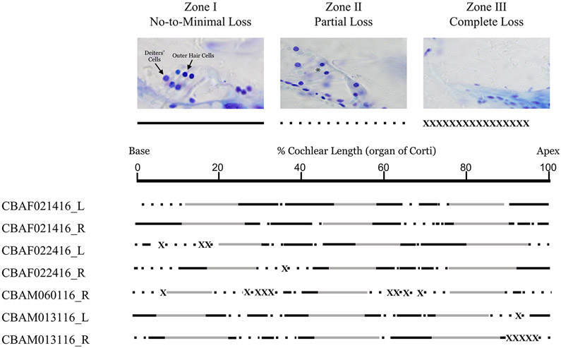
Age-related hearing loss is a very common sensory disability, affecting one in three older adults. Establishing a link between anatomical, physiological, and behavioral markers of presbycusis in a mouse model can improve the understanding of this disorder in humans. We measured age-related hearing loss for a variety of acoustic signals in quiet and noisy environments using an operant conditioning procedure and investigated the status of peripheral structures in CBA/CaJ mice. Mice showed the greatest degree of hearing loss in the last third of their lifespan, with higher thresholds in noisy than in quiet conditions. Changes in auditory brainstem response thresholds and waveform morphology preceded behavioral hearing loss onset. Loss of hair cells, auditory nerve fibers, and signs of stria vascularis degeneration were observed in old mice. The present work underscores the difficulty in ascribing the primary cause of age-related hearing loss to any particular type of cellular degeneration. Revealing these complex structure-function relationships is critical for establishing successful intervention strategies to restore hearing or prevent presbycusis.

Keywords: ARHL; Animal psychoacoustics; Auditory brainstem response; Cochlear degeneration; Cochlear nucleus; Olivocochlear.


Conflict of interest statement

Declaration of interest

The authors declare no conflict of interest.

Figures

Figure 1.
Figure 1.
Schematic of the operant conditioning setup used in the behavioral sound detection experiments. Mice were trained to nose-poke in an observation hole in quiet or background noise conditions. Detection of a target sound was indicated by nose-poking in a report hole. Correct detection of the target sounds was reinforced with chocolate Ensure from a dipper. False alarms were punished with a 3-5 s time out period.
Figure 2.
Figure 2.
Spectrograms of the four ultrasonic vocalizations used as stimuli in the behavioral detection experiments.
Figure 3.
Figure 3.
Age-related deterioration of behavioral detection of sounds in quiet. Performance becomes especially poor after 650 d.o. for USVs (A), and after 700 d.o. for 42 kHz tones (B). Age-related hearing loss occurs progressively up to 450 d.o. and after 650 d.o. for 56 kHz pure tones, with a stable plateau between those ages (C). Each plot depicts thresholds from multiple mice across their lifespans (males = filled blue symbols, females = open symbols). Lines represent the best cubic data fits. The amount of variability in the data explained by aging is expressed in the form of r2 for males and females separately. Dashed lines indicate the onset of hearing loss for USVs and 42 kHz tones, and plateau boundaries for 56 kHz tones.
Figure 4.
Figure 4.
Age-related deterioration of behavioral detection of sounds in white noise. Performance becomes especially poor after about 600 d.o. for USVs (A), and after 700 d.o. for 42 kHz tones (B). Age-related hearing loss progressed continuously for 56 kHz tones up to 750 d.o., with a ceiling boundary late in life (C). Each plot depicts thresholds from multiple mice across their lifespans (males = filled blue symbols, females = open symbols). Lines represent the best cubic data fits. The amount of variability in the data explained by aging is expressed in the form of r2 for males and females separately. Dashed lines indicate the onset of hearing loss for UVSs (A), and 42 kHz tones (B), and a ceiling boundary for 56 kHz tones (C).
Figure 5.
Figure 5.
Threshold stability across multiple days of testing as a function of age for A) four mice 425-550 d.o. or B) three mice 875-1000 d.o.. Each line depicts thresholds for one subject in quiet (open symbols) or in white noise (filled symbols).
Figure 6.
Figure 6.
Auditory brainstem responses in young and old mice show substantial deficits in function by 600 d.o.. Average waveforms in response to 90 dB (A) and 50 dB (B) clicks in young (60-90 d.o.; black) and old (600-800 d.o.; red) mice, respectively. Standard error is depicted in gray and pink shading for young and old mice, respectively. Legend in A applies to panel B. Thresholds in response to clicks (C), and 8 kHz, 16 kHz, and 32 kHz tones (D-F) in female (red) and male (blue) mice. Amplitudes for peak 1 (p1), peak 2 (p2), peak 3 (p3), and peak 4 (p4) (G-J) and corresponding latencies for p1, p2, p3, and p4 of responses to 70 dB pSPL clicks plotted as a function of age (K-N). Inter-peak intervals for responses to 70 dB pSPL clicks are plotted as a function of age (O-Q). Legend in panel C applies to panels D-Q. In panels C-Q, data points for 60-90 d.o. mice depict means, error bars depict standard errors. Note that error bars in panels K-Q are obscured by the markers.
Figure 7.
Figure 7.
Percent surviving outer hair cells (OHC) and inner hair cells (IHC) in 600-800 d.o. mice (9 female; 6 male) relative to 60-90 d.o. mice (7 female; 5 male) (A). Representative maximum-intensity projections of confocal z-stacks of young (120 d.o.) and old (751 d.o.) female mouse organ of Corti labeled with antibodies against myosin 6 (hair cells), CTBP2 (presynaptic ribbons), and neurofilament (nerve fibers) (B). Number of surviving presynaptic ribbons per IHC in young (1 female; 2 male) and old (3 female; 4 male) mice (C). Data points in (C) depict means, error bars depict standard errors. Stars indicate significant differences.
Figure 8.
Figure 8.
Quantification of efferent synapses labeled with antibodies against synaptic vesicle protein (SV2) in young (60-90 d.o.) and old (600-800 d.o.) mice (A-D). Representative maximum-intensity projections of confocal z-stacks of young (120 d.o.) and old (751 d.o.) mouse organ of Corti labeled with antibodies against myosin 6 (hair cells) and SV2 (efferent synapses) (A). Total area of labeling per IHC in young (black) and old (red) mice (B). Number of efferent terminals (C) and total area of labeling per OHC (D). For all panels, young mice consist of 5 females and 3 males; old mice consist of 6 females and 2 males. All points depict means, error bars depict standard errors. Stars indicate significant differences.
Figure 9.
Figure 9.
Cochlear histology in old (900-975 d.o.) female (F, 1-4) and male (M, 5-7) mice for left (L) and right (R) cochlea. The lines represent the percentage of degeneration along the length of the organ of Corti from base to apex. The solid black lines represent areas with no-to-minimal hair cell loss, the areas with dashed lines represent areas with partial hair cell loss, and the crossed out portions of the line represent areas with complete hair cell loss. The blank areas represent areas where the hair cell loss could not be determined due to mechanical damage from the plastic embedding process or the angle at which the cochlea was sectioned.
Figure 10.
Figure 10.
Examination of stria vascularis (A-B) and spiral ganglion neurons (C-D) in young male (A, C) and old female (B, D) mice. The stria vascularis in old mice contained more melanin and lipofusin granules, indicating degeneration (B). Stria vascularis is thinner at the apical turn in the aged mouse cochlea (B) than in the young mouse cochlea (A). Spiral ganglion in young mouse appears intact (C), while holes are present in the spiral ganglion of the aged cochlea (D). The population of spiral ganglion neurons in the aged cochlea (D) is diminished compared to that of the young cochlea (C), which is another visible indication of degeneration.

References

    1. Abdala C, Dhar S, Ahmadi M, Luo P. Aging of the medial olivocochlear reflex and associations with speech perception. J. Acoust. Soc. Am, 135 (2014), pp. 754–765. doi: 10.1121/1.4861841 - DOI - PMC - PubMed
    1. Abel SM, Krever EM, Alberti PW. Auditory detection, discrimination and speech processing in ageing, noise-sensitive and hearing-impaired listeners. Scand. Audiol, 19 (1990), pp. 43–54. doi: 10.3109/01050399009070751 - DOI - PubMed
    1. Alvarado JC, Fuentes-Santamaría V, Gabaldón-Ull MC, Blanco JL, Juiz JM. Wistar rats: a forgotten model of age-related hearing loss. Front. Aging Neurosci, 6 (2014), pp. 29. doi: 10.3389/fnagi.2014.00029 - DOI - PMC - PubMed
    1. Anderson S, Parbery-Clark A, Yi HG, Kraus N. A neural basis of speech-in-noise perception in older adults. Ear Hear., 32 (2011), pp. 750–757. doi: 10.1097/AUD.0b013e31822229d3 - DOI - PMC - PubMed
    1. Bacon SP, Opie JM, Montoya DY. The effects of hearing loss and noise masking on the masking release for speech in temporally complex backgrounds. J. Speech Lang. Hear. Res, 41 (1998), pp. 549–563. doi: 10.1044/jslhr.4103.549 - DOI - PubMed

Publication types