. 2020 Dec;147(14):1678-1688.
doi: 10.1017/S0031182020001778. Epub 2020 Sep 21.

Acanthamoeba spp. monoclonal antibody against a CPA2 transporter: a promising molecular tool for acanthamoebiasis diagnosis and encystment study

Affiliations

Acanthamoeba spp. monoclonal antibody against a CPA2 transporter: a promising molecular tool for acanthamoebiasis diagnosis and encystment study

Michele Martha Weber-Lima et al. Parasitology. 2020 Dec.

Abstract

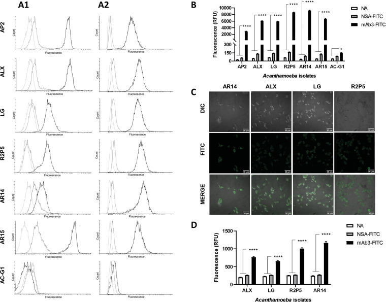
Free-living amoeba of the genus Acanthamoeba are ubiquitous protozoa involved in opportunistic and non-opportunistic infection in humans, such as granulomatous amoebic encephalitis and amoebic keratitis. Both infections have challenging characteristics such as the formation of the resistant cysts in infected tissues, hampering the treatment and most usual diagnosis depending on time-consuming and/or low sensitivity techniques. The use of monoclonal antibodies presents itself as an opportunity for the development of more effective alternative diagnostic methods, as well as an important and useful tool in the search for new therapeutic targets. This study investigated the possibility of using a previously produced monoclonal antibody (mAb3), as a diagnostic tool for the detection of Acanthamoeba trophozoites by direct and indirect flow cytometry and immunofluorescence. Immunoprecipitation assay and mass spectrometry allowed the isolation of the antibody's target and suggested it is a transporter part of the CPA (cation: proton antiporter) superfamily. In vitro tests indicate an important role of this target in Acanthamoeba's encystment physiology. Our results support the importance of studying the role of CPA2 transporters in the context of acanthamoebiasis, as this may be a way to identify new therapeutic candidates.

Keywords: Acanthamoeba; CPA2 transporters; diagnosis; encystment; flow cytometry; monoclonal antibody.


Figures

Fig. 1.
Fig. 1.
mAb3V-domains. (A) Agarose gel electrophoresis (1.5%) showing amplification of the antibody's variable heavy chain sequence (VH) with 400 bp and of the lambda light chain sequence (Vƛ) with 350 bp and their respective negative controls (CVH and CVƛ) without the addition of cDNA. (B) Secondary structure representation of the variable region sequences in the ‘collier de perles’ analysed by the IMGT and coloured following their colour menu. Heavy (H) and light (L) chain CDRs in red (H1), orange (H2), purple (H3), blue (L1), light green (L2) and forest green (L3); positions in light blue show amino acids with positive values in the hydropathy index or a tryptophan (W); amino acids shown in red letters correspond to conserved positions of a V-domain; anchor positions are shown in squares; hatched circles indicate gaps according to the unique numbering for V-domains by the IMGT; positions in light yellow show prolines (P); β-strands and their directions indicated by the arrows. (C) Stereo view of three-dimensional structure of the heavy and light variable regions, with coloured CDRs as specified by the IMGT colour menu, the same as previously described.
Fig. 2.
Fig. 2.
mAb3 characterization. (A1) Indirect ELISA showing reactivity of mAb3 antibody (5 μg mL−1) or irrelevant monoclonal antibody and polyclonal antibodies (1:100) against ALX, AP2, AR14, AR15, LG, R2P5 and AC-G1 Acanthamoeba strains sonicated antigens (10 μg mL−1), immunocomplexes were revealed using HRP-conjugated anti-mouse IgG (1:4000). (A2) Indirect ELISA showing different concentrations of mAb3 antibody reactivity against strain AP2 (10 μg mL−1). (B) WB of AP2, ALX, LG (clinical samples from corneal scrapings of AK patients), R2P5, AR14, AR15 (environmental samples from dust) and AC-G1 (environmental sample from soil) protein extract (5 μg) (obtained with lysis buffer) in nitrocellulose membrane stained by Ponceau (B1), followed by incubation with mAb3 (1.5 μg mL−1) (B2), immunocomplexes were revealed using HRP-conjugated anti-mouse IgG (1: 10 000) and DAB/chloronaphthol staining. (C) Reactivity of mAb3 against 1.5 μg of Fusarium sp., Aspergillus sp., Candida sp. and Acanthamoeba sp. sonicated antigens assayed by WB (C1) and indirect ELISA (C2). In WB, proteins were transferred to a PVDF membrane, incubated with 5 μg mL−1 of mAb3 and HRP-conjugated anti-mouse IgG (1:4000); reaction was revealed by chemiluminescence detection). Indirect ELISA was performed with 10 μg mL−1 of antigen against 5 μg mL−1 of mAb3. (D1) Indirect flow cytometry histogram of strain ALX previously treated with tunicamycin incubated with mAb3 (dark grey), untreated ALX incubated with mAb3 (black), untreated ALX without mAb3 (light grey). (D2) Mean fluorescence intensity of events detected by indirect flow cytometry of ALX with (+) or without (−) tunicamycin pre-treatment and mAb3 incubation, quantified in relative fluorescence units (RFU). (E1) Schematic representation of mAb3 immunoprecipitation of target protein. (E2) WB of Acanthamoeba sp. sonicated total protein extract (1.5 μg) (1); flow through fraction (2) and eluted fraction of immunoprecipitation assay (3). Antigens were transferred to PVDF membrane incubated with mAb3 (5 μg mL−1) and HRP-conjugated anti-mouse IgG (1:4000), and the reaction was revealed by chemiluminescence detection.
Fig. 3.
Fig. 3.
Multiple amino acid sequence alignment of CPA2 family antiporters, L8HBJ5_ACACA, L8H7S3_ACACA and L8H9U2_ACACA from Acanthamoeba castellanii, Q5SIA2_THET8 from Thermus thermophilus and A0A4S5A442_ECOLI from Escherichia coli. Conserved motif (grey box) involved in antiporter function. The last column shows per cent identity of each sequence when compared to L8H7S3_ACACA, the supposed mAb3 target. Peptide (TVSLPR) found in the mass spectrometry analysis of mAb3's purified target protein (transparent box). Asterisks (*) indicate positions with a fully conserved residue, colons (:) indicate conservation of residues belonging to groups with highly similar properties scoring >0.5 in the Gonnet PAM 250 matrix and periods (.) indicate conservation of residues belonging to groups with weakly similar properties scoring ⩽0.5 in the Gonnet PAM 250 matrix.
Fig. 4.
Fig. 4.
Reactivity of mAb3 to Acanthamoeba strains analysed by flow cytometry and immunofluorescence. (A) Histograms of indirect (A1) and direct (A2) flow cytometry of trophozoites from Acanthamoeba strains. Curves in light grey represent samples without antibodies, in dark grey are those treated only with secondary anti-mouse antibody (indirect assay) or FITC-conjugated non-specific IgG (direct assay) and in black are the samples treated with mAb3. (B) Mean fluorescence intensity of events detected by direct flow cytometry of Acanthamoeba samples treated with no antibodies (NA), with FITC-conjugated non-specific antibody (NSA-FITC) and with FITC-conjugated mAb3 (mAb3-FITC), quantified in relative fluorescence units (RFU), bars represent mean ± s.e.m. (5453 ⩽ n ⩽ 10 785), ****P < 0.0001, *P < 0.05. (C) Direct Immunofluorescence of Acanthamoeba trophozoites incubated with FITC-conjugated mAb3, images shown were generated using differential interference contrast (DIC) and fluorescence (FITC) illumination. Autofluorescence of untreated trophozoites was subtracted from the fluorescence intensity observed in the images. (D) Mean fluorescence intensity, detected by direct immunofluorescence, of untreated Acanthamoeba trophozoites (NA), treated with FITC-conjugated non-specific antibody (NSA) and with FITC-conjugated mAb3 (mAb3-FITC), quantified in relative fluorescence units (RFU). Bars represent mean ± s.e.m. (n = 100), ****P < 0.0001.
Fig. 5.
Fig. 5.
Effect of mAb3 on Acanthamoeba encystation. (A) Kinetics of encystment of Acanthamoeba isolate AP2 in Neff's encystation saline, showing the percentage of trophozoites, precyst and mature cysts. (B, C) Effect of mAb3 (30 μg mL−1), non-specific antibodies (NSA) (30 μg mL−1) and vehicle (PBS) treatment in the amount of trophozoites and precyst. Points in graph represent mean ± s.e.m. (n = 4), *P < 0.01 mAb3 vs PBS, #P < 0.01 mAb3 vs NSA. (D) Effect of mAb3 (30 μg mL−1), non-specific antibodies (30 μg mL−1) and vehicle (PBS) treatment on precyst (PC) formation rate. Bars represent mean ± s.e.m. (n = 4), *P < 0.05, ***P < 0.001.

References

    1. Alsam S, Sissons J, Jayasekera S and Khan NA (2005) Extracellular proteases of Acanthamoeba castellanii (encephalitis isolate belonging to T1 genotype) contribute to increased permeability in an in vitro model of the human blood–brain barrier. Journal of Infection 51, 150–156. - PubMed
    1. Andreotti PE, Ludwig GV, Peruski AH, Tuite JJ, Morse SS and Peruski LF (2003) Immunoassay of infectious agents. BioTechniques 35, 850–859. - PubMed
    1. Becker-Finco A, Costa AO, Silva SK, Ramada JS, Furst C, Stinghen AE, De Figueiredo BC, De Moura J and Alvarenga LM (2013) Physiological, morphological, and immunochemical parameters used for the characterization of clinical and environmental isolates of Acanthamoeba. Parasitology 140, 396–405. - PubMed
    1. Bradbury A and Plückthun A (2015) Reproducibility: standardize antibodies used in research. Nature 518, 27–29. - PubMed
    1. Bunsuwansakul C, Mahboob T, Hounkong K, Laohaprapanon S, Chitapornpan S, Jawjit S, Yasiri A, Barusrux S, Bunluepuech K, Sawangjaroen N, Salibay CC, Kaewjai C, de Pereira ML and Nissapatorn V (2019) Acanthamoeba in Southeast Asia – overview and challenges. The Korean Journal of Parasitology 57, 341–357. - PMC - PubMed

Publication types

MeSH terms