

Four Deep Brain Stimulation Targets for Obsessive-Compulsive Disorder: Are They Different?
- PMID: 32951818
- PMCID: PMC9569132
- DOI: 10.1016/j.biopsych.2020.06.031
Four Deep Brain Stimulation Targets for Obsessive-Compulsive Disorder: Are They Different?
Abstract
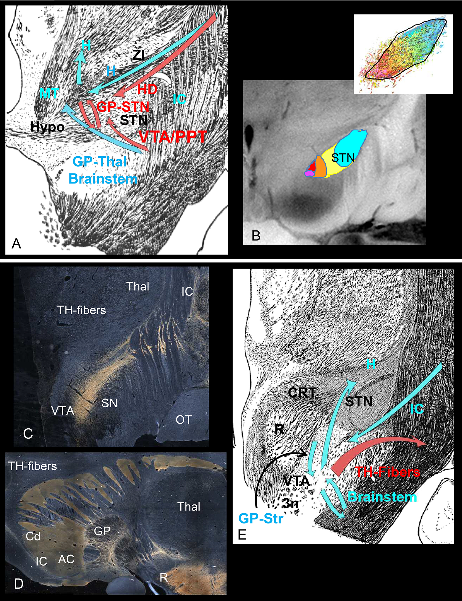
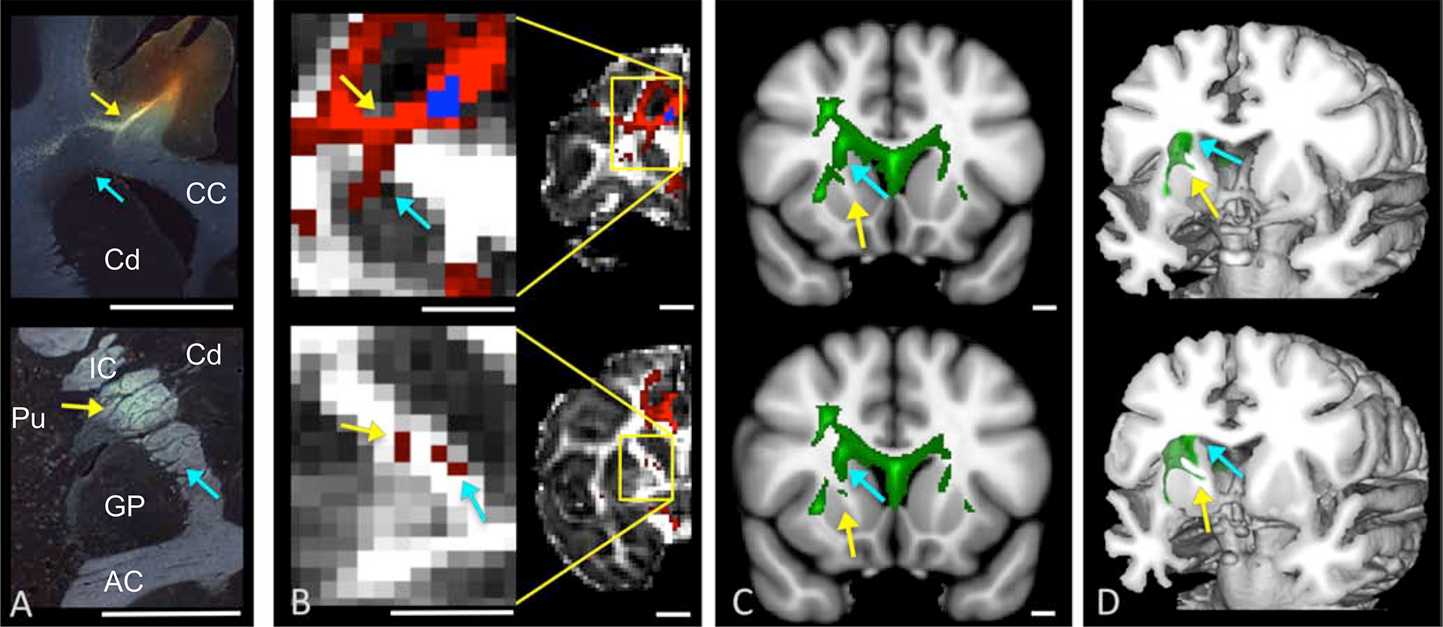
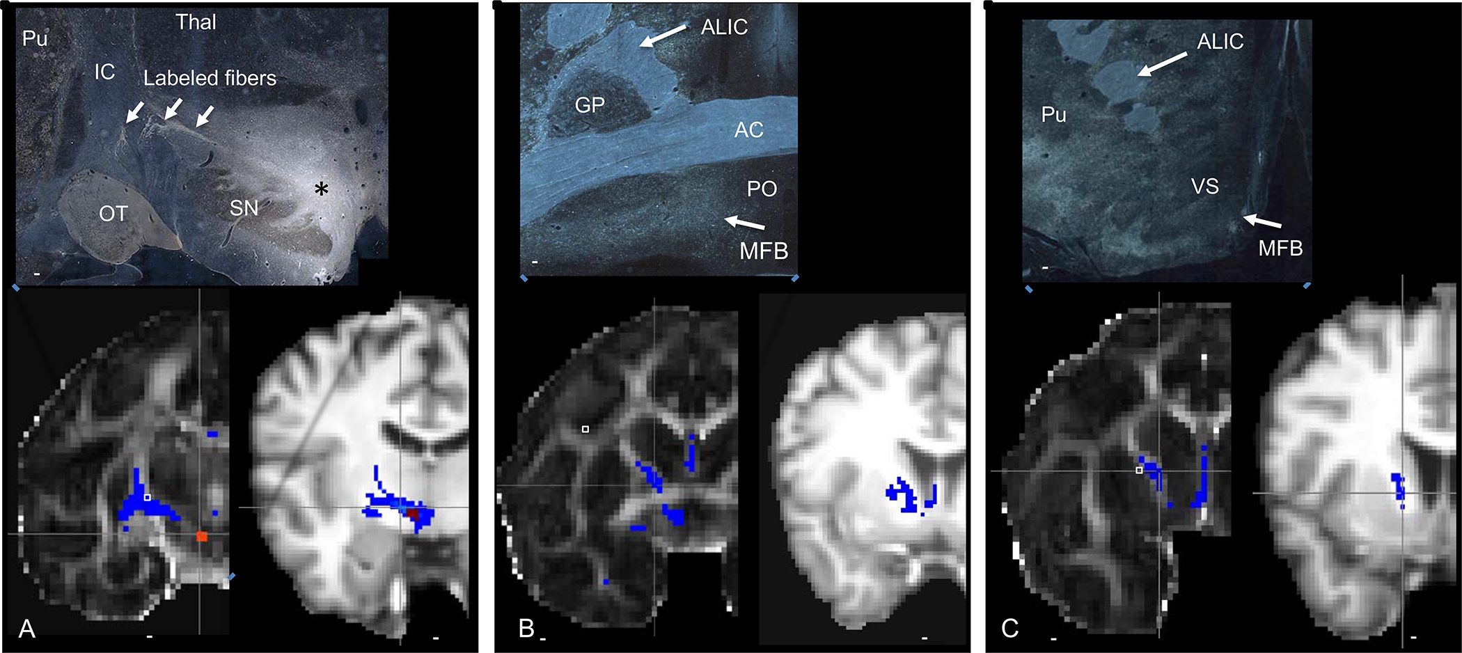
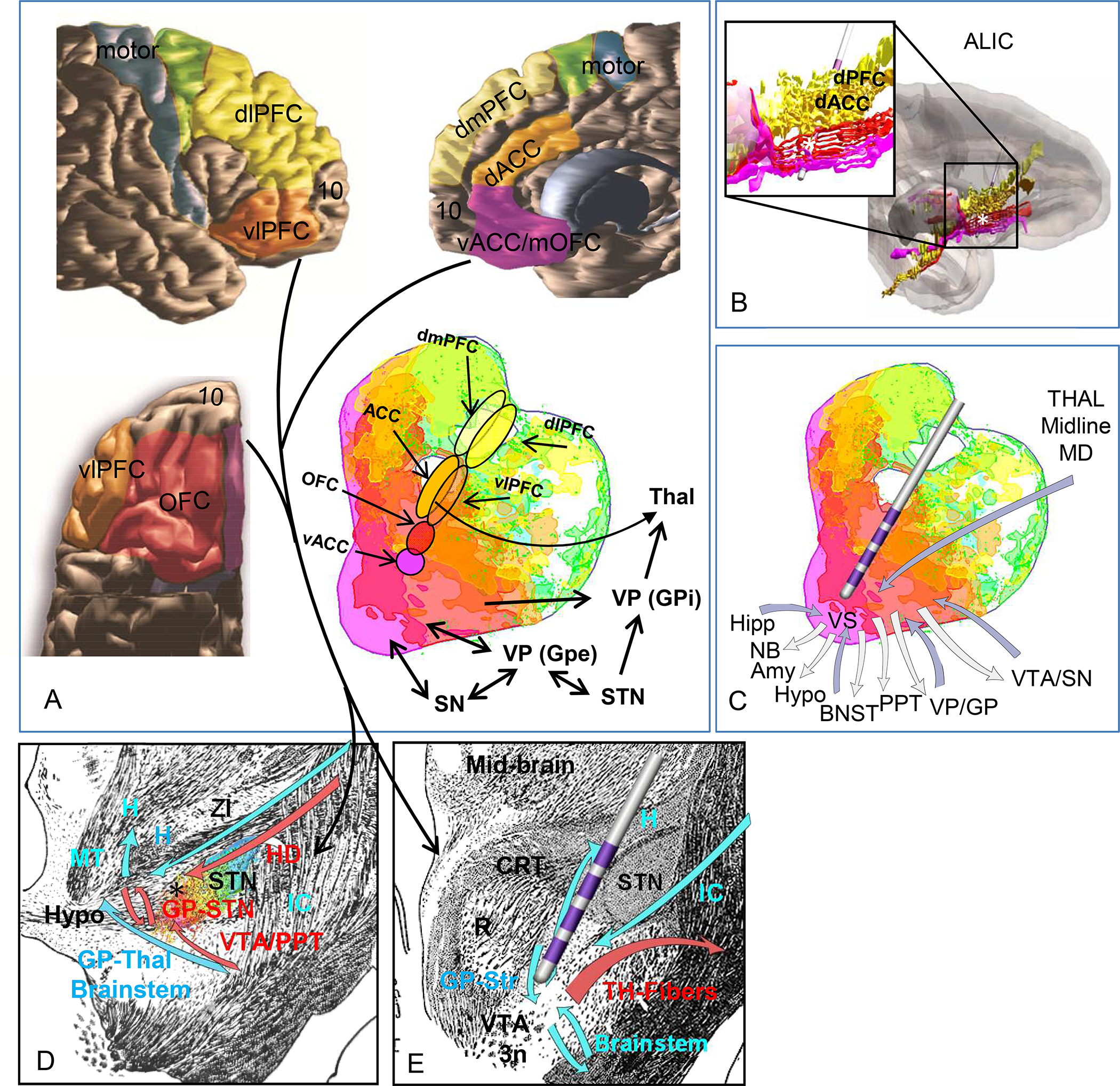
Deep brain stimulation is a promising therapeutic approach for patients with treatment-resistant obsessive-compulsive disorder, a condition linked to abnormalities in corticobasal ganglia networks. Effective targets are placed in one of four subcortical areas with the goal of capturing prefrontal, anterior cingulate, and basal ganglia connections linked to the limbic system. These include the anterior limb of the internal capsule, the ventral striatum, the subthalamic nucleus, and a midbrain target. The goal of this review is to examine these 4 targets with respect to the similarities and differences of their connections. Following a review of the connections for each target based on anatomic studies in nonhuman primates, we examine the accuracy of diffusion magnetic resonance imaging tractography to replicate those connections in nonhuman primates, before evaluating the connections in the human brain based on diffusion magnetic resonance imaging tractography. Results demonstrate that the four targets generally involve similar connections, all of which are part of the internal capsule. Nonetheless, some connections are unique to each site. Delineating the similarities and differences across targets is a critical step for evaluating and comparing the effectiveness of each and how circuits contribute to the therapeutic outcome. It also underscores the importance that the terminology used for each target accurately reflects its position and its anatomic connections, so as to enable comparisons across clinical studies and for basic scientists to probe mechanisms underlying deep brain stimulation.
Keywords: Anatomic tracing; Anterior limb of the internal capsule; Diffusion magnetic resonance imaging; Medial forebrain bundle; Subthalamic nucleus; Ventral striatum; White matter pathways.
Copyright © 2020 Society of Biological Psychiatry. Published by Elsevier Inc. All rights reserved.
Conflict of interest statement
Financial Disclosures.
Drs. Haber, Yendiki, and Jbabdi reported no biomedical financial interests or potential conflicts of interest.
Figures

Comment in
-
Deep Brain Stimulation for Obsessive-Compulsive Disorder: Why Anatomy Matters.Biol Psychiatry. 2021 Nov 15;90(10):662-663. doi: 10.1016/j.biopsych.2021.06.011. Biol Psychiatry. 2021. PMID: 34674799 No abstract available.
Similar articles
-
Anatomic Review of the Ventral Capsule/Ventral Striatum and the Nucleus Accumbens to Guide Target Selection for Deep Brain Stimulation for Obsessive-Compulsive Disorder.World Neurosurg. 2019 Jun;126:1-10. doi: 10.1016/j.wneu.2019.01.254. Epub 2019 Feb 18. World Neurosurg. 2019. PMID: 30790738 Review.
-
The Rostral Zona Incerta: A Subcortical Integrative Hub and Potential Deep Brain Stimulation Target for Obsessive-Compulsive Disorder.Biol Psychiatry. 2023 Jun 1;93(11):1010-1022. doi: 10.1016/j.biopsych.2023.01.006. Epub 2023 Jan 18. Biol Psychiatry. 2023. PMID: 37055285
-
A Randomized Trial Directly Comparing Ventral Capsule and Anteromedial Subthalamic Nucleus Stimulation in Obsessive-Compulsive Disorder: Clinical and Imaging Evidence for Dissociable Effects.Biol Psychiatry. 2019 May 1;85(9):726-734. doi: 10.1016/j.biopsych.2019.01.017. Epub 2019 Jan 30. Biol Psychiatry. 2019. PMID: 30853111 Free PMC article. Clinical Trial.
-
Deep Brain Stimulation of the Nucleus Accumbens, Ventral Striatum, or Internal Capsule Targets for Medication-Resistant Obsessive-Compulsive Disorder: A Multicenter Study.World Neurosurg. 2021 Nov;155:e168-e176. doi: 10.1016/j.wneu.2021.08.039. Epub 2021 Aug 14. World Neurosurg. 2021. PMID: 34403796
-
Current status of deep brain stimulation for obsessive-compulsive disorder: a clinical review of different targets.Curr Psychiatry Rep. 2011 Aug;13(4):274-82. doi: 10.1007/s11920-011-0200-8. Curr Psychiatry Rep. 2011. PMID: 21505875 Review.
Cited by
-
Stereo-EEG-guided network modulation for psychiatric disorders: Surgical considerations.Brain Stimul. 2023 Nov-Dec;16(6):1792-1798. doi: 10.1016/j.brs.2023.07.057. Epub 2023 Dec 20. Brain Stimul. 2023. PMID: 38135358 Free PMC article.
-
White Matter Tracts Associated With Deep Brain Stimulation Targets in Major Depressive Disorder: A Systematic Review.Front Psychiatry. 2022 Apr 28;13:806916. doi: 10.3389/fpsyt.2022.806916. eCollection 2022. Front Psychiatry. 2022. PMID: 35573379 Free PMC article. Review.
-
Closed-loop neurostimulation for the treatment of psychiatric disorders.Neuropsychopharmacology. 2024 Jan;49(1):163-178. doi: 10.1038/s41386-023-01631-2. Epub 2023 Jun 27. Neuropsychopharmacology. 2024. PMID: 37369777 Free PMC article. Review.
-
The anterior perforated substance (APS) revisited: Commented anatomical and imagenological views.Brain Behav. 2023 Dec;13(12):e3029. doi: 10.1002/brb3.3029. Epub 2023 Nov 27. Brain Behav. 2023. PMID: 38010896 Free PMC article.
-
Functional Approaches to the Surgery of Brain Gliomas.Adv Tech Stand Neurosurg. 2022;45:35-96. doi: 10.1007/978-3-030-99166-1_2. Adv Tech Stand Neurosurg. 2022. PMID: 35976447 Review.
References
-
- Mallet L, Polosan M, Jaafari N, Baup N, Welter ML, Fontaine D, et al. (2008): Subthalamic nucleus stimulation in severe obsessive-compulsive disorder. N Engl J Med. 359:2121–2134. - PubMed
-
- Denys D, Mantione M, Figee M, van den Munckhof P, Koerselman F, Westenberg H, et al. (2010): Deep brain stimulation of the nucleus accumbens for treatment-refractory obsessive-compulsive disorder. Arch Gen Psychiatry. 67:1061–1068. - PubMed
-
- Admon R, Bleich-Cohen M, Weizmant R, Poyurovsky M, Faragian S, Hendler T (2012): Functional and structural neural indices of risk aversion in obsessive-compulsive disorder (OCD). Psychiatry Res. 203:207–213. - PubMed
Publication types
MeSH terms
Grants and funding
LinkOut - more resources
Full Text Sources
Medical
