Review
. 2021 Jan-Feb;15(1):27-36.
doi: 10.1016/j.jcct.2020.08.013. Epub 2020 Sep 4.

Role of computed tomography in COVID-19

Affiliations
Review

Role of computed tomography in COVID-19

Gianluca Pontone et al. J Cardiovasc Comput Tomogr. 2021 Jan-Feb.

Abstract

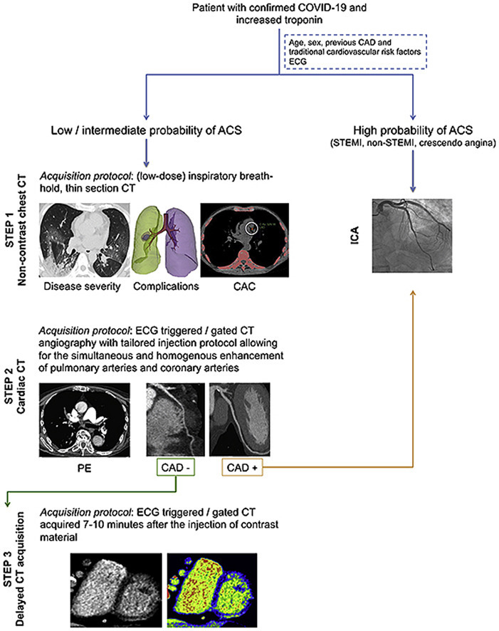
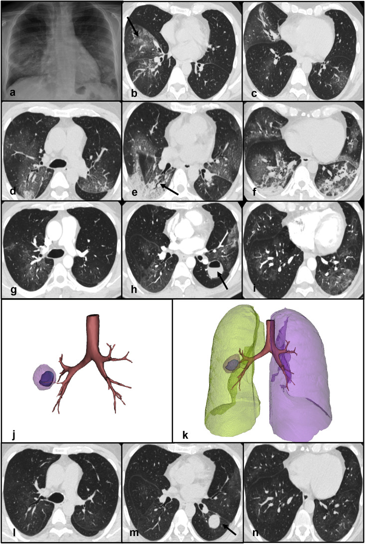
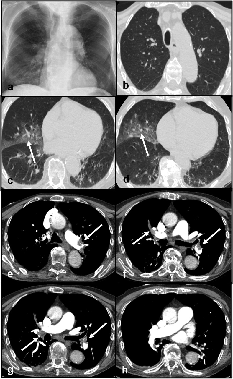
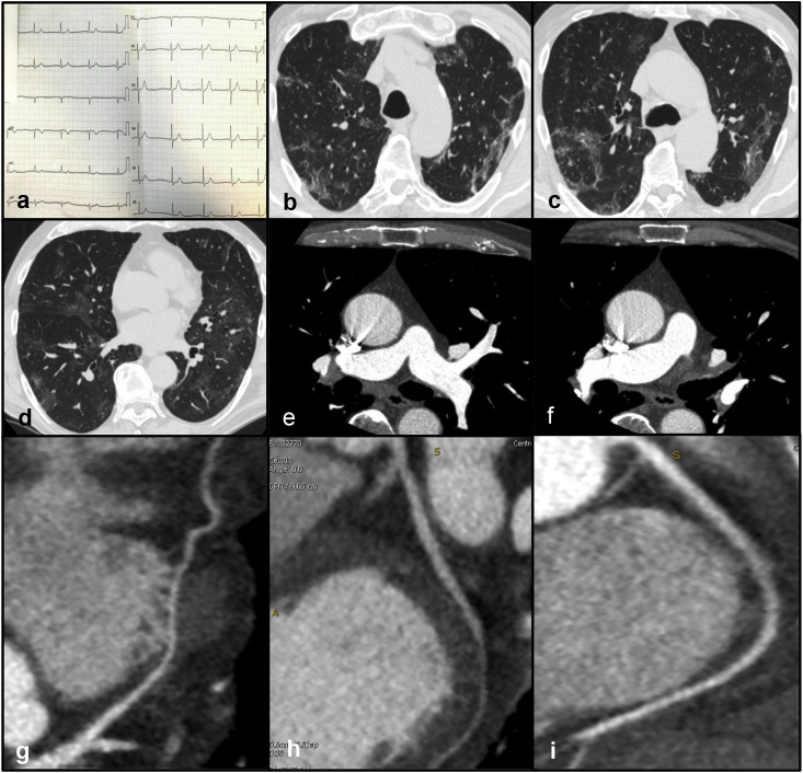
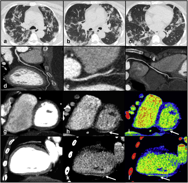
Coronavirus disease 2019 (COVID-19) has become a rapid worldwide pandemic. While COVID-19 primarily manifests as an interstitial pneumonia and severe acute respiratory distress syndrome, severe involvement of other organs has been documented. In this article, we will review the role of non-contrast chest computed tomography in the diagnosis, follow-up and prognosis of patients affected by COVID-19 pneumonia with a detailed description of the imaging findings that may be encountered. Given that patients with COVID-19 may also suffer from coagulopathy, we will discuss the role of CT pulmonary angiography in the detection of acute pulmonary embolism. Finally, we will describe more advanced applications of CT in the differential diagnosis of myocardial injury with an emphasis on ruling out acute coronary syndrome and myocarditis.

Keywords: COVID-19; Computed tomography; Myocardial injury; Pneumonia; Pulmonary embolism.


Conflict of interest statement

Declaration of competing interest The authors declare that they have no known competing financial interests or personal relationships that could have appeared to influence the work reported in this paper.

Figures

Image 1
Graphical abstract
Fig. 1
Fig. 1
Panel a - Frontal chest X-ray demonstrating subtle, bilateral opacities mainly involving the mid and lower zones of the lungs. Panels b, c - Non-contrast chest CT scan showing multiple ground glass opacities and “crazy-paving” pattern (arrow - panel b) in the right middle and lower lobes. Panels d, e, f - Non-contrast chest CT scan documenting increased extent of GGOs mixed with new areas of consolidation in the lower pulmonary lobes. An air bronchogram is visible in the right lower lobe (arrow - panel e). Panels g, h, i - Contrast CT scan showing a lung abscess containing an air-fluid level in the superior segment of the left upper lobe (arrow - panel h). GGOs and consolidative areas are significantly decreased compared to the previous CT scan. Panels j, k - 3D reconstructions of the lung abscess. Panels l, m, n - Non-contrast chest CT demonstrating the lung abscess (arrow - panel m) and the near resolution of the parenchymal abnormalitiesAbbreviations CT: computed tomography; GGO: ground-glass opacities.
Fig. 2
Fig. 2
Panel a - Frontal chest X-ray demonstrating a consolidation in the lower zone of the right lung. Panels b, c, d - Non-contrast CT scan showing multiple GGOs in both lungs (arrows). Panels e, f, g, h - CT pulmonary angiography documenting multiple filling defects in the pulmonary arteries (arrows). Abbreviations: CT: computed tomography; GGO: ground-glass opacities.
Fig. 3
Fig. 3
Panel a - Normal ECG. Panels b, c, d - Non-contrast CT documenting multiple, patchy GGOs in both lungs consistent with viral pneumonia. Panels e, f - CCTA excluding acute pulmonary embolism. Panels g, h, d – CCTA showing absence of coronary artery disease in the left anterior descending artery (panel g), left circumflex (panel h) and right coronary artery (panel i) Abbreviations: CCTA: coronary computed tomography angiography; ECG: electrocardiogram.
Fig. 4
Fig. 4
Panels a, b, c - Non-contrast chest CT demonstrating the presence of multiple, bilateral GGOs mixed with areas of consolidation with a predominant sub-pleural distribution in both lungs. Panels d, e, f - CCTA showing absence of coronary artery disease in the left anterior descending artery (panel d), left circumflex (panel e) and right coronary artery (panel f). Panels g, j - CCTA excluding perfusion defects in the left ventricular myocardium. Panels h, k, i, l - Delayed post-contrast scan (panels h, k) and corresponding color-maps (panels i, l) documenting a sub-epicardial area of hyperdensity in the basal, infero-lateral wall of the left ventricleAbbreviations CCTA: coronary computed tomography angiography; CT: computed tomography; GGO: ground-glass opacities. (For interpretation of the references to color in this figure legend, the reader is referred to the Web version of this article.)

References

    1. Guzik T.J., Mohiddin S.A., Dimarco A., et al. COVID-19 and the cardiovascular system: Implications for risk assessment, diagnosis, and treatment options. Cardiovasc Res. 2020;116:1666–1687. - PMC - PubMed
    1. Huang C., Wang Y., Li X., et al. Clinical features of patients infected with 2019 novel coronavirus in Wuhan, China. Lancet. 2020;395:497–506. - PMC - PubMed
    1. Wu Z., McGoogan J.M. Characteristics of and important lessons from the coronavirus disease 2019 (COVID-19) outbreak in China: Summary of a report of 72314 cases from the Chinese center for disease control and prevention. J Am Med Assoc. 2020 doi: 10.1001/jama.2020.2648. In press. - DOI - PubMed
    1. General Office of the National Health Commission and the Office of the National Administration of Traditional Chinese Medicine . 2020. Diagnosis and Treatment Protocol for COVID-19 (Trial Version 7)
    1. ACR . 2020. ACR Recommendations for the Use of Chest Radiography and Computed Tomography (CT) for Suspected COVID-19 Infection.