

Tumor necrosis factor alpha receptor 1 deficiency in hepatocytes does not protect from non-alcoholic steatohepatitis, but attenuates insulin resistance in mice
- PMID: 32952340
- PMCID: PMC7476178
- DOI: 10.3748/wjg.v26.i33.4933
Tumor necrosis factor alpha receptor 1 deficiency in hepatocytes does not protect from non-alcoholic steatohepatitis, but attenuates insulin resistance in mice
Abstract
Background: End-stage liver disease caused by non-alcoholic steatohepatitis (NASH) is the second leading indication for liver transplantation. To date, only moderately effective pharmacotherapies exist to treat NASH. Understanding the pathogenesis of NASH is therefore crucial for the development of new therapies. The inflammatory cytokine tumor necrosis factor alpha (TNF-α) is important for the progression of liver disease. TNF signaling via TNF receptor 1 (TNFR1) has been hypothesized to be important for the development of NASH and hepatocellular carcinoma in whole-body knockout animal models.
Aim: To investigate the role of TNFR1 signaling in hepatocytes for steatohepatitis development in a mouse model of diet-induced NASH.
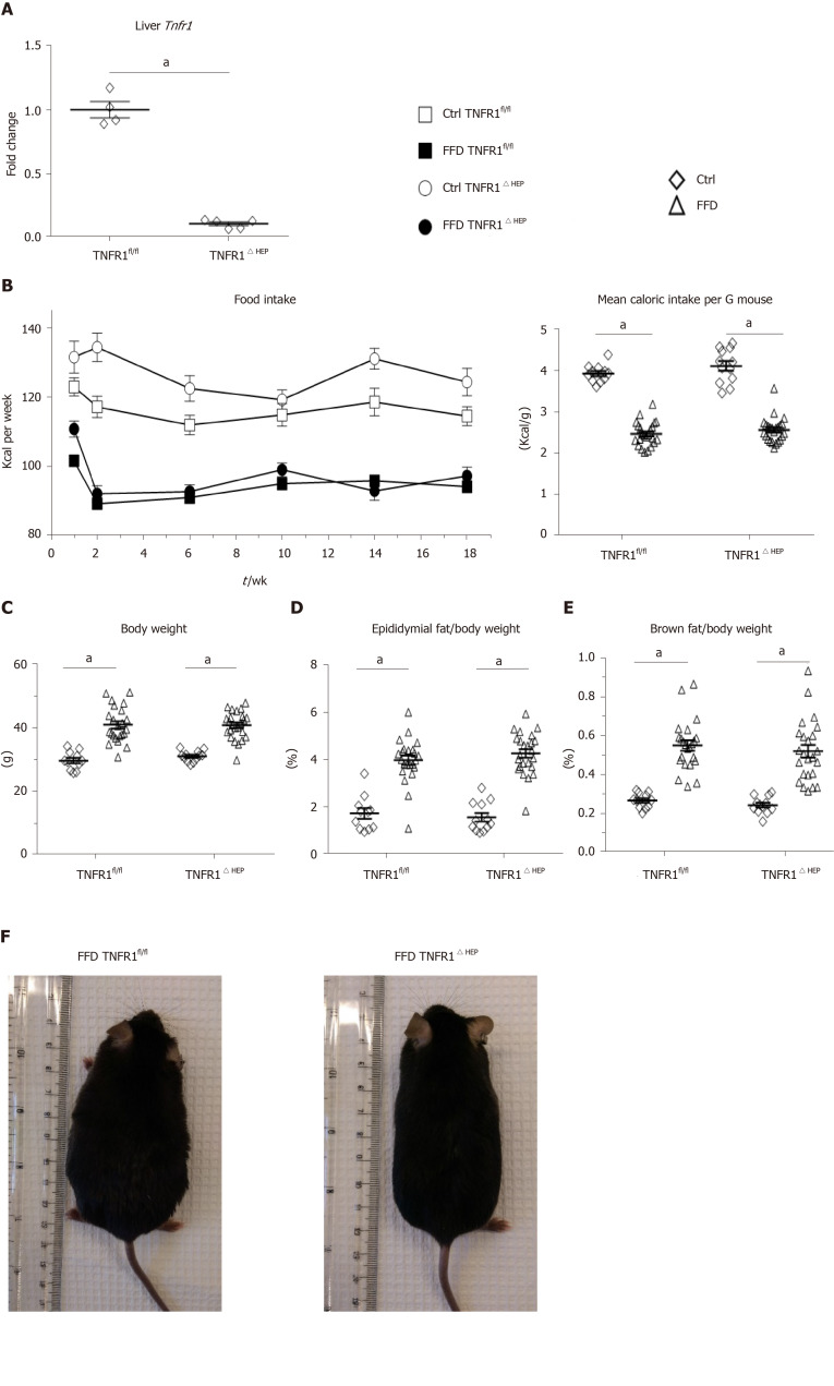
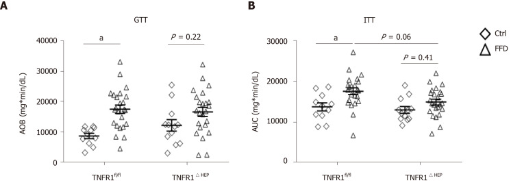
Methods: NASH was induced by a western-style fast-food diet in mice deficient for TNFR1 in hepatocytes (TNFR1ΔHEP) and their wild-type littermates (TNFR1fl/fl). Glucose tolerance was assessed after 18 wk and insulin resistance after 19 wk of feeding. After 20 wk mice were assessed for features of NASH and the metabolic syndrome such as liver weight, liver steatosis, liver fibrosis and markers of liver inflammation.
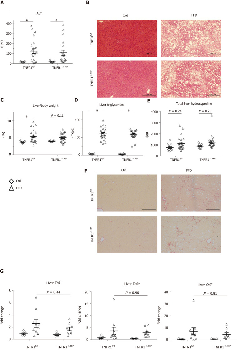
Results: Obesity, liver injury, inflammation, steatosis and fibrosis was not different between TNFR1ΔHEP and TNFR1fl/fl mice. However, Tnfr1 deficiency in hepatocytes protected against glucose intolerance and insulin resistance.
Conclusion: Our results indicate that deficiency of TNFR1 signaling in hepatocytes does not protect from diet-induced NASH. However, improved insulin resistance in this model strengthens the role of the liver in glucose homeostasis.
Keywords: Glucose intolerance; Insulin resistance; Non-alcoholic fatty liver disease; Non-alcoholic steatohepatitis; Tumor necrosis factor alpha receptor 1; Type 2 diabetes.
©The Author(s) 2020. Published by Baishideng Publishing Group Inc. All rights reserved.
Conflict of interest statement
Conflict-of-interest statement: Schnabl B has been consulting for Ferring Research Institute, HOST Therabiomics, Intercept Pharmaceuticals and Patara Pharmaceuticals. Schnabl B’s institution UC San Diego has received research support from Axial Biotherapeutics, BiomX, CymaBay Therapeutics, NGM Biopharmaceuticals, and Synlogic Operating Company.
Figures
Similar articles
-
Macrophage p38α promotes nutritional steatohepatitis through M1 polarization.J Hepatol. 2019 Jul;71(1):163-174. doi: 10.1016/j.jhep.2019.03.014. Epub 2019 Mar 23. J Hepatol. 2019. PMID: 30914267
-
Role of XBP1 in regulating the progression of non-alcoholic steatohepatitis.J Hepatol. 2022 Aug;77(2):312-325. doi: 10.1016/j.jhep.2022.02.031. Epub 2022 Mar 12. J Hepatol. 2022. PMID: 35292349
-
Disruption of tumor necrosis factor alpha receptor 1 signaling accelerates NAFLD progression in mice upon a high-fat diet.J Nutr Biochem. 2018 Aug;58:17-27. doi: 10.1016/j.jnutbio.2018.04.013. Epub 2018 May 2. J Nutr Biochem. 2018. PMID: 29860102
-
A Comparison of the Gene Expression Profiles of Non-Alcoholic Fatty Liver Disease between Animal Models of a High-Fat Diet and Methionine-Choline-Deficient Diet.Molecules. 2022 Jan 27;27(3):858. doi: 10.3390/molecules27030858. Molecules. 2022. PMID: 35164140 Free PMC article. Review.
-
[VNUT Is a Therapeutic Target for Type 2 Diabetes and NASH].Yakugaku Zasshi. 2021;141(4):517-526. doi: 10.1248/yakushi.20-00204-4. Yakugaku Zasshi. 2021. PMID: 33790119 Review. Japanese.
Cited by
-
Apoptotic cell death in disease-Current understanding of the NCCD 2023.Cell Death Differ. 2023 May;30(5):1097-1154. doi: 10.1038/s41418-023-01153-w. Epub 2023 Apr 26. Cell Death Differ. 2023. PMID: 37100955 Free PMC article. Review.
-
Metabolic Messengers: tumour necrosis factor.Nat Metab. 2021 Oct;3(10):1302-1312. doi: 10.1038/s42255-021-00470-z. Epub 2021 Oct 14. Nat Metab. 2021. PMID: 34650277 Review.
-
Overview of Cellular and Soluble Mediators in Systemic Inflammation Associated with Non-Alcoholic Fatty Liver Disease.Int J Mol Sci. 2023 Jan 24;24(3):2313. doi: 10.3390/ijms24032313. Int J Mol Sci. 2023. PMID: 36768637 Free PMC article. Review.
-
The Roles of TNF Signaling Pathways in Metabolism of Bone Tumors.Front Pharmacol. 2022 Jun 29;13:907629. doi: 10.3389/fphar.2022.907629. eCollection 2022. Front Pharmacol. 2022. PMID: 35847045 Free PMC article.
-
The TNFR1 Antagonist Atrosimab Is Therapeutic in Mouse Models of Acute and Chronic Inflammation.Front Immunol. 2021 Jul 7;12:705485. doi: 10.3389/fimmu.2021.705485. eCollection 2021. Front Immunol. 2021. PMID: 34305946 Free PMC article.
References
-
- Angulo P. Nonalcoholic fatty liver disease. N Engl J Med. 2002;346:1221–1231. - PubMed
-
- Vernon G, Baranova A, Younossi ZM. Systematic review: the epidemiology and natural history of non-alcoholic fatty liver disease and non-alcoholic steatohepatitis in adults. Aliment Pharmacol Ther. 2011;34:274–285. - PubMed
-
- Charlton M, Krishnan A, Viker K, Sanderson S, Cazanave S, McConico A, Masuoko H, Gores G. Fast food diet mouse: novel small animal model of NASH with ballooning, progressive fibrosis, and high physiological fidelity to the human condition. Am J Physiol Gastrointest Liver Physiol. 2011;301:G825–G834. - PMC - PubMed
-
- Patel SS, Siddiqui MS. Current and Emerging Therapies for Non-alcoholic Fatty Liver Disease. Drugs. 2019;79:75–84. - PubMed
-
- Abdou RM, Zhu L, Baker RD, Baker SS. Gut Microbiota of Nonalcoholic Fatty Liver Disease. Dig Dis Sci. 2016;61:1268–1281. - PubMed
MeSH terms
Substances
Grants and funding
LinkOut - more resources
Full Text Sources
Medical
Molecular Biology Databases
