Review
. 2020 Sep;36(5):409-415.
doi: 10.6515/ACS.202009_36(5).20200516A.

Delayed Coronary Obstruction after Transcatheter Aortic Valve Replacement - An Uncommon But Serious Complication

Affiliations
Review

Delayed Coronary Obstruction after Transcatheter Aortic Valve Replacement - An Uncommon But Serious Complication

Wei-Hsian Yin et al. Acta Cardiol Sin. 2020 Sep.

Abstract

As transcatheter aortic valve replacement (TAVR) becomes the mainstream treatment for valvular aortic stenosis, it is vitally important to recognize its associated procedural complications. Among the clinically relevant but uncommonly seen complications, the development of delayed coronary obstruction (DCO) occurring during the early post-procedural phase or even later following the index TAVR procedure, has been reported. These reports have raised concerns as TAVR comes more common in lower-risk patients. In this review article, we explored the implications of DCO for pre-procedural computed tomography evaluation, valve selection and sizing, intra-procedural manipulation, and approaches to post-procedural management.

Keywords: Complication; Delayed coronary obstruction; Transcatheter aortic valve replacement.


Figures

Figure 1
Figure 1
An 84-year-old woman was admitted with symptomatic, severe aortic stenosis for transcatheter aortic valve replacement. Her aortic valve annulus measured 19.4 mm on three-dimensional computed tomography imaging. The left and right coronary ostial height was 9 mm and 10 mm, respectively. A 26 mm CoreValve (Medtronic) was deployed in a good anatomical position. Aortic root angiography suggested the presence of bioprosthetic valve struts and tissue close to, but not obstructing, the left coronary ostium (A, arrow). As the patient remained hemodynamically stable, sheaths were removed and the arteriotomy at the femoral artery was closed. However, about 1 hour later, she became abruptly hypotensive, and severe ST-segment elevation appeared on electrocardiograph monitor was noted. Hemodynamic status deteriorated rapidly and cardiopulmonary resuscitation was initiated. Emergency percutaneous coronary intervention was planned, but total occlusion of the left main stem (LMS) by the CoreValve with obliteration of the space between the bioprosthesis and the coronary orifice (B, arrow), which precluded the engagement of guiding catheter and wiring of the coronary arteries, was demonstrated (C, arrow). Therefore, emergency coronary artery bypass grafting was performed. Post-operative aortogram showed improved left ventricular function, patent LMS (D, arrow) and left internal mammary artery graft to left anterior descending artery (D, small arrow).
Figure 2
Figure 2
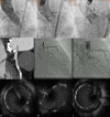
A 90-year-old woman was admitted with symptomatic, severe aortic stenosis for transcatheter aortic valve replacement (TAVR). Pre-implantation computed tomography indicated left and right coronary ostial heights of 13.4 mm and 17.6 mm, respectively. During balloon valvuloplasty for pre-dilatation with a 25 mm × 40 mm balloon, the left aortic leaflet was displaced toward the left coronary ostium (A, arrow); therefore, we decided to perform coronary protection. A 29 mm SAPIEN XT valve (Edwards Lifesciences) was then implanted. Aortography suggested the presence of stent frames of TAVR device close to, but not obstructing, the left coronary ostium (B, arrow). A post-TAVR selective angiography also showed a patent left main stem (LMS) with TIMI 3 flow (C, arrow). The patient was discharged uneventfully but a computed tomography follow-up six month after TAVR showed new tissue growth near the ostium of LMS (D, arrow). The patient declined percutaneous coronary intervention at that time because she continued to do well. Another 5 months passed, the patient was admitted for unstable angina and the LMS was stented with a 4.0 mm × 12 mm integrity resolute stent (Medtronic) (E, arrow). Post-dilatation with a 4.5 mm non-compliant balloon led to improvement in the angiographic appearance of the stent (F, arrows). Intravascular ultrasound examination demonstrated improvement in minimum lumen area and diameter, but residual ovoid-shaped stented segment discovered at the site of the impingement of stent frames of the TAVR device (G-I).

References

    1. Baumgartner H, Falk V, Bax JJ, et al. 2017 ESC/EACTS Guidelines for the management of valvular heart disease. Eur Heart J. 2017;38:2739–2791. - PubMed
    1. Cahill TJ, Chen M, Hayashida K, et al. Transcatheter aortic valve implantation: current status and future perspectives. Eur Heart J. 2018;39:2625–2634. - PubMed
    1. Leon MB, Smith CR, Mack MJ, et al. Transcatheter or surgical aortic-valve replacement in intermediate-risk patients. N Engl J Med. 2016;374:1609–1620. - PubMed
    1. Reardon MJ, Van Mieghem NM, Popma JJ, et al. Surgical or transcatheter aortic-valve replacement in intermediate-risk patients. N Engl J Med. 2017;376:1321–1331. - PubMed
    1. Thyregod HG, Steinbrüchel DA, Ihlemann N, et al. Transcatheter versus surgical aortic valve replacement in patients with severe aortic valve stenosis: 1-year results from the all-comers NOTION randomized clinical trial. J Am Coll Cardiol. 2015;65:2184–2194. - PubMed

LinkOut - more resources