

Naringin and bone marrow mesenchymal stem cells repair articular cartilage defects in rabbit knees through the transforming growth factor-β superfamily signaling pathway
- PMID: 32952649
- PMCID: PMC7485297
- DOI: 10.3892/etm.2020.9187
Naringin and bone marrow mesenchymal stem cells repair articular cartilage defects in rabbit knees through the transforming growth factor-β superfamily signaling pathway
Abstract
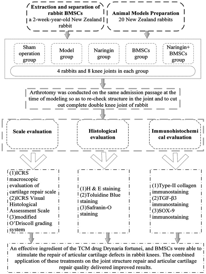
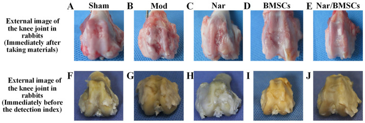
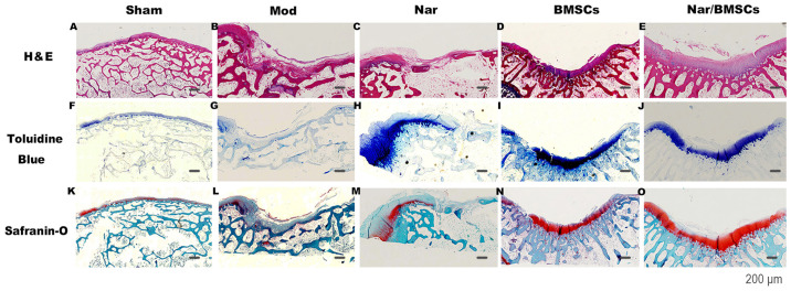
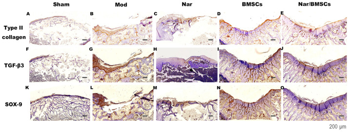
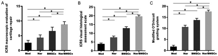
The present study aimed to assess the effect of a combination of naringin and rabbit bone marrow mesenchymal stem cells (BMSCs) on the repair of cartilage defects in rabbit knee joints and to assess possible involvement of the transforming growth factor-β (TGF-β) signaling pathway in this process. After establishing an articular cartilage defect model in rabbit knees, 20 New Zealand rabbits were divided into a sham operation group (Sham), a model group (Mod), a naringin treatment group (Nar), a BMSC group (BMSCs) and a naringin + BMSC group (Nar/BMSCs). At 12 weeks after treatment, the cartilage was evaluated using the International Cartilage Repair Society (ICRS)'s macroscopic evaluation of cartilage repair scale, the ICRS's visual histological assessment scale, the Modified O'Driscoll grading system, histological staining (hematoxylin and eosin staining, toluidine blue staining and safranin O staining) and immunohistochemical staining (type-II collagen, TGF-β3 and SOX-9 immunostaining). Using the above grading systems to quantify the extent of repair, histological quantification and macro quantification of joint tissue repair showed that the Nar/BMSCs group displayed repair after treatment in comparison to the untreated Mod group. Among the injury model groups (Mod, Nar, BMSCs and Nar/BMSCs), the Nar/BMSCs group displayed the highest degree of morphological repair. The results of histological and immunohistochemical staining of the repaired region of the joint defect indicated that the BMSCs had a satisfactory effect on the repair of the joint structure but had a poor effect on the repair of cartilage quality. The Nar/BMSCs group displayed satisfactory therapeutic effects on both repair of the joint structure and cartilage quality. The expression level of type-II collagen was high in the Nar/BMSCs group. Additionally, staining of TGF-β3 and SOX-9 in the Nar/BMSCs group was the strongest compared with that of any other group in the present study. Naringin and/BMSCs together demonstrated a more efficient repair effect on articular cartilage defects in rabbit knees than the use of either treatment alone in terms of joint structure and cartilage quality. One potential mechanism of naringin action may be through activation and continuous regulation of the TGF-β superfamily signaling pathway, which can promote BMSCs to differentiate into chondrocytes.
Keywords: bone mesenchymal stem cells; cartilage defect; knee joint; naringin; rabbit; superfamily signaling pathway; transforming growth factor-β.
Copyright: © Ye et al.
Figures
Similar articles
-
Naringin in the repair of knee cartilage injury via the TGF-β/ALK5/Smad2/3 signal transduction pathway combined with an acellular dermal matrix.J Orthop Translat. 2021 Aug 6;32:1-11. doi: 10.1016/j.jot.2021.06.004. eCollection 2022 Jan. J Orthop Translat. 2021. PMID: 35591936 Free PMC article.
-
Demineralized bone matrix combined bone marrow mesenchymal stem cells, bone morphogenetic protein-2 and transforming growth factor-β3 gene promoted pig cartilage defect repair.PLoS One. 2014 Dec 29;9(12):e116061. doi: 10.1371/journal.pone.0116061. eCollection 2014. PLoS One. 2014. PMID: 25545777 Free PMC article.
-
Naringin in repairing articular cartilage injury by activating TGF-β/Smad signaling pathway to attenuate inflammatory response.Arch Biochem Biophys. 2025 Jun;768:110396. doi: 10.1016/j.abb.2025.110396. Epub 2025 Mar 20. Arch Biochem Biophys. 2025. PMID: 40120921
-
New Perspectives in the Pharmacological Potential of Naringin in Medicine.Curr Med Chem. 2021;28(10):1987-2007. doi: 10.2174/0929867327666200604171351. Curr Med Chem. 2021. PMID: 32496985 Review.
-
Chondrodysplasias and TGFβ signaling.Bonekey Rep. 2015 Mar 11;4:642. doi: 10.1038/bonekey.2015.9. eCollection 2015. Bonekey Rep. 2015. PMID: 25798233 Free PMC article. Review.
Cited by
-
Protective effects of naringin on glucocorticoid-induced osteoporosis through regulating the PI3K/Akt/mTOR signaling pathway.Am J Transl Res. 2021 Jun 15;13(6):6330-6341. eCollection 2021. Am J Transl Res. 2021. PMID: 34306372 Free PMC article.
-
TP8, A Novel Chondroinductive Peptide, Significantly Promoted Neo-Cartilage Repair without Activating Bone Formation.Adv Healthc Mater. 2025 Mar;14(6):e2401752. doi: 10.1002/adhm.202401752. Epub 2024 Dec 17. Adv Healthc Mater. 2025. PMID: 39690790 Free PMC article.
-
The Development of Naringin for Use against Bone and Cartilage Disorders.Molecules. 2023 Apr 25;28(9):3716. doi: 10.3390/molecules28093716. Molecules. 2023. PMID: 37175126 Free PMC article. Review.
-
The role of the immune microenvironment in bone, cartilage, and soft tissue regeneration: from mechanism to therapeutic opportunity.Mil Med Res. 2022 Nov 19;9(1):65. doi: 10.1186/s40779-022-00426-8. Mil Med Res. 2022. PMID: 36401295 Free PMC article. Review.
-
Three-Dimensional Bioprinting: A Comprehensive Review for Applications in Tissue Engineering and Regenerative Medicine.Bioengineering (Basel). 2024 Jul 31;11(8):777. doi: 10.3390/bioengineering11080777. Bioengineering (Basel). 2024. PMID: 39199735 Free PMC article. Review.
References
LinkOut - more resources
Full Text Sources
Research Materials
Miscellaneous