

Morphological Alteration and TGF-β1 Expression in Multifidus with Lumbar Disc Herniation
- PMID: 32952922
- PMCID: PMC7474038
- DOI: 10.1007/s43465-020-00213-4
Morphological Alteration and TGF-β1 Expression in Multifidus with Lumbar Disc Herniation
Abstract
Background: Lumbar disc herniation (LDH) can cause lumbar nerve root compression, which can lead to denervated atrophy of paraspinal muscles theoretically, however, the conclusions of morphological alteration in multifidus with LDH remain controversial. Transforming growth factor-beta 1 (TGF-β1) plays an essential role in the development of tissue fibrosis and is a molecular marker in the study of muscle fibrosis, but no relevant studies on TGF-β1 expression in multifidus have been reported so far. This study is to observe altered morphology of multifidus in patients with LDH, and to explore the correlation between multifidus fibrosis and TGF-β1 expression.
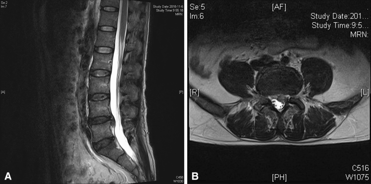
Materials and methods: 46 LDH patients with low back pain combined with unilateral leg radiation pain and/or numbness were selected. Patients were divided into four groups according to their medical histories. Group 1: medical history less than 6 months (15 cases); group 2: a medical history of 6-12 months (10 cases); group 3: a medical history of 12-24 months (13 cases); and group 4: medical history > 24 months (8 cases). Bilateral multifidus specimens were taken from compressed nerve root segments, and morphological changes in multifidus were determined. Multi-parameter changes in TGF-β1 expression in multifidus were observed by immunohistochemistry and immunofluorescence.
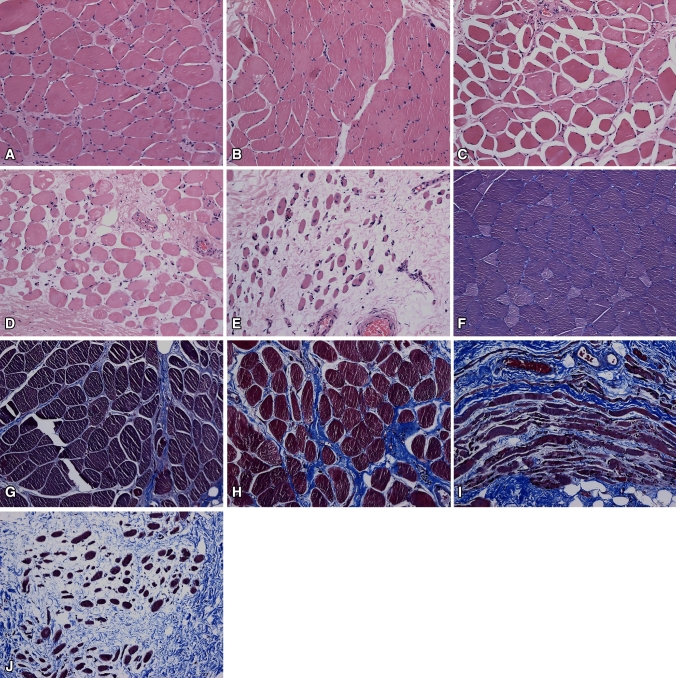
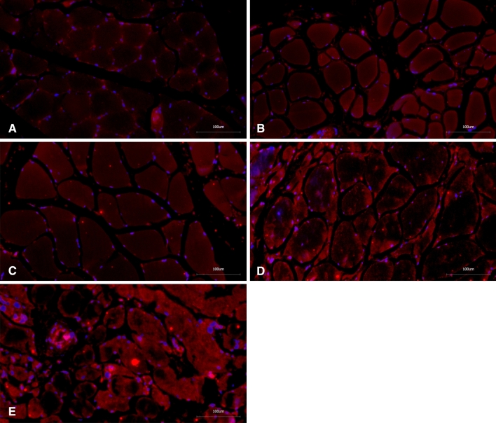
Results: HE staining showed that the cross-sectional area (CSA) of multifidus in the involved sides decreased and muscle fibers atrophied. Masson's trichrome staining showed a decrease in the sectional area ratio of myofibers to collagen fibers in the involved side. In groups 1 and 2, there were no significant differences in the aforementioned parameters. In groups 3 and 4, statistically significant differences in the sectional area ratio of myofibers to collagen fibers in both sides were seen (P < 0.05). TGF-β1 expression was significantly enhanced in both muscle cells and the matrix of the involved side, while no expression or a little expression was found in the matrix in the uninvolved side. In group 1, there was no statistically significant difference in TGF-β1 expression in both sides. In the remaining three groups, TGF-β1 expression in the involved sides was higher than were found in the uninvolved sides.
Conclusions: Nerve root compression by LDH leads to multifidus atrophy, fibrosis, and increased TGF-β1 expression, which might promote multifidus fibrosis.Trials registration All Clinical Trials done in India should preferably be registered with the Clinical Trials Registry of India, set up by the Indian Council of Medical Research (website: http://ctri.nic.in). Authors should provide the CTRI number along with the manuscript.
Keywords: Fibrosis; Intervertebral disc degeneration; Lumbar disc herniation; Lumbar vertebrae; Microscopic findings; Morphological; Multifidus; Paraspinal muscles; Transforming growth factor beta 1.
© Indian Orthopaedics Association 2020.
Conflict of interest statement
Conflict of InterestThe authors declare that they have no conflict of interest.
Figures

Similar articles
-
Correlation between inflammatory cytokine expression in paraspinal tissues and severity of disc degeneration in individuals with lumbar disc herniation.BMC Musculoskelet Disord. 2023 Mar 14;24(1):193. doi: 10.1186/s12891-023-06295-z. BMC Musculoskelet Disord. 2023. PMID: 36918849 Free PMC article.
-
Unilateral changes of the multifidus in persons with lumbar disc herniation: a systematic review and meta-analysis.Spine J. 2020 Oct;20(10):1573-1585. doi: 10.1016/j.spinee.2020.04.007. Epub 2020 Apr 20. Spine J. 2020. PMID: 32325246
-
Histochemistry and morphology of the multifidus muscle in lumbar disc herniation: comparative study between diseased and normal sides.Spine (Phila Pa 1976). 2000 Sep 1;25(17):2191-9. doi: 10.1097/00007632-200009010-00009. Spine (Phila Pa 1976). 2000. PMID: 10973402
-
Correlation of multifidus degeneration with sex, age and side of herniation in patients with lumbar disc herniation.BMC Musculoskelet Disord. 2023 Aug 16;24(1):652. doi: 10.1186/s12891-023-06783-2. BMC Musculoskelet Disord. 2023. PMID: 37587417 Free PMC article.
-
Relationships between paraspinal muscle morphology and neurocompressive conditions of the lumbar spine: a systematic review with meta-analysis.BMC Musculoskelet Disord. 2018 Sep 27;19(1):351. doi: 10.1186/s12891-018-2266-5. BMC Musculoskelet Disord. 2018. PMID: 30261870 Free PMC article.
Cited by
-
Correlation between inflammatory cytokine expression in paraspinal tissues and severity of disc degeneration in individuals with lumbar disc herniation.BMC Musculoskelet Disord. 2023 Mar 14;24(1):193. doi: 10.1186/s12891-023-06295-z. BMC Musculoskelet Disord. 2023. PMID: 36918849 Free PMC article.
-
Spinal degeneration is associated with lumbar multifidus morphology in secondary care patients with low back or leg pain.Sci Rep. 2022 Aug 29;12(1):14676. doi: 10.1038/s41598-022-18984-1. Sci Rep. 2022. PMID: 36038653 Free PMC article.
References
-
- Ploumis A, Michailidis N, Christodoulou P, Kalaitzoglou I, Gouvas G, Beris A. Ipsilateral atrophy of paraspinal and psoas muscle in unilateral back pain patients with monosegmental degenerative disc disease. British Journal of Radiology. 2011;84:709–713. doi: 10.1259/bjr/58136533. - DOI - PMC - PubMed
LinkOut - more resources
Full Text Sources