. 2019 Sep 27;10(11):1948-1957.
doi: 10.1039/c9md00372j. eCollection 2019 Nov 1.

Characterization of the genomically encoded fosfomycin resistance enzyme from Mycobacterium abscessus

Affiliations

Characterization of the genomically encoded fosfomycin resistance enzyme from Mycobacterium abscessus

Skye Travis et al. Medchemcomm. .

Abstract

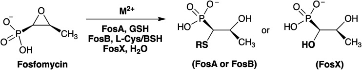
Mycobacterium abscessus belongs to a group of rapidly growing mycobacteria (RGM) and accounts for approximately 65-80% of lung disease caused by RGM. It is highly pathogenic and is considered the prominent Mycobacterium involved in pulmonary infection in patients with cystic fibrosis and chronic pulmonary disease (CPD). FosM is a putative 134 amino acid fosfomycin resistance enzyme from M. abscessus subsp. bolletii that shares approximately 30-55% sequence identity with other vicinal oxygen chelate (VOC) fosfomycin resistance enzymes and represents the first of its type found in any Mycobacterium species. Genes encoding VOC fosfomycin resistance enzymes have been found in both Gram-positive and Gram-negative pathogens. Given that FosA enzymes from Gram-negative bacteria have evolved optimum activity towards glutathione (GSH) and FosB enzymes from Gram-positive bacteria have evolved optimum activity towards bacillithiol (BSH), it was originally suggested that FosM might represent a fourth class of enzyme that has evolved to utilize mycothiol (MSH). However, a sequence similarity network (SSN) analysis identifies FosM as a member of the FosX subfamily, indicating that it may utilize water as a substrate. Here we have synthesized MSH and characterized FosM with respect to divalent metal ion activation and nucleophile selectivity. Our results indicate that FosM is a Mn2+-dependent FosX-type hydrase with no selectivity toward MSH or other thiols as analyzed by NMR and mass spectroscopy.


Figures

Fig. 1
Fig. 1. Reactions catalyzed by fosfomycin resistance enzymes.
Fig. 2
Fig. 2. Structures of low molecular weight thiols found in Gram-negative bacteria, Gram-positive bacteria, and Mycobacteria. Gram-negative bacteria biosynthesize GSH, Gram-positive bacteria biosynthesize BSH, and Mycobacteria biosynthesize MSH.
Fig. 3
Fig. 3. Sequence alignment of fosfomycin resistance enzymes. Alignment was performed on the sequences of FosM from Mycobacterium abscessus, FosX from Listeria monocytogenes, FosB from Bacillus cereus, FosB from Staphylococcus aureus, and FosA from Pseudomonas aeruginosa. Residues marked with (*) are residues used in binding metals in the active site. Residues marked with (#) are residues important in coordinating fosfomycin. Red blocks depict residues that are conserved throughout all sequences. Blue boxes and red text depict residues or motifs that are highly, but not universally, conserved.
Fig. 4
Fig. 4. Sequence similarity networks of the IPR037434 family. The sequence similarity networks were visualized using Cytoscape as described in materials and methods. To visualize how the clusters develop, networks constructed at alignment score cutoff of 10–45 (A), 10–50 (B), and 10–55 (C) are shown. Nodes are colored by taxonomic phylum as described in the legend. Nodes with a diamond shape have structural data reported in the PDB databank. The network in panel C has 223 nodes and 2066 edges with an average of 84% sequence identity over 135 residues.
Fig. 5
Fig. 5. Circular dichroism (CD) spectra of the fosfomycin resistance enzymes, FosA from P. aeruginosa, FosB from B. cereus, FosX from L. monocytogenes, and FosM from M. abscessus. The proteins were stored in 20 mM HEPES pH 7.0 and diluted in MQ water for the scan. The distinct secondary structure of FosM (black) is richer in β-sheet character than that shown for FosA (green) and FosX (red) enzymes as indicated by the relatively sharper peak at ∼210 nm.
Fig. 6
Fig. 6. TOF MS ES data of FosM with various substrates and product. Reactions were carried out in water at 25 °C with: 8 mM fosfomycin (A); 0.6 μM FosM and 8 mM fosfomycin (B); 0.6 μM FosM, 8 mM fosfomycin, and 4 mM l-Cys (C); 0.6 μM FosM, 8 mM fosfomycin, and 4 mM GSH (D); 0.6 μM FosM, 8 mM fosfomycin, and 4 mM MSH (E). Other masses present in (E) are due to residual impurities from the MSH synthesis.
Fig. 7
Fig. 7. Time-trace kinetics for the FosM-catalyzed addition of water to fosfomycin in the presence of Mn2+ (▲), Mg2+ (■), or Zn2+ ([black circle]). Reactions were carried out at 25 °C with 8 mM fosfomycin and 0.6 μM enzyme in 20 mM HEPES, pH 7.5. In the absence of FosM, no hydrated fosfomycin product was formed under otherwise identical conditions (Fig. S4†).
Fig. 8
Fig. 8. (Left) FosM homology model based on FosX with the two subunits illustrated in blue and red. The positions of the Mn2+ ions are shown in purple. This image was generated using the program Chimera. (Right) Overlay of the active site residues of FosA (blue PDB entry ; 1LQP), FosB (green PDB entry ; 4JH6), FosX (purple; PDB entry ; 1R9C), and FosM (grey) with bound fosfomycin from FosA and FosB and Mn2+.

References

    1. Griffith D. E., Aksamit T., Brown-Elliot B. A., Catanzaro A., Daley C., Gordin F., Holland S. M., Horsburgh R., Huitt G., Iademarco M. F., Iseman M., Olivier K., Ruoss S., von Reyn C. F., Wallace, Jr. R. J., Winthrop K. Am. J. Respir. Crit. Care Med. 2007;175:367–416. - PubMed
    1. Koh W.-J., Kwon O. J., Lee N. Y., Kook Y.-H., Lee H.-K., Kim B.-J. J. Clin. Microbiol. 2009;47:3362–3366. - PMC - PubMed
    1. Zelazny A. M., Root J. M., Shea Y. R., Colombo R. E., Shamputa I. C., Stock F., Conlan S., McNulty S., Brown-Elliott B. A., Wallace, Jr. R. J., Olivier K. N., Holland S. M., Sampaio E. P. J. Clin. Microbiol. 2009;47:1985–1995. - PMC - PubMed
    1. Adekambi T., Berger P., Raoult D., Drancourt M. Int. J. Syst. Evol. Microbiol. 2006;56:133–143. - PubMed
    1. Griffith D. E., Girard W. M., Wallace, Jr. R. J. Am. Rev. Respir. Dis. 1993;147:1271–1278. - PubMed