

The diagnostic value of MRI for architectural distortion categorized as BI-RADS category 3-4 by mammography
- PMID: 32953609
- PMCID: PMC7475343
- DOI: 10.21037/gs-20-505
The diagnostic value of MRI for architectural distortion categorized as BI-RADS category 3-4 by mammography
Abstract
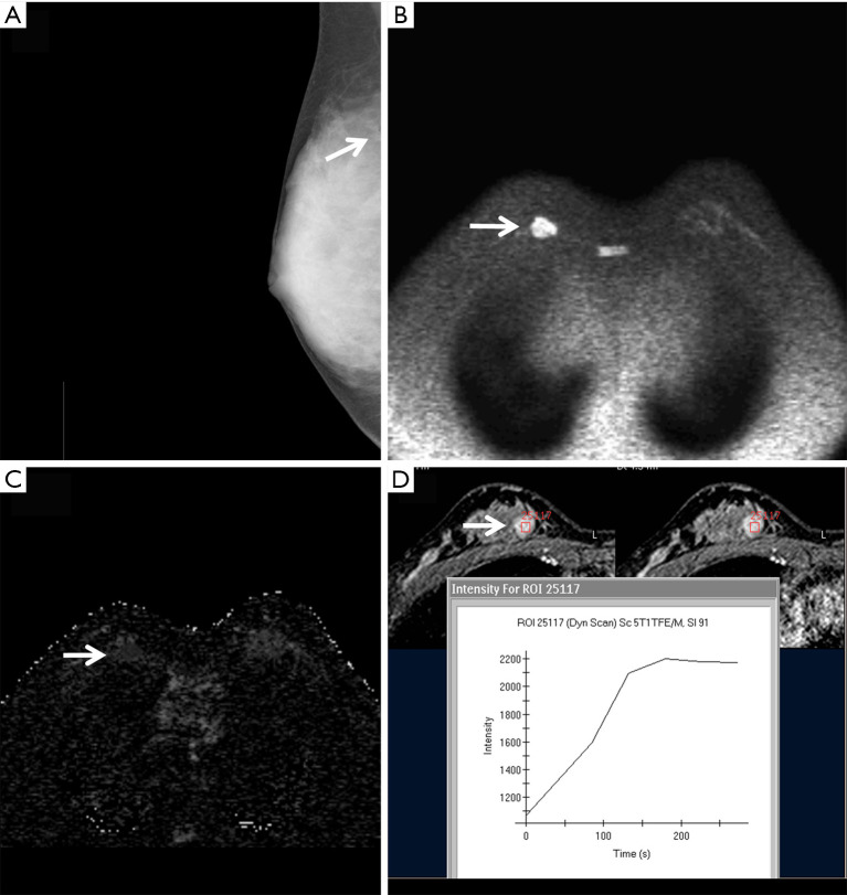
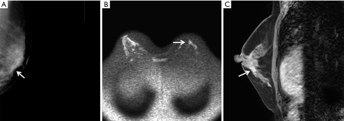
Background: Architectural distortion is a common mammographic sign that can be benign or malignant. This study investigated the diagnostic value of magnetic resonance imaging (MRI) for architectural distortions that were category 3-4 under the breast imaging reporting and data system (BI-RADS) by mammography.
Methods: We retrospectively analyzed 219 pathologically confirmed lesions in 208 patients who had BI-RADS category 3-4 architectural distortion in mammography images. Two radiologists described and categorized the architectural distortion and assigned the BI-RADS categories to the corresponding lesions on MRI images. Using the postoperative pathological diagnosis as the gold standard, we performed receiver operating characteristic (ROC) analysis for the efficacy of mammography and MRI in differentiating patients with benign or malignant lesions.
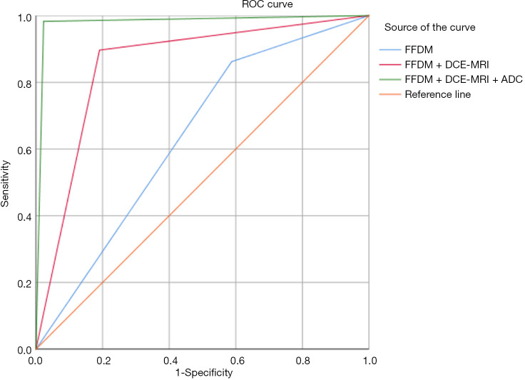
Results: Totally 151 benign lesions and 68 malignant lesions were confirmed. According to the full-field digital mammography (FFDM), 82 lesions were in BI-RADS category 3, 104 lesions in 4A, 29 lesions in 4B, and 4 lesions in 4C. The positive predictive values of FFDM for BI-RADS categories 3, 4A, 4B, and 4C were 13.4% (11/82), 27.9% (29/104), 82.8% (24/29), and 100.0% (4/4), respectively. According to MRI, 59 lesions were in BI-RADS categories 1-2, 87 lesions in 3, 39 lesions in 4, and 34 lesions in 5, with their positive predictive values being 0.0% (0/58), 2.3% (2/87), 89.7% (35/39), and 100.0% (34/34), respectively. The area under the ROC curve (AUC) of breast benign and malignant lesions differentiated by FFDM was 0.647, and the diagnostic sensitivity, specificity, and Youden index were 86.3%, 41.7%, and 0.280, respectively. The AUC of FFDM combined with dynamic contrast-enhanced MRI (DCE-MRI) in differentiating breast benign vs. malignant lesions was 0.851, and the diagnostic sensitivity, specificity, and Youden index were 89.2%, 80.7%, and 0.699, respectively. The AUC of FFDM combined with DCE-MRI and the apparent diffusion coefficient (ADC) in differentiating benign vs. malignant lesions was 0.983, and the diagnostic sensitivity, specificity, and Youden index were 98.1%, 97.5%, and 0.956, respectively.
Conclusions: MRI can improve the diagnostic efficiency of mammography in diagnosing BI-RADS category 3-4 architectural distortions and can help in the qualitative diagnosis of architectural distortion lesions.
Keywords: Mammography; architectural distortion; magnetic resonance imaging (MRI).
2020 Gland Surgery. All rights reserved.
Conflict of interest statement
Conflicts of Interest: All authors have completed the ICMJE uniform disclosure form (available at http://dx.doi.org/10.21037/gs-20-505). The authors have no conflicts of interest to declare.
Figures
Similar articles
-
MRI in the differential diagnosis of primary architectural distortion detected by mammography.Diagn Interv Radiol. 2016 Mar-Apr;22(2):141-50. doi: 10.5152/dir.2016.15017. Diagn Interv Radiol. 2016. PMID: 26899149 Free PMC article.
-
The value of noncontrast MRI in evaluating breast imaging reporting and data system category 0 lesions on digital mammograms.Quant Imaging Med Surg. 2022 Aug;12(8):4069-4080. doi: 10.21037/qims-21-968. Quant Imaging Med Surg. 2022. PMID: 35919041 Free PMC article.
-
Value of breast MRI omics features and clinical characteristics in Breast Imaging Reporting and Data System (BI-RADS) category 4 breast lesions: an analysis of radiomics-based diagnosis.Ann Transl Med. 2021 Nov;9(22):1677. doi: 10.21037/atm-21-5441. Ann Transl Med. 2021. PMID: 34988186 Free PMC article.
-
Breast magnetic resonance imaging as a problem-solving modality in mammographic BI-RADS 3 lesions.Cancer Imaging. 2010 Oct 4;10 Spec no A(1A):S54-8. doi: 10.1102/1470-7330.2010.9020. Cancer Imaging. 2010. PMID: 20880790 Free PMC article. Review.
-
BI-RADS 3: Current and Future Use of Probably Benign.Curr Radiol Rep. 2018;6(2):5. doi: 10.1007/s40134-018-0266-8. Epub 2018 Jan 27. Curr Radiol Rep. 2018. PMID: 29399419 Free PMC article. Review.
Cited by
-
A comprehensive analysis of imaging features and clinical characteristics to differentiate malignant from non-malignant mammographic architectural distortion.Gland Surg. 2024 May 30;13(5):669-683. doi: 10.21037/gs-24-110. Epub 2024 May 27. Gland Surg. 2024. PMID: 38845839 Free PMC article.
-
Prediction of pathological complete response to neoadjuvant chemotherapy in patients with breast cancer using a combination of contrast-enhanced ultrasound and dynamic contrast-enhanced magnetic resonance imaging.Cancer Med. 2023 Jan;12(2):1389-1398. doi: 10.1002/cam4.5019. Epub 2022 Jul 13. Cancer Med. 2023. PMID: 35822639 Free PMC article.
-
Contrast-enhanced mammography in the management of breast architectural distortions and avoidance of unnecessary biopsies.Breast Cancer. 2024 Sep;31(5):851-857. doi: 10.1007/s12282-024-01599-x. Epub 2024 May 29. Breast Cancer. 2024. PMID: 38811515
-
Unveiling the potential of breast MRI: a game changer for BI-RADS 4A microcalcifications.Breast Cancer Res Treat. 2024 Jul;206(2):425-435. doi: 10.1007/s10549-024-07320-y. Epub 2024 Apr 26. Breast Cancer Res Treat. 2024. PMID: 38664289
-
Visualization positioning-guided biopsy of suspicious breast microcalcifications: a retrospective cohort study.Ann Transl Med. 2021 Nov;9(21):1620. doi: 10.21037/atm-21-4496. Ann Transl Med. 2021. PMID: 34926664 Free PMC article.
References
LinkOut - more resources
Full Text Sources