

Assessing Low Skeletal Mass in Patients Undergoing Hip Surgery: The Role of Sonoelastography
- PMID: 32953705
- PMCID: PMC7476788
- DOI: 10.5371/hp.2020.32.3.132
Assessing Low Skeletal Mass in Patients Undergoing Hip Surgery: The Role of Sonoelastography
Abstract
Purpose: To analyze the utility of sonoelastography-a radiation-free procedure to characterize muscle properties-as an instrument to qualitatively and quantitatively assess the rectus femoris muscle.
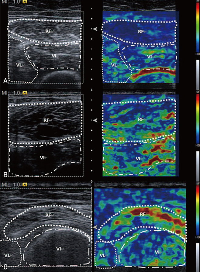
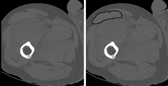
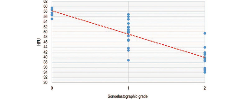
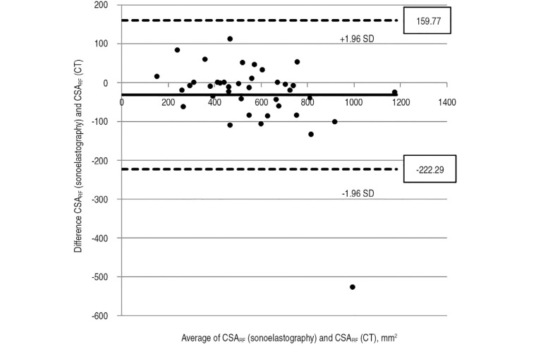
Materials and methods: Fifty-one consecutive patients who underwent a pelvic computed tomography (CT) exam were enrolled prospectively. The final analysis was conducted using data from 39 patients after 12 were removed due to exclusion criteria (muscle strength could not be measured due to poor cognition [n=11]; too young [n=1]). The potential correlation between average Hounsfield unit (HFU) at the rectus femoris muscle (measured by CT) and muscle quality grade (determined by sonoelastography) was assessed along with a retrospective analysis of the relationship between hand grip strength, knee extensor power, history of intensive care unit stay, length of hospital day and sonoelastographic grade.
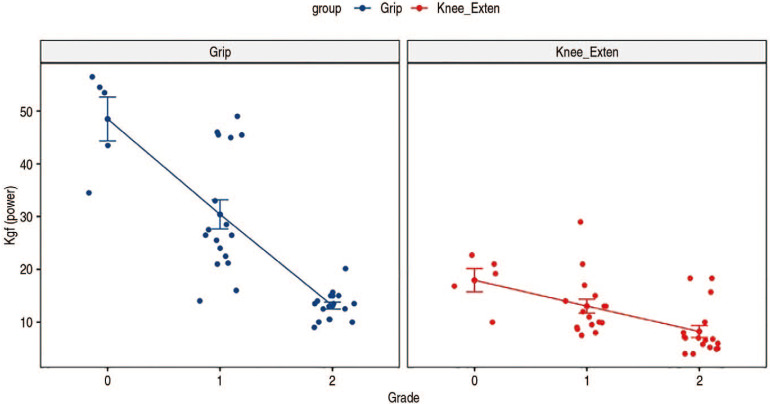
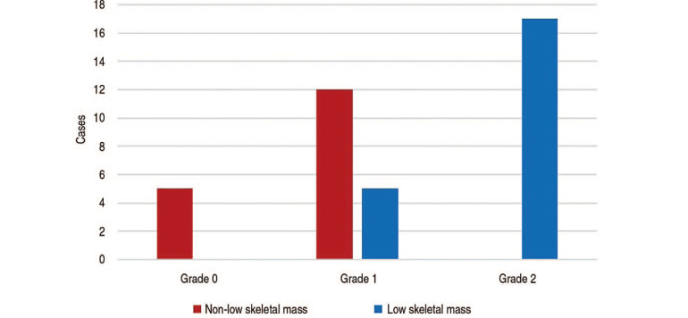
Results: There was a significant correlation between sonoelastographic grade and the average HFU (P<0.001). Furthermore, hand grip strength (P<0.001) and knee extensor power (P<0.001) decreased significantly as the sonoelastographic grade increased. The likelihood of an intensive care unit stay and prevalence of low skeletal mass increased significantly with an increase in sonoelastography grade (P=0.037, P<0.001, respectively). The sensitivity, specificity, and accuracy of sonoelastographic images for predicting low skeletal mass were 77.3%, 100%, and 87.5%, respectively.
Conclusion: Sonoelastography advantages, including the lack of radiation and greater accessibility, may make it a valuable alternative to qualitatively and quantitatively identify sarcopenia and low skeletal mass.
Keywords: Femur; Muscle strength; Sarcopenia; Sonoelastography.
Copyright © 2020 by Korean Hip Society.
Conflict of interest statement
CONFLICT OF INTEREST: The authors declare that there is no potential conflict of interest relevant to this article.
Figures
Similar articles
-
Clinical Outcomes of Living Liver Transplantation According to the Presence of Sarcopenia as Defined by Skeletal Muscle Mass, Hand Grip, and Gait Speed.Transplant Proc. 2017 Nov;49(9):2144-2152. doi: 10.1016/j.transproceed.2017.09.017. Transplant Proc. 2017. PMID: 29149975
-
Computed tomography-derived skeletal muscle index: A novel predictor of frailty and hospital length of stay after transcatheter aortic valve replacement.Am Heart J. 2016 Dec;182:21-27. doi: 10.1016/j.ahj.2016.08.016. Epub 2016 Sep 12. Am Heart J. 2016. PMID: 27914496
-
The relationship between the degree of skin fibrosis by sonoelastography and the degree of pulmonary involvement in scleroderma.Turk J Med Sci. 2017 Nov 13;47(5):1555-1559. doi: 10.3906/sag-1702-44. Turk J Med Sci. 2017. PMID: 29151332
-
Sonoelastography to Assess Muscular Stiffness Among Older Adults and its Use for the Diagnosis of Sarcopenia: A Systematic Review.Ultraschall Med. 2021 Dec;42(6):634-642. doi: 10.1055/a-1293-8057. Epub 2020 Nov 13. Ultraschall Med. 2021. PMID: 33187010 English.
-
Computed Tomography-Assessed Skeletal Muscle Mass as a Predictor of Outcomes in Lung Cancer Surgery.Ann Thorac Surg. 2019 Nov;108(5):1555-1564. doi: 10.1016/j.athoracsur.2019.04.090. Epub 2019 Jun 19. Ann Thorac Surg. 2019. PMID: 31228408
Cited by
-
Biomarkers and Nutrients in Musculoskeletal Disorders.Nutrients. 2021 Jan 20;13(2):283. doi: 10.3390/nu13020283. Nutrients. 2021. PMID: 33498342 Free PMC article.
References
-
- Rosenberg IH. Summary comments. Am J Clin Nutr. 1989;50:1231–1233.
-
- Gariballa S, Alessa A. Sarcopenia: prevalence and prognostic significance in hospitalized patients. Clin Nutr. 2013;32:772–776. - PubMed
-
- Jones KI, Doleman B, Scott S, Lund JN, Williams JP. Simple psoas cross-sectional area measurement is a quick and easy method to assess sarcopenia and predicts major surgical complications. Colorectal Dis. 2015;17:O20–O26. - PubMed
LinkOut - more resources
Full Text Sources
