

Tetralogy of Fallot: cardiac imaging evaluation
- PMID: 32953766
- PMCID: PMC7475417
- DOI: 10.21037/atm.2020.02.18
Tetralogy of Fallot: cardiac imaging evaluation
Abstract
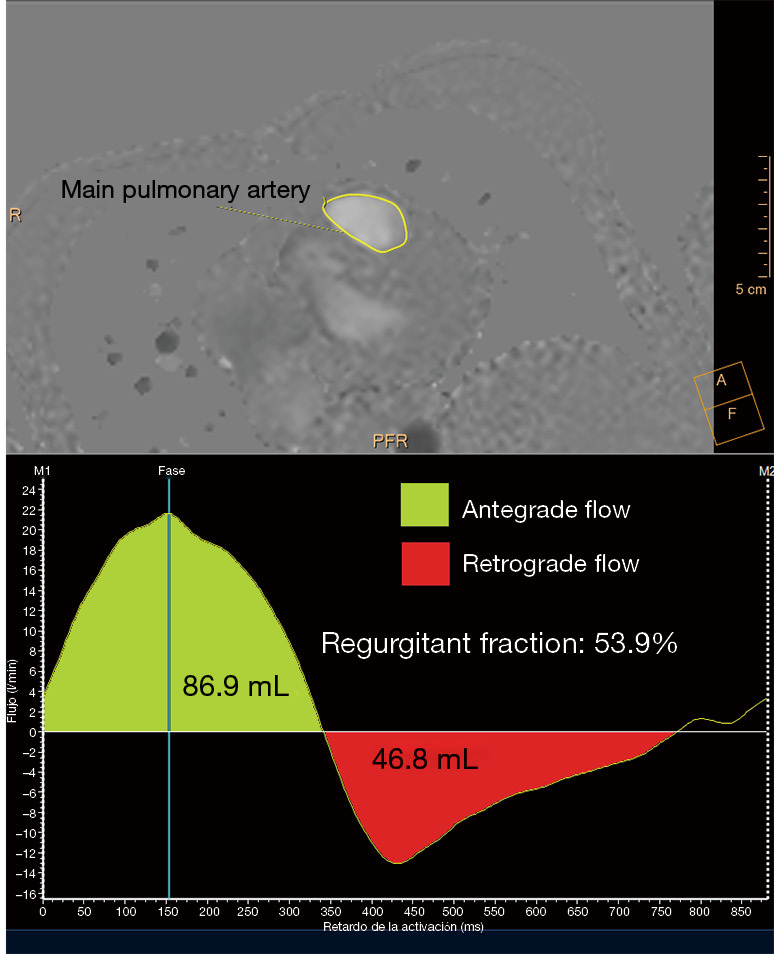
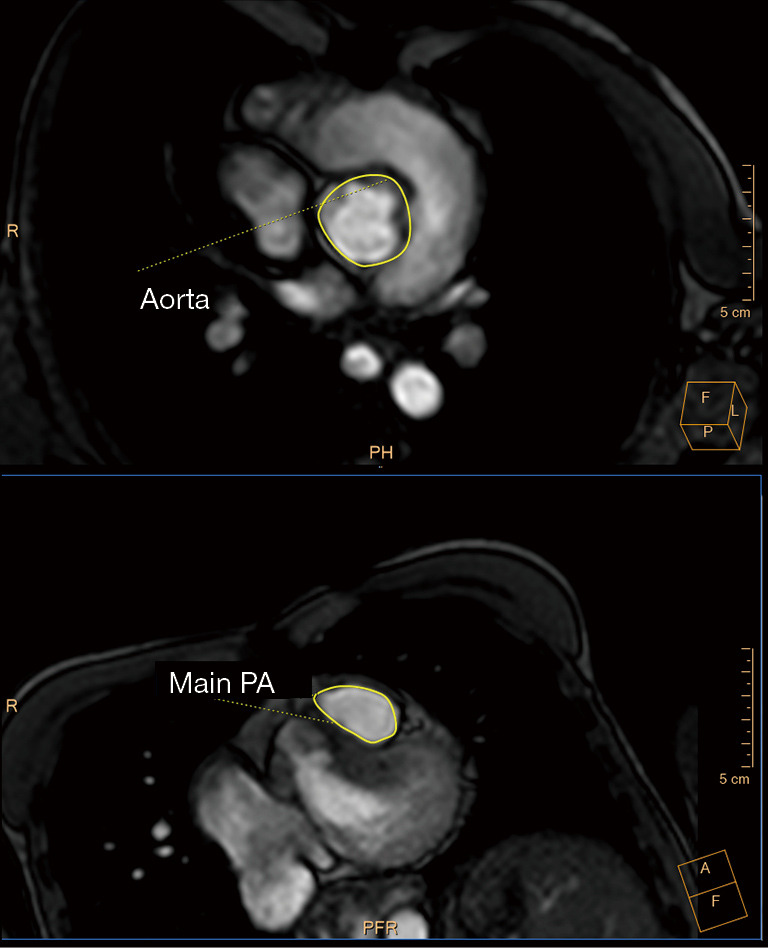
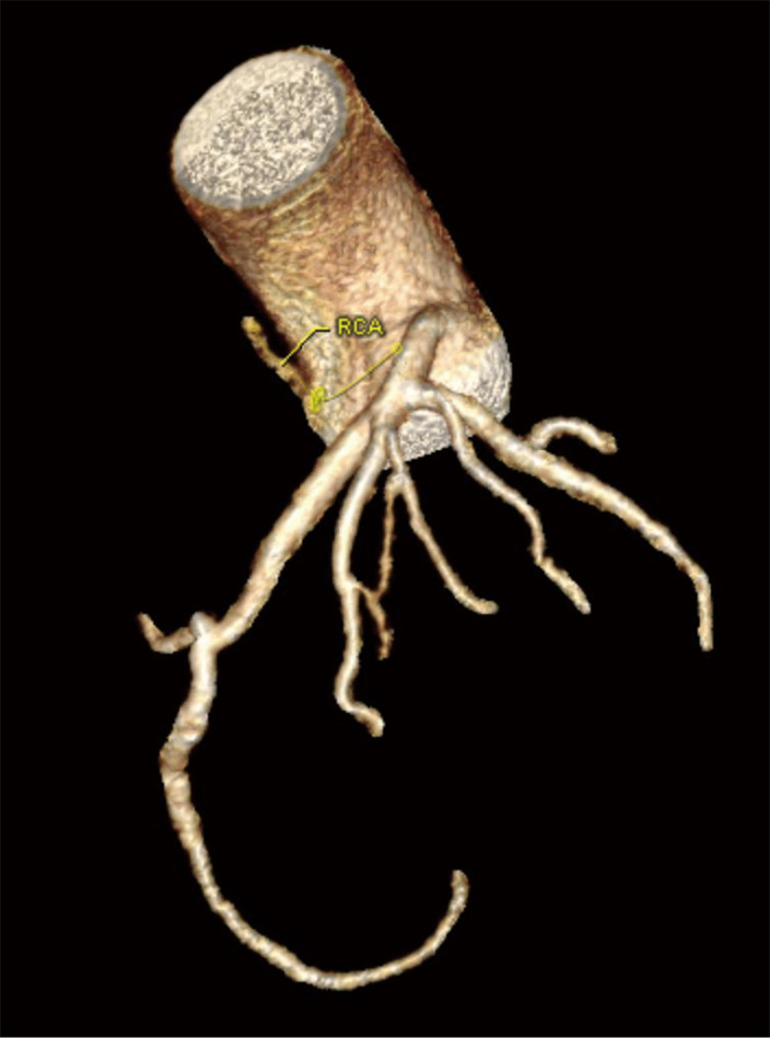
Thanks to advances in pediatric cardiology, most infants with tetralogy of Fallot (TOF) now survive into adulthood. This relatively new population of adult patients may face long-term complications, including pulmonary regurgitation (PR), right ventricular (RV) tract obstruction, residual shunts, RV dysfunction, and arrythmias. They will often need to undergo pulmonary valve (PV) replacement and other invasive re-interventions. However, the optimal timing for these procedures is challenging, largely due to the complexity of evaluating RV volume and function. The options for the follow-up of these patients have rapidly evolved from an angiography-based approach to the surge of advanced imaging techniques, mainly echocardiography, cardiac magnetic resonance (CMR), and computer tomography (CT). In this review, we outline the indications, strengths and limitations of these modalities in the adult TOF population.
Keywords: Computed tomography; cardiac magnetic resonance (CMR); echocardiography; tetralogy of Fallot (TOF).
2020 Annals of Translational Medicine. All rights reserved.
Conflict of interest statement
Conflicts of Interest: Both authors have completed the ICMJE uniform disclosure form (available at http://dx.doi.org/10.21037/atm.2020.02.18). The series “Structural Heart Disease: The Revolution” was commissioned by the editorial office without any funding or sponsorship. The authors have no other conflicts of interest to declare.
Figures
Similar articles
-
Hot topics in tetralogy of Fallot.J Am Coll Cardiol. 2013 Dec 10;62(23):2155-66. doi: 10.1016/j.jacc.2013.07.100. Epub 2013 Sep 27. J Am Coll Cardiol. 2013. PMID: 24076489
-
A comparative study: right ventricular assessment in post-repaired tetralogy of Fallot patients by echocardiogram with cardiac magnetic resonance imaging.J Med Assoc Thai. 2014 Jun;97 Suppl 6:S232-8. J Med Assoc Thai. 2014. PMID: 25391198
-
Impact of residual right ventricular outflow tract obstruction on biventricular strain and synchrony in patients after repair of tetralogy of Fallot: a cardiac magnetic resonance feature tracking study.Eur J Cardiothorac Surg. 2015 Jul;48(1):83-90. doi: 10.1093/ejcts/ezu396. Epub 2014 Nov 5. Eur J Cardiothorac Surg. 2015. PMID: 25378364
-
Pulmonary Regurgitation after Tetralogy of Fallot Repair: A Diagnostic and Therapeutic Challenge.J Cardiovasc Echogr. 2013 Jan-Mar;23(1):1-9. doi: 10.4103/2211-4122.117975. J Cardiovasc Echogr. 2013. PMID: 28465877 Free PMC article. Review.
-
Defining and refining indications for transcatheter pulmonary valve replacement in patients with repaired tetralogy of Fallot: Contributions from anatomical and functional imaging.Int J Cardiol. 2016 Oct 15;221:916-25. doi: 10.1016/j.ijcard.2016.07.120. Epub 2016 Jul 9. Int J Cardiol. 2016. PMID: 27441469 Review.
Cited by
-
Multimodality Imaging Assessment of Tetralogy of Fallot: From Diagnosis to Long-Term Follow-Up.Children (Basel). 2023 Oct 27;10(11):1747. doi: 10.3390/children10111747. Children (Basel). 2023. PMID: 38002838 Free PMC article. Review.
-
Management of Fallot's Uncorrected Tetralogy in Adulthood: A Narrative Review.Cureus. 2024 Aug 17;16(8):e67063. doi: 10.7759/cureus.67063. eCollection 2024 Aug. Cureus. 2024. PMID: 39286683 Free PMC article. Review.
-
Greater than the sum of its parts: multimodality imaging in adults with congenital heart disease.Cardiovasc Diagn Ther. 2024 Dec 31;14(6):1176-1185. doi: 10.21037/cdt-24-363. Epub 2024 Dec 19. Cardiovasc Diagn Ther. 2024. PMID: 39790212 Free PMC article. Review.
-
Multimodality Imaging of the Neglected Valve: Role of Echocardiography, Cardiac Magnetic Resonance and Cardiac Computed Tomography in Pulmonary Stenosis and Regurgitation.J Imaging. 2022 Oct 10;8(10):278. doi: 10.3390/jimaging8100278. J Imaging. 2022. PMID: 36286372 Free PMC article. Review.
-
Navigating Complex Cardiac Defects: A Neonatal Case of Pentalogy of Fallot With Vascular Anomalies Identified via Cardiac CT Angiography.Cureus. 2025 Mar 10;17(3):e80338. doi: 10.7759/cureus.80338. eCollection 2025 Mar. Cureus. 2025. PMID: 40206902 Free PMC article.
References
-
- Kochav, J. Adult congenital heart disease in clinical practice. In: DeFariah Yeh D, Bhatt A. Tetralogy of Fallot New York, NY: Springer Berlin Heidelberg; 2018. Chapter 23:295-318.
Publication types
LinkOut - more resources
Full Text Sources
Research Materials