

Tetralogy of Fallot: cardiac imaging evaluation
- PMID: 32953766
- PMCID: PMC7475417
- DOI: 10.21037/atm.2020.02.18
Tetralogy of Fallot: cardiac imaging evaluation
Abstract
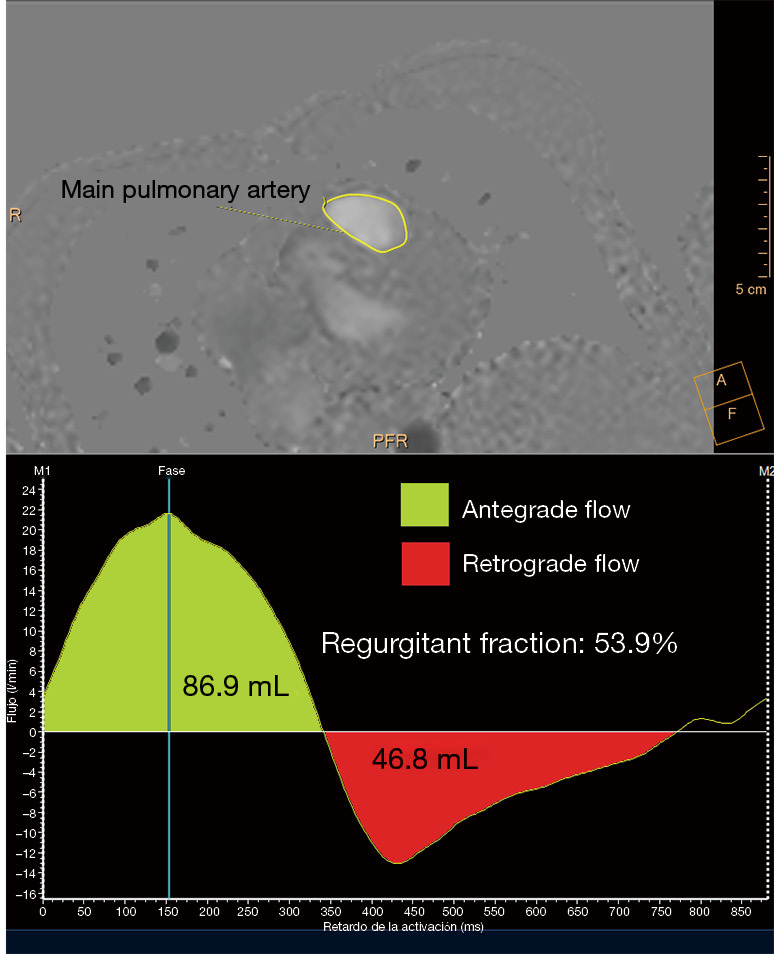
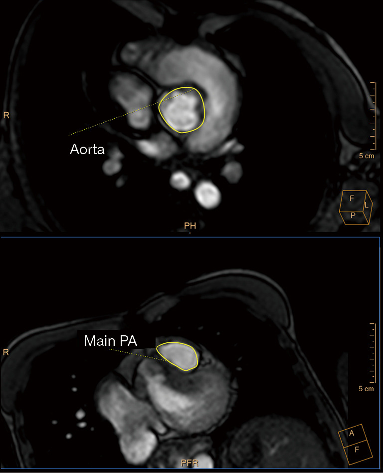
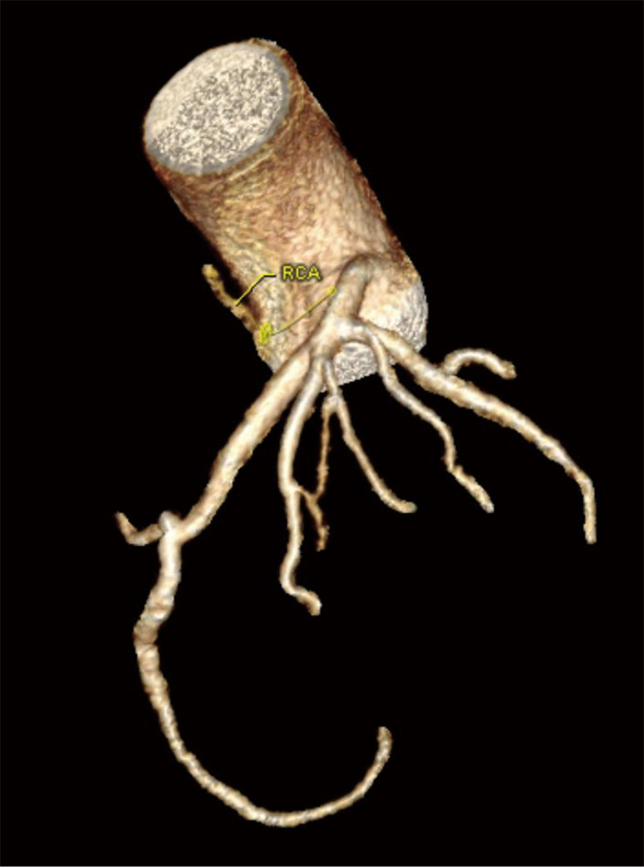
Thanks to advances in pediatric cardiology, most infants with tetralogy of Fallot (TOF) now survive into adulthood. This relatively new population of adult patients may face long-term complications, including pulmonary regurgitation (PR), right ventricular (RV) tract obstruction, residual shunts, RV dysfunction, and arrythmias. They will often need to undergo pulmonary valve (PV) replacement and other invasive re-interventions. However, the optimal timing for these procedures is challenging, largely due to the complexity of evaluating RV volume and function. The options for the follow-up of these patients have rapidly evolved from an angiography-based approach to the surge of advanced imaging techniques, mainly echocardiography, cardiac magnetic resonance (CMR), and computer tomography (CT). In this review, we outline the indications, strengths and limitations of these modalities in the adult TOF population.
Keywords: Computed tomography; cardiac magnetic resonance (CMR); echocardiography; tetralogy of Fallot (TOF).
2020 Annals of Translational Medicine. All rights reserved.
Conflict of interest statement
Conflicts of Interest: Both authors have completed the ICMJE uniform disclosure form (available at http://dx.doi.org/10.21037/atm.2020.02.18). The series “Structural Heart Disease: The Revolution” was commissioned by the editorial office without any funding or sponsorship. The authors have no other conflicts of interest to declare.
Figures
References
-
- Kochav, J. Adult congenital heart disease in clinical practice. In: DeFariah Yeh D, Bhatt A. Tetralogy of Fallot New York, NY: Springer Berlin Heidelberg; 2018. Chapter 23:295-318.
Publication types
LinkOut - more resources
Full Text Sources
Research Materials