

COVID-19 detection in CT images with deep learning: A voting-based scheme and cross-datasets analysis
- PMID: 32953971
- PMCID: PMC7487744
- DOI: 10.1016/j.imu.2020.100427
COVID-19 detection in CT images with deep learning: A voting-based scheme and cross-datasets analysis
Abstract
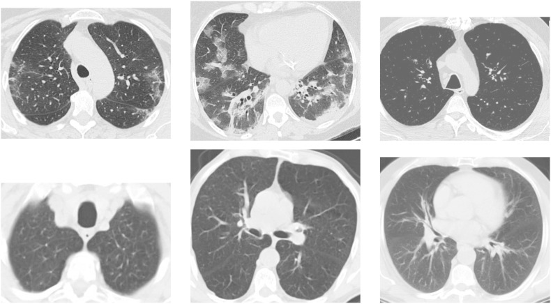
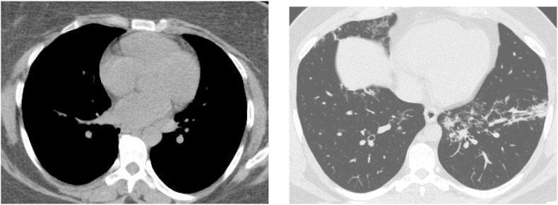
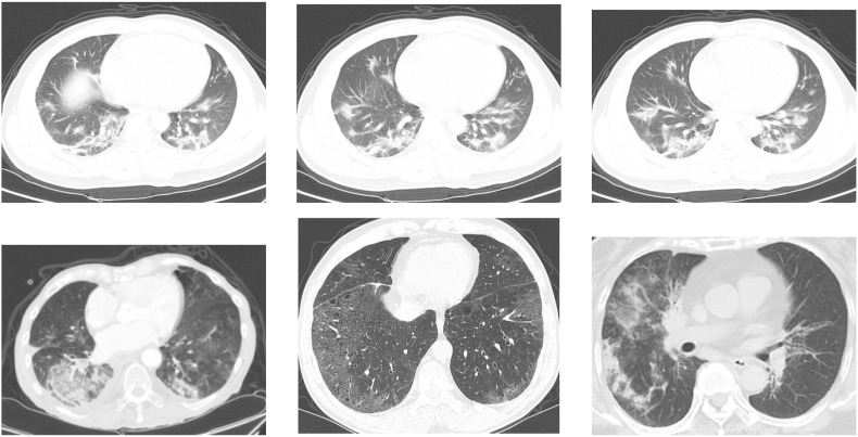
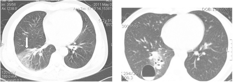
Early detection and diagnosis are critical factors to control the COVID-19 spreading. A number of deep learning-based methodologies have been recently proposed for COVID-19 screening in CT scans as a tool to automate and help with the diagnosis. These approaches, however, suffer from at least one of the following problems: (i) they treat each CT scan slice independently and (ii) the methods are trained and tested with sets of images from the same dataset. Treating the slices independently means that the same patient may appear in the training and test sets at the same time which may produce misleading results. It also raises the question of whether the scans from the same patient should be evaluated as a group or not. Moreover, using a single dataset raises concerns about the generalization of the methods. Different datasets tend to present images of varying quality which may come from different types of CT machines reflecting the conditions of the countries and cities from where they come from. In order to address these two problems, in this work, we propose an Efficient Deep Learning Technique for the screening of COVID-19 with a voting-based approach. In this approach, the images from a given patient are classified as group in a voting system. The approach is tested in the two biggest datasets of COVID-19 CT analysis with a patient-based split. A cross dataset study is also presented to assess the robustness of the models in a more realistic scenario in which data comes from different distributions. The cross-dataset analysis has shown that the generalization power of deep learning models is far from acceptable for the task since accuracy drops from 87.68% to 56.16% on the best evaluation scenario. These results highlighted that the methods that aim at COVID-19 detection in CT-images have to improve significantly to be considered as a clinical option and larger and more diverse datasets are needed to evaluate the methods in a realistic scenario.
Keywords: COVID-19; Chest radiography; Deep learning; EfficientNet; Pneumonia.
© 2020 The Author(s).
Conflict of interest statement
The authors declare that they have no known competing financial interests or personal relationships that could have appeared to influence the work reported in this paper.
Figures
References
-
- Hemdan E.E.-D., Shouman M.A., Karar M.E. 2020. Covidx-net: a framework of deep learning classifiers to diagnose covid-19 in x-ray images; p. 11055. arXiv preprint arXiv:2003.
-
- Farooq M., Hafeez A. 2020. Covid-resnet: a deep learning framework for screening of covid19 from radiographs; p. 14395. arXiv preprint arXiv:2003.
-
- Li T., Han Z., Wei B., Zheng Y., Hong Y., Cong J. 2020. Robust screening of covid-19 from chest x-ray via discriminative cost-sensitive learning; p. 12592. ArXiv abs/2004.
LinkOut - more resources
Full Text Sources