. 2019 Jul 1;5(2):189.
doi: 10.18063/ijb.v5i2.189. eCollection 2019.

Development and characterization of a photocurable alginate bioink for three-dimensional bioprinting

Affiliations

Development and characterization of a photocurable alginate bioink for three-dimensional bioprinting

H H Mishbak et al. Int J Bioprint. .

Erratum in

  • ERRATUM.
    [No authors listed] [No authors listed] Int J Bioprint. 2020 Sep 17;6(4):309. doi: 10.18063/ijb.v6i4.309. eCollection 2020. Int J Bioprint. 2020. PMID: 33102924 Free PMC article.

Abstract

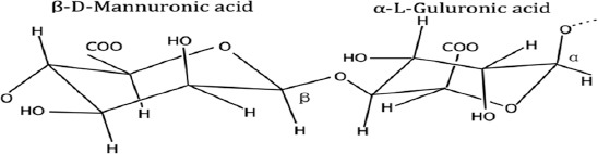
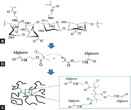
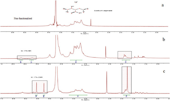
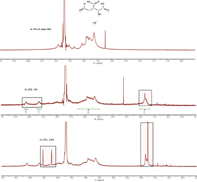
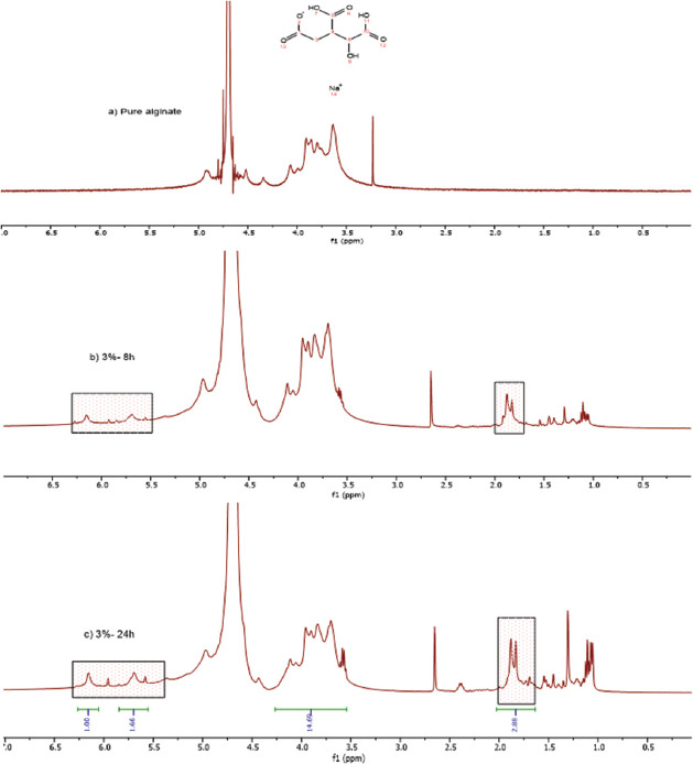
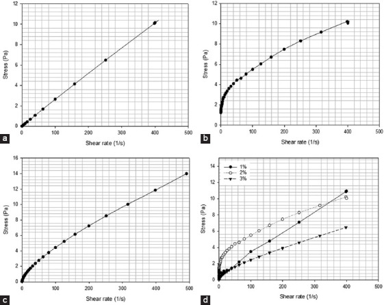
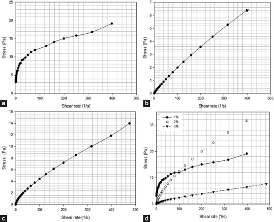
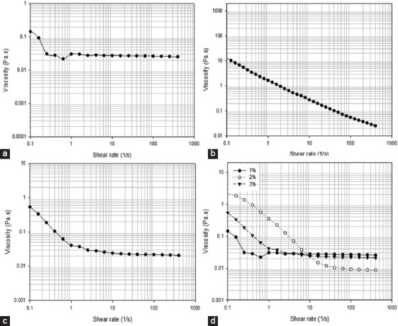
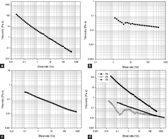
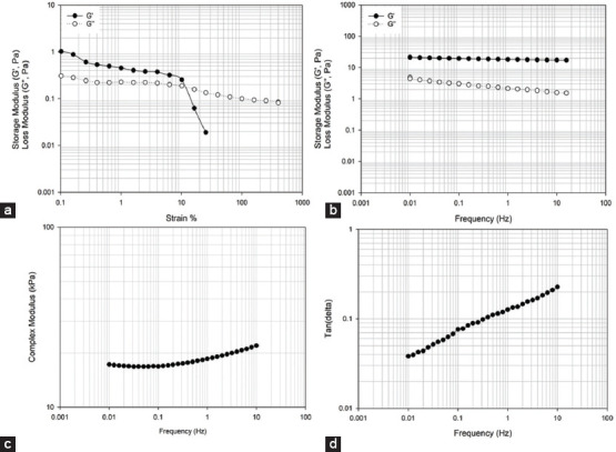
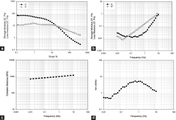
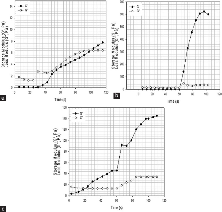
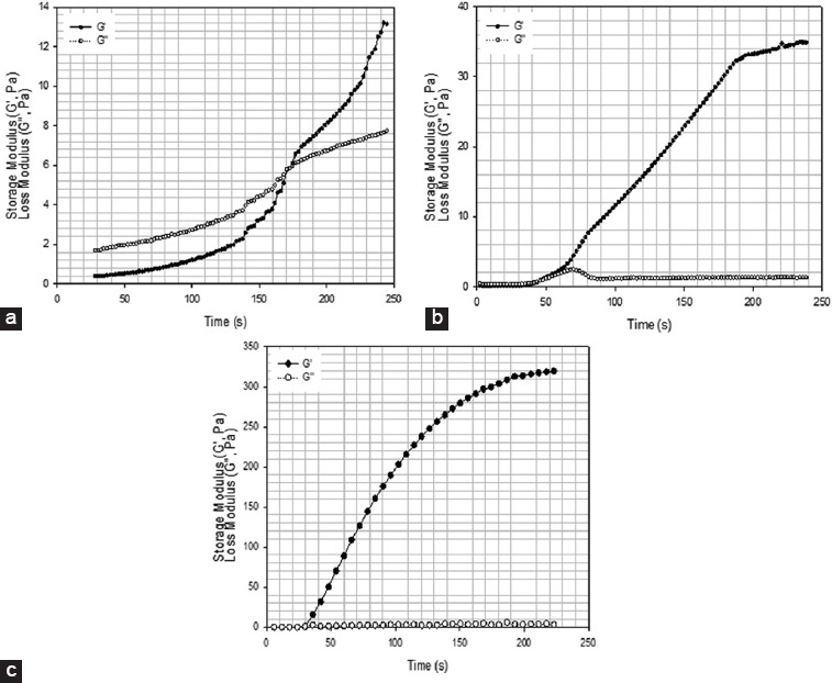
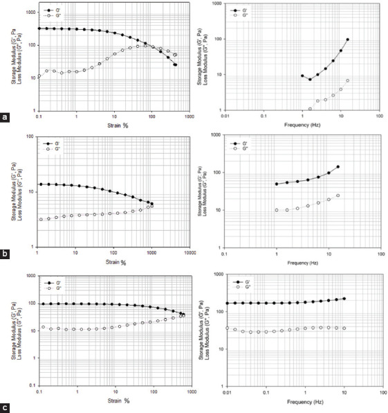
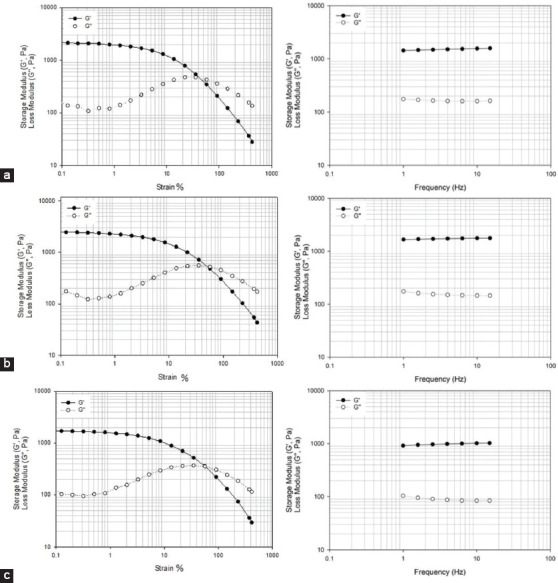
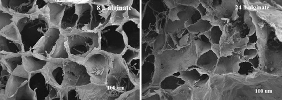
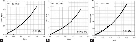
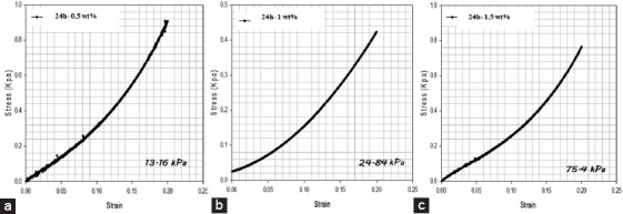
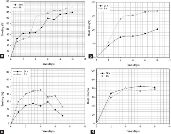
Alginate is a biocompatible material suitable for biomedical applications, which can be processed under mild conditions on irradiation. This paper investigates the preparation and the rheological behavior of different pre-polymerized and polymerized alginate methacrylate systems for three-dimensional photopolymerization bioprinting. The effect of the functionalization time on the mechanical, morphological, swelling, and degradation characteristics of cross-linked alginate hydrogel is also discussed. Alginate was chemically-modified with methacrylate groups and different reaction times considered. Photocurable alginate systems were prepared by dissolving functionalized alginate with 0.5- 1.5% w/v photoinitiator solutions and cross-linked by ultraviolet light (8 mW/cm2 for 8 minutes).

Keywords: 3D bioprinting; Alginate hydrogel; Functionalization; Photopolymerization; Rheology.


Figures

Figure 1
Figure 1
Alginate showing a linkage between the mannuronic and guluronic acid[25].
Figure 2
Figure 2
Schematic representation of the photopolymerization process of alginate methacrylate. (a) After exposing the polymer solution to ultraviolet radiation, the photoinitiators generated free radicals that react with the vinyl methylene starting the crosslinking reaction; (b) the reaction propagates with macroradicals reacting with unreacted carbon-carbon double bonds. (c) At the end through a bimolecular termination mechanism, a three-dimensional network of the cross-linked hydrogel is formed [30].
Figure 3
Figure 3
(a) Non-functionalized alginate (1% w/v), (b) functionalized alginate (1% w/v) after 8 h of reaction, (c) functionalized alginate (1% w/v) after 24 h of reaction. The functionalization is confirmed by the presence of new peaks in the spectra at 5.63 ppm and 6.09 ppm attributed to the methylene group and a peak at 1.82 ppm that corresponds to the methyl group.
Figure 4
Figure 4
(a) Non-functionalized alginate (2% w/v), (b) functionalized alginate (2% w/v) after 8 h of reaction, (c) functionalized alginate (2% w/v) after 24 h of reaction. The functionalization is confirmed by the presence of new peaks in the spectra at 5.63 ppm and 6.09 ppm attributed to the methylene group and a peak at 1.82 ppm that corresponds to the methyl group.
Figure 5
Figure 5
(a) Non-functionalized alginate (3% w/v), (b) functionalized alginate (2% w/v) after 8 h of reaction, (c) functionalized alginate (2% w/v) after 24 h of reaction. The functionalization is confirmed by the presence of new peaks in the spectra at 5.63 ppm and 6.09 ppm attributed to the methylene group and a peak at 1.82 ppm that corresponds to the methyl group.
Figure 6
Figure 6
Stress versus shear rate profiles for alginate methacrylate solutions containing different alginate methacrylate concentrations, reacted for 8 h. (a) 1% w/v of alginate, (b) 2% w/v of alginate, (c) 3% w/v of alginate, (d) comparison of all compositions.
Figure 7
Figure 7
Stress versus shear rate for solutions containing different alginate methacrylate concentrations reacted for 24 h. (a) 1% w/v of alginate, (b) 2% w/v of alginate, (c) 3% w/v of alginate, (d) comparison of all compositions.
Figure 8
Figure 8
Viscosity versus shear rate for solutions containing different alginate methacrylate concentrations reacted for 8 h. (a) 1% w/v, (b) 2% w/v, (c) 3% w/v, (d) comparison of all compositions.
Figure 9
Figure 9
Viscosity versus shear rate for solutions containing different alginate methacrylate concentrations reacted for 24 h. (a) 1% w/v, (b) 2% w/v, (c) 3% w/v, (d) comparison of all compositions.
Figure 10
Figure 10
2% wt. alginate solution reacted for 8 h. (a) storage and loss modulus versus strain (b) storage and loss modulus versus frequency, (c) complex modulus versus frequency, (d) tan δ versus frequency.
Figure 11
Figure 11
2% wt. alginate solution reacted for 24 h. (a) storage and loss modulus versus strain, (b) storage and loss modulus versus frequency, (c) complex modulus versus frequency, (d) tan δ versus frequency.
Figure 12
Figure 12
2% wt. methacrylate alginate with different concentration of VA-086 photoinitiators functionalized for 8 h (a) 0.5% w/v of VA-086, (b) 1% w/v of VA-086, (c) 1.5% w/v of VA-086, d) 0.05%.
Figure 13
Figure 13
2% wt. methacrylate alginate with different concentration of VA-086 photoinitiators functionalized for 8 h (a) 0.5% w/v of VA-086, (b) 1% w/v of VA-086, (c) 1.5% w/v of VA-086, (d) 0.05%.
Figure 14
Figure 14
2% wt. methacrylate alginate hydrogel with different concentration of VA-086 functionalized for 8 h. (a) 0.5 w/v % of VA-086, (b) 1 w/v % of VA-086, (c) 1.5 w/v % of VA-086.
Figure 15
Figure 15
2% wt. methacrylate alginate with different concentration of VA-086 functionalized for 24 h. (a) 0.5 w/v % of VA-086, (b) 1 w/v % of VA-086, (c) 1.5 w/v % of VA-086.
Figure 16
Figure 16
Scanning electron microscopy images of cross-linked methacrylate alginate hydrogel structures obtained from alginate-methacrylate at different reaction times: 8 and 24 h.
Figure 17
Figure 17
Compression tests for alginate methacrylate samples (8 h) with different photoinitiator concentrations (a) 0.5% w/v VA-086, (b) 1% w/v VA-086, (c) 1.5% w/v VA-086.
Figure 18
Figure 18
Compression tests for alginate methacrylate samples (24 h) with different photoinitiator concentrations (a) 0.5% w/v VA-086, (b) 1% w/v VA-086, (c) 1.5% w/v VA-086.
Figure 19
Figure 19
Swelling and degradation rate for 2% wt. functionalized alginate (24 and 8 h) reaction time in (a) distilled water (diH2O), (b) degradation rate in diH2O, (c) Dulbecco’s Modified Eagle’s Medium (DMEM), (d) degradation rate in DMEM.

Similar articles

Cited by

References

    1. Pereira R, Bártolo PJF. 3D Bioprinting of Photocrosslinkable Hydrogel Constructs. Appl Polym Sci. DOI 10.1002/app42458 2015;132(48):132A4.
    1. Zhou D, Yoshihiro I. Visible Light-curable Polymers for Biomedical Applications. Sci China Chem. 2014;57(4):510–21. DOI 10.1007/s11426-014-5069-z.
    1. Pereira RF, Sousa A, Barrias CC, et al. A Single-component Hydrogel Bioink for Bioprinting of Bioengineered 3D Constructs for Dermal Tissue Engineering. Mater Horiz. 2018;5(6):1100–11. DOI 10.1039/c8mh00525g.
    1. Vyas C, Pereira R, Huang B, et al. Engineering the Vasculature with Additive Manufacturing. Curr Opin Biomed Eng. 2017;2:1–13.
    1. Wang W, Caetano G, Ambler W S, et al. Engineering. 12. Vol. 9. Materials (Basel, Switzerland): 2016. Enhancing the Hydrophilicity and Cell Attachment of 3D Printed PCL/Graphene Scaffolds for Bone Tissue; p. 992. DOI 10.3390/ma9120992. - PMC - PubMed

LinkOut - more resources