

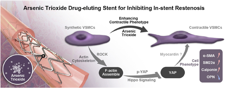
A novel mechanism of inhibiting in-stent restenosis with arsenic trioxide drug-eluting stent: Enhancing contractile phenotype of vascular smooth muscle cells via YAP pathway
- PMID: 32954055
- PMCID: PMC7484501
- DOI: 10.1016/j.bioactmat.2020.08.018
A novel mechanism of inhibiting in-stent restenosis with arsenic trioxide drug-eluting stent: Enhancing contractile phenotype of vascular smooth muscle cells via YAP pathway
Erratum in
-
Corrigendum to "A novel mechanism of inhibiting in-stent restenosis with arsenic trioxide drug-eluting stent: Enhancing contractile phenotype of vascular smooth muscle cells via YAP pathway" [Bioact. Mater. 6 2 (February 2021) 375-385].Bioact Mater. 2021 Aug 2;8:574. doi: 10.1016/j.bioactmat.2021.07.032. eCollection 2022 Feb. Bioact Mater. 2021. PMID: 34786522 Free PMC article.
Abstract
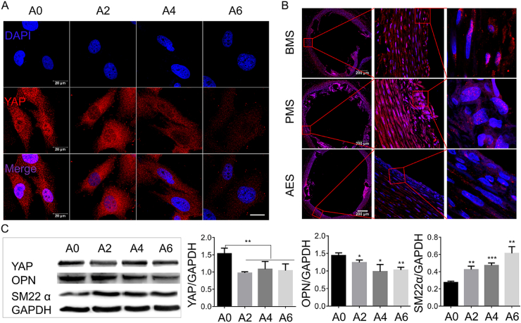
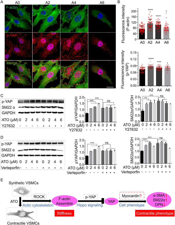
Objective: Arsenic trioxide (ATO or As2O3) has beneficial effects on suppressing neointimal hyperplasia and restenosis, but the mechanism is still unclear. The goal of this study is to further understand the mechanism of ATO's inhibitory effect on vascular smooth muscle cells (VSMCs).
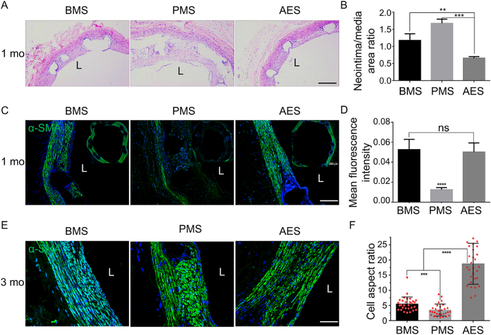
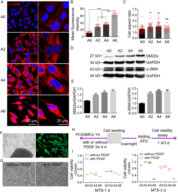
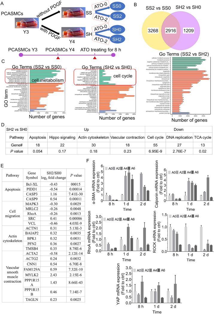
Methods and results: Through in vitro cell culture and in vivo stent implanting into the carotid arteries of rabbit, a synthetic-to-contractile phenotypic transition was induced and the proliferation of VSMCs was inhibited by ATO. F-actin filaments were clustered and the elasticity modulus was increased within the phenotypic modulation of VSMCs induced by ATO in vitro. Meanwhile, Yes-associated protein (YAP) nuclear translocation was inhibited by ATO both in vivo and in vitro. It was found that ROCK inhibitor or YAP inactivator could partially mask the phenotype modulation of ATO on VSMCs.
Conclusions: The interaction of YAP with the ROCK pathway through ATO seems to mediate the contractile phenotype of VSMCs. This provides an indication of the clinical therapeutic mechanism for the beneficial bioactive effect of ATO-drug eluting stent (AES) on in-stent restenosis (ISR).
Keywords: Arsenic trioxide (ATO); Bioactive; In-stent restenosis (ISR); Yes-associated protein (YAP).
© 2020 The Authors. Publishing services by Elsevier B.V. on behalf of KeAi Communications Co., Ltd.
Conflict of interest statement
The authors declare that they have no known competing financial interests or personal relationships that could have appeared to influence the work reported in this paper.
Figures

Similar articles
-
Drug-eluting stent specifically designed to target vascular smooth muscle cell phenotypic modulation attenuated restenosis through the YAP pathway.Am J Physiol Heart Circ Physiol. 2019 Sep 1;317(3):H541-H551. doi: 10.1152/ajpheart.00089.2019. Epub 2019 Jul 12. Am J Physiol Heart Circ Physiol. 2019. PMID: 31298560
-
Arsenic trioxide activates yes-associated protein by lysophosphatidic acid metabolism to selectively induce apoptosis of vascular smooth muscle cells.Biochim Biophys Acta Mol Cell Res. 2022 Apr;1869(4):119211. doi: 10.1016/j.bbamcr.2022.119211. Epub 2022 Jan 15. Biochim Biophys Acta Mol Cell Res. 2022. PMID: 35041860
-
Drug-Eluting Stent Targeting Sp-1-Attenuated Restenosis by Engaging YAP-Mediated Vascular Smooth Muscle Cell Phenotypic Modulation.J Am Heart Assoc. 2020 Jan 7;9(1):e014103. doi: 10.1161/JAHA.119.014103. Epub 2019 Dec 27. J Am Heart Assoc. 2020. PMID: 31880978 Free PMC article.
-
Vascular Smooth Muscle Cells Phenotypic Switching in Cardiovascular Diseases.Cells. 2022 Dec 15;11(24):4060. doi: 10.3390/cells11244060. Cells. 2022. PMID: 36552822 Free PMC article. Review.
-
Arsenic trioxide preferentially induces nonapoptotic cell deaths as well as actin cytoskeleton rearrangement in the CHO AA8 cell line.Postepy Hig Med Dosw (Online). 2014 Dec 21;68:1492-500. doi: 10.5604/17322693.1133098. Postepy Hig Med Dosw (Online). 2014. PMID: 25531713 Review.
Cited by
-
[Hot Topics and Emerging Trends in Mechanobiology Research].Sichuan Da Xue Xue Bao Yi Xue Ban. 2024 Jan 20;55(1):1-5. doi: 10.12182/20240160104. Sichuan Da Xue Xue Bao Yi Xue Ban. 2024. PMID: 38322522 Free PMC article. Chinese.
-
Arsenic trioxide: applications, mechanisms of action, toxicity and rescue strategies to date.Arch Pharm Res. 2024 Mar;47(3):249-271. doi: 10.1007/s12272-023-01481-y. Epub 2023 Dec 26. Arch Pharm Res. 2024. PMID: 38147202 Review.
-
Recent advances in surface functionalization of cardiovascular stents.Bioact Mater. 2024 Oct 28;44:389-410. doi: 10.1016/j.bioactmat.2024.10.025. eCollection 2025 Feb. Bioact Mater. 2024. PMID: 39539518 Free PMC article. Review.
-
Multilevel Assessment of Stent-Induced Inflammation in the Adjacent Vascular Tissue.ACS Biomater Sci Eng. 2023 Aug 14;9(8):4747-4760. doi: 10.1021/acsbiomaterials.3c00540. Epub 2023 Jul 21. ACS Biomater Sci Eng. 2023. PMID: 37480152 Free PMC article.
-
Comparative Study of Porous Iron Foams for Biodegradable Implants: Structural Analysis and In Vitro Assessment.J Funct Biomater. 2023 May 24;14(6):293. doi: 10.3390/jfb14060293. J Funct Biomater. 2023. PMID: 37367257 Free PMC article.
References
-
- Waxman S., Anderson K.C. History of the development of arsenic derivatives in cancer therapy. Oncol. 2001;6(S2):3–10. - PubMed
-
- Guo Z.Z., Meng M.J., Geng S.N. The optimal dose of arsenic trioxide induced opposite efficacy in autophagy between K562 cells and their initiating cells to eradicate human myelogenous leukemia. J. Ethnopharmacol. 2017;196:29–38. - PubMed
-
- Chandy M., George B., Mathews V. Treatment of children with newly diagnosed acute promyelocytic leukemia (APML) using intravenous arsenic trioxide (As2O3) Blood. 2003;102(11):620A.
-
- Zhu J., Chen Z., Lallemand-Breitenbach V. How acute promyelocytic leukaemia revived arsenic. Nat. Rev. Canc. 2002;2(9):705–713. - PubMed
LinkOut - more resources
Full Text Sources
Other Literature Sources
Research Materials
