

Brachial and lumbosacral plexopathies: A review
- PMID: 32954064
- PMCID: PMC7484503
- DOI: 10.1016/j.cnp.2020.07.005
Brachial and lumbosacral plexopathies: A review
Abstract
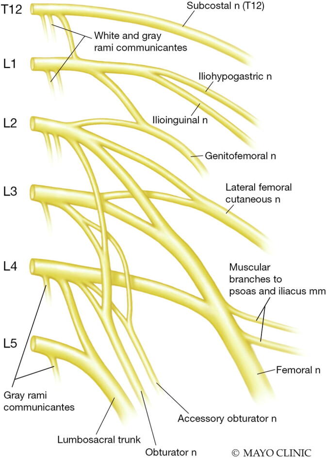
Diseases of the brachial and lumbosacral plexus are uncommon and complex. The diagnosis of plexopathies is often challenging for the clinician, both in terms of localizing a patient's symptoms to the plexus as well as determining the etiology. The non-specific clinical features and similar presentations to other root, nerve, and non-neurologic disorders emphasize the importance of a high clinical index of suspicion for a plexopathy and comprehensive clinical evaluation. Various diagnostic tests, including electrodiagnostic (EDX) studies, neuroimaging (including ultrasound, MRI, or PET), serologic studies, and genetic testing, may be used to confirm a plexopathy and assist in identifying the underlying etiology. EDX testing plays an important role in confirming a plexopathy defining the localization, pathophysiology, chronicity, severity, and prognosis. Given the complexity of the plexus anatomy, multiple common and uncommon NCS and an extensive needle examination is often required, and a comprehensive, individualized approach to each patient is necessary. Treatment of plexopathies often focuses on symptomatic management although, depending on the etiology, specific targeted treatments may improve outcome. This article reviews the clinical features, EDX approaches, and evaluation and treatment of brachial and lumbosacral plexopathies.
Keywords: Brachial plexus; Electrodiagnosis; Imaging; Inflammatory; Lumbosacral plexus; Neoplastic; Radiation; Thoracic outlet syndrome; Trauma.
© 2020 International Federation of Clinical Neurophysiology. Published by Elsevier B.V.
Figures


References
-
- Abel N.A., Januszewski J., Vivas A.C., Uribe J.S. Femoral nerve and lumbar plexus injury after minimally invasive lateral retroperitoneal transpsoas approach: electrodiagnostic prognostic indicators and a roadmap to recovery. Neurosurg. Rev. 2018;41(2):457–464. - PubMed
-
- Ahearn B.M., Starr H.M., Seiler J.G. Traumatic brachial plexopathy in athletes. J. Am. Acad. Orthop. Surg. 2019;27(18):677–684. - PubMed
-
- Aho K., Sainio K. Late irradiation-induced lesions of the lumbosacral plexus. Neurology. 1983;33(7):953–955. - PubMed
-
- Aval S.M., Durand P., Shankwiler J.A. Neurovascular injuries to the athlete’s shoulder: Part I. J. Am. Acad. Orthop. Surg. 2007;15(4):249–256. - PubMed
-
- Bastron J.A., Thomas J.E. Diabetic polyradiculopathy: clinical and electromyographic findings in 105 patients. Mayo Clin. Proc. 1981;56(12):725–732. - PubMed
Publication types
LinkOut - more resources
Full Text Sources
