

Structural brain networks and functional motor outcome after stroke-a prospective cohort study
- PMID: 32954275
- PMCID: PMC7425342
- DOI: 10.1093/braincomms/fcaa001
Structural brain networks and functional motor outcome after stroke-a prospective cohort study
Abstract
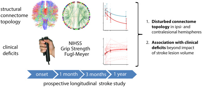
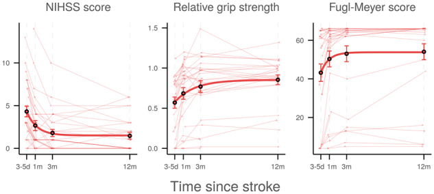
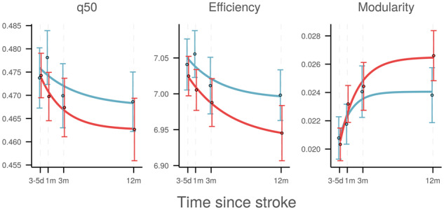
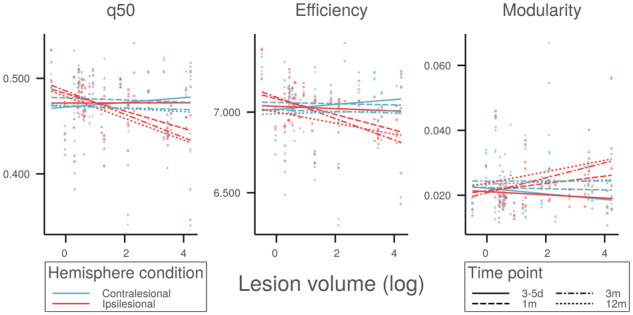
The time course of topological reorganization that occurs in the structural connectome after an ischaemic stroke is currently not well understood. We aimed to determine the evolution of structural brain networks in stroke patients with motor deficits and relate changes in their global topology to residual symptom burden and functional impairment. In this prospective cohort study, ischaemic stroke patients with supratentorial infarcts and motor symptoms were assessed longitudinally by advanced diffusion MRI and detailed clinical testing of upper extremity motor function at four time points from the acute to the chronic stage. For each time point, structural connectomes were reconstructed, and whole-hemisphere global network topology was quantified in terms of integration and segregation parameters. Using non-linear joint mixed-effects regression modelling, network evolution was related to lesion volume and clinical outcome. Thirty patients were included for analysis. Graph-theoretical analysis demonstrated that, over time, brain networks became less integrated and more segregated with decreasing global efficiency and increasing modularity. Changes occurred in both stroke and intact hemispheres and, in the latter, were positively associated with lesion volume. Greater change in topology was associated with larger residual symptom burden and greater motor impairment 1, 3 and 12 months after stroke. After ischaemic stroke, brain networks underwent characteristic changes in both ipsi- and contralesional hemispheres. Topological network changes reflect the severity of damage to the structural network and are associated with functional outcome beyond the impact of lesion volume.
Keywords: graph theory; ischaemic stroke; motor function; recovery; structural connectivity.
© The Author(s) (2020). Published by Oxford University Press on behalf of the Guarantors of Brain.
Figures
Comment on
-
Brain responsivity provides an individual readout for motor recovery after stroke.Brain. 2020 Jun 1;143(6):1873-1888. doi: 10.1093/brain/awaa127. Brain. 2020. PMID: 32375172 Free PMC article.
References
-
- Aerts H, Fias W, Caeyenberghs K, Marinazzo D. Brain networks under attack: robustness properties and the impact of lesions. Brain 2016; 139: 3063–83. - PubMed
-
- Akaike H. A new look at the statistical model identification. IEEE Trans Automat Contr 1974; 19: 716–23.
Publication types
LinkOut - more resources
Full Text Sources