

Differential effects of deep brain stimulation and levodopa on brain activity in Parkinson's disease
- PMID: 32954278
- PMCID: PMC7425344
- DOI: 10.1093/braincomms/fcaa005
Differential effects of deep brain stimulation and levodopa on brain activity in Parkinson's disease
Abstract
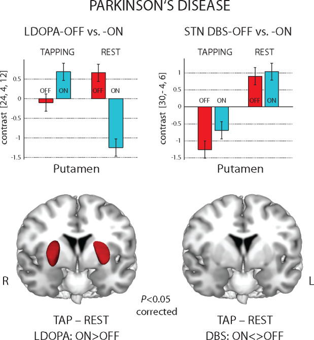
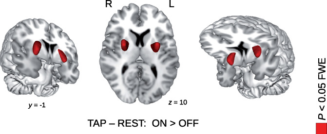
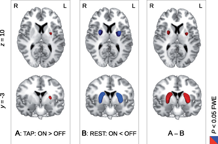
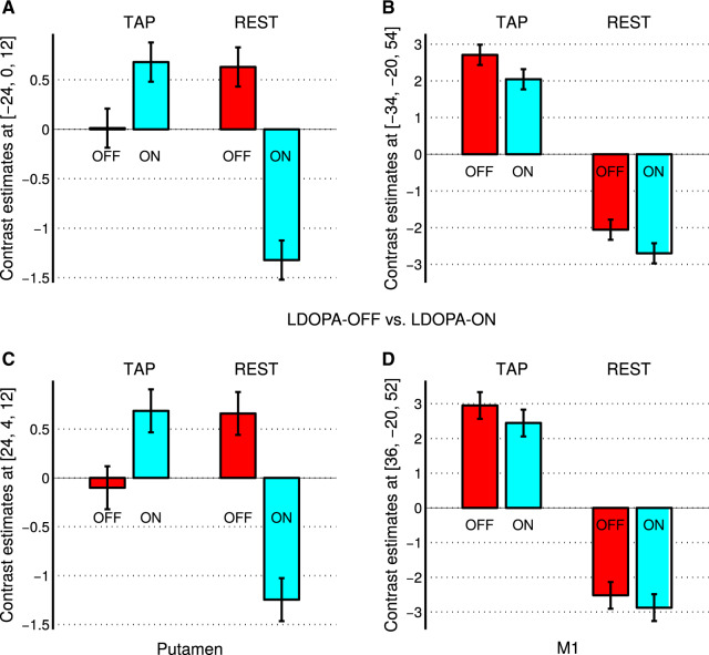
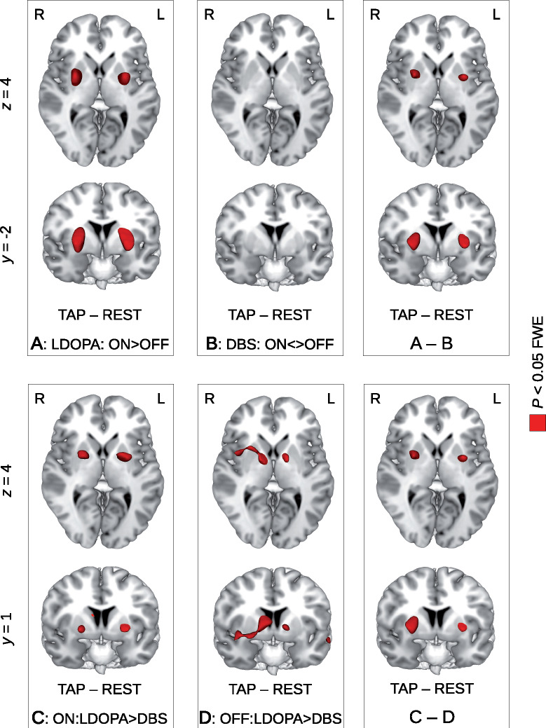
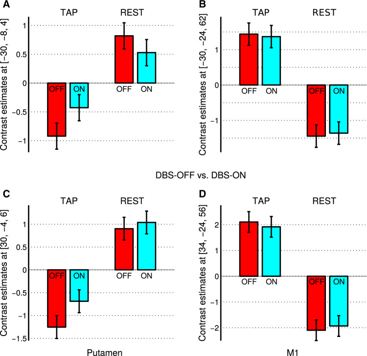
Levodopa is the first-line treatment for Parkinson's disease, although the precise mechanisms mediating its efficacy remain elusive. We aimed to elucidate treatment effects of levodopa on brain activity during the execution of fine movements and to compare them with deep brain stimulation of the subthalamic nuclei. We studied 32 patients with Parkinson's disease using functional MRI during the execution of finger-tapping task, alternating epochs of movement and rest. The task was performed after withdrawal and administration of a single levodopa dose. A subgroup of patients (n = 18) repeated the experiment after electrode implantation with stimulator on and off. Investigating levodopa treatment, we found a significant interaction between both factors of treatment state (off, on) and experimental task (finger tapping, rest) in bilateral putamen, but not in other motor regions. Specifically, during the off state of levodopa medication, activity in the putamen at rest was higher than during tapping. This represents an aberrant activity pattern probably indicating the derangement of basal ganglia network activity due to the lack of dopaminergic input. Levodopa medication reverted this pattern, so that putaminal activity during finger tapping was higher than during rest, as previously described in healthy controls. Within-group comparison with deep brain stimulation underlines the specificity of our findings with levodopa treatment. Indeed, a significant interaction was observed between treatment approach (levodopa, deep brain stimulation) and treatment state (off, on) in bilateral putamen. Our functional MRI study compared for the first time the differential effects of levodopa treatment and deep brain stimulation on brain motor activity. We showed modulatory effects of levodopa on brain activity of the putamen during finger movement execution, which were not observed with deep brain stimulation.
Keywords: Parkinson’s disease; deep brain stimulation; dopaminergic treatment; functional magnetic resonance imaging; levodopa.
© The Author(s) (2020). Published by Oxford University Press on behalf of the Guarantors of Brain.
Figures
Similar articles
-
Subthalamic deep brain stimulation can improve gastric emptying in Parkinson's disease.Brain. 2012 May;135(Pt 5):1478-85. doi: 10.1093/brain/aws086. Epub 2012 Apr 19. Brain. 2012. PMID: 22522940
-
Functional segregation of basal ganglia pathways in Parkinson's disease.Brain. 2018 Sep 1;141(9):2655-2669. doi: 10.1093/brain/awy206. Brain. 2018. PMID: 30084974
-
Different effects of levodopa and subthalamic stimulation on emotional conflict in Parkinson's disease.Hum Brain Mapp. 2018 Dec;39(12):5014-5027. doi: 10.1002/hbm.24341. Epub 2018 Sep 26. Hum Brain Mapp. 2018. PMID: 30259598 Free PMC article.
-
Subthalamic deep brain stimulation and levodopa in Parkinson's disease: a meta-analysis of combined effects.J Neurol. 2019 Feb;266(2):289-297. doi: 10.1007/s00415-018-8936-2. Epub 2018 Jun 16. J Neurol. 2019. PMID: 29909467 Review.
-
[Deep brain stimulation].Rev Neurol (Paris). 2004 May;160(5 Pt 1):511-21. doi: 10.1016/s0035-3787(04)70980-7. Rev Neurol (Paris). 2004. PMID: 15269668 Review. French.
Cited by
-
A Multiscale, Systems-Level, Neuropharmacological Model of Cortico-Basal Ganglia System for Arm Reaching Under Normal, Parkinsonian, and Levodopa Medication Conditions.Front Comput Neurosci. 2022 Jan 3;15:756881. doi: 10.3389/fncom.2021.756881. eCollection 2021. Front Comput Neurosci. 2022. PMID: 35046787 Free PMC article.
-
Use of Functional MRI in Deep Brain Stimulation in Parkinson's Diseases: A Systematic Review.Front Neurol. 2022 Mar 23;13:849918. doi: 10.3389/fneur.2022.849918. eCollection 2022. Front Neurol. 2022. PMID: 35401406 Free PMC article.
-
Predicting Motor Outcome and Quality of Life After Subthalamic Deep Brain Stimulation for Parkinson's Disease: The Role of Standard Screening Measures and Wearable-Data.J Parkinsons Dis. 2023;13(4):575-588. doi: 10.3233/JPD-225101. J Parkinsons Dis. 2023. PMID: 37182900 Free PMC article.
-
Medication versus globus pallidus internus deep brain stimulation in Parkinson's disease with deep learning video analysis of finger tapping.Sci Rep. 2025 May 21;15(1):17557. doi: 10.1038/s41598-025-02098-5. Sci Rep. 2025. PMID: 40394036 Free PMC article.
-
Predicting Motor Outcome of Subthalamic Nucleus Deep Brain Stimulation for Parkinson's Disease Using Quantitative Susceptibility Mapping and Radiomics: A Pilot Study.Front Neurosci. 2021 Sep 7;15:731109. doi: 10.3389/fnins.2021.731109. eCollection 2021. Front Neurosci. 2021. PMID: 34557069 Free PMC article.
References
-
- Agostino R, Berardelli A, Currà A, Accornero N, Manfredi M.. Clinical impairment of sequential finger movements in Parkinson’s disease. Mov Disord 1998; 13: 418–21. - PubMed
-
- Agostino R, Currà A, Giovannelli M, Modugno N, Manfredi M, Berardelli A.. Impairment of individual finger movements in Parkinson’s disease. Mov Disord 2003; 18: 560–5. - PubMed
-
- Ashburner J, Friston KJ.. Unified segmentation. Neuroimage 2005; 26: 839–51. - PubMed
LinkOut - more resources
Full Text Sources