. 2020 Jun 8;2(2):fcaa076.
doi: 10.1093/braincomms/fcaa076. eCollection 2020.

Respiratory-related brain pulsations are increased in epilepsy-a two-centre functional MRI study

Affiliations

Respiratory-related brain pulsations are increased in epilepsy-a two-centre functional MRI study

Janne Kananen et al. Brain Commun. .

Abstract

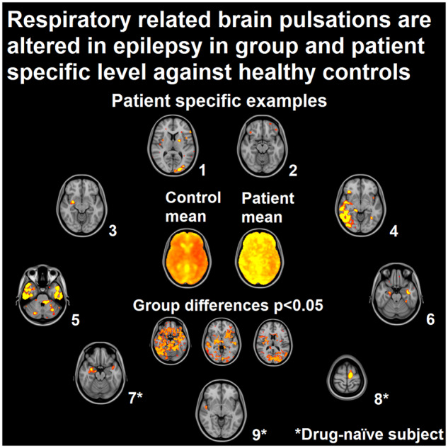
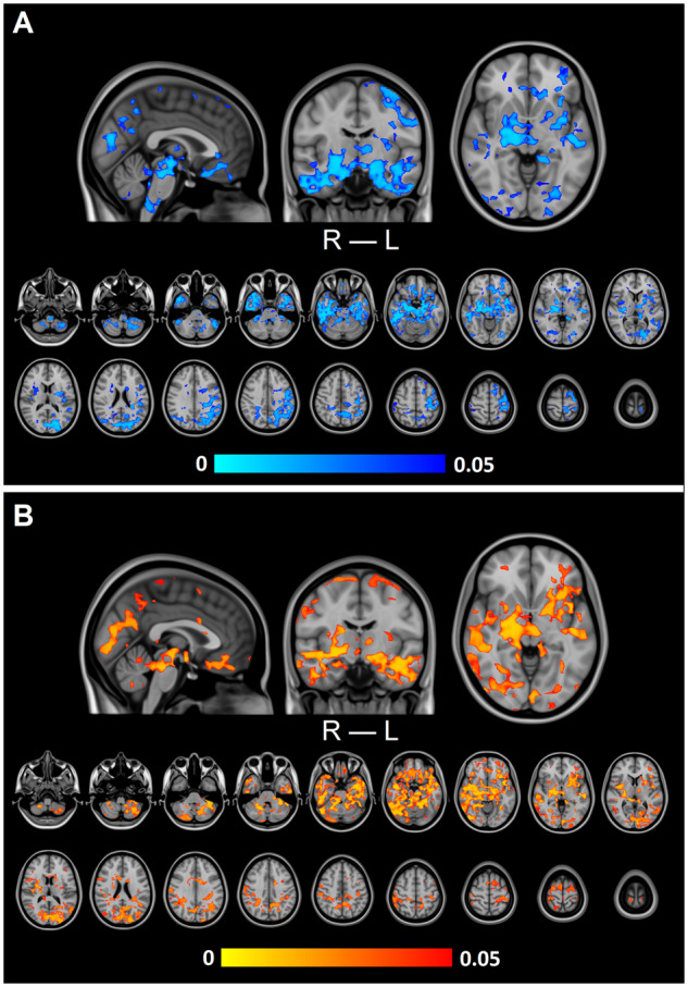
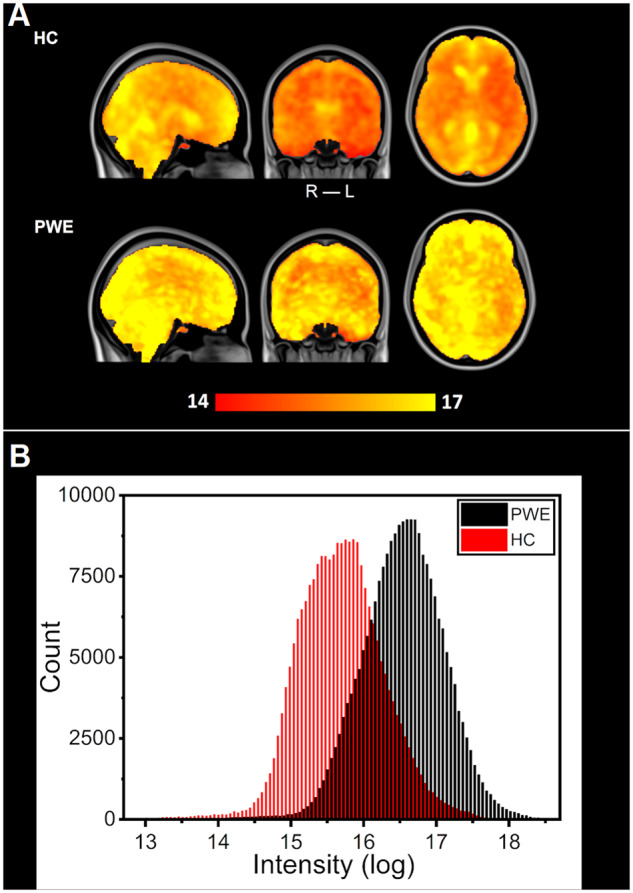
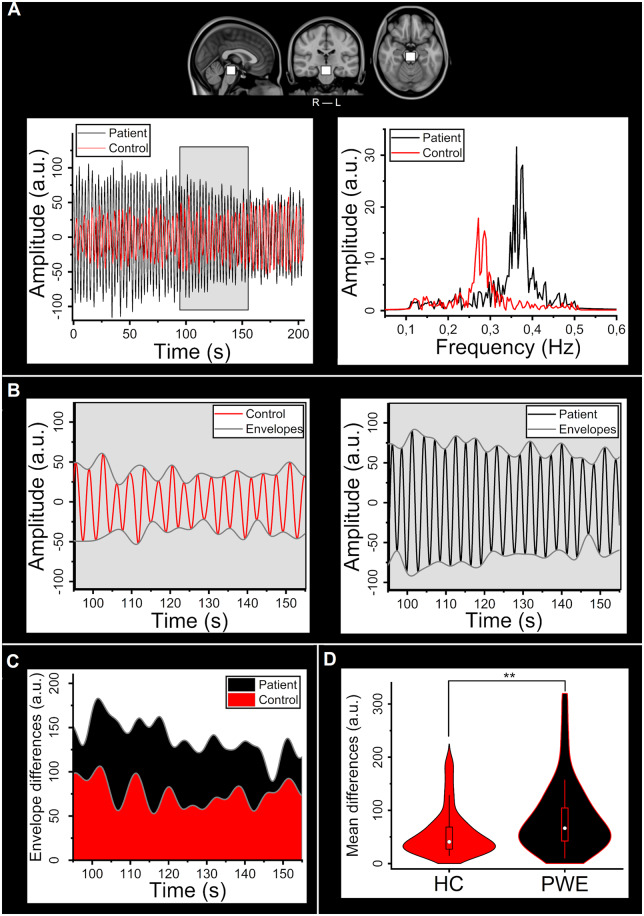
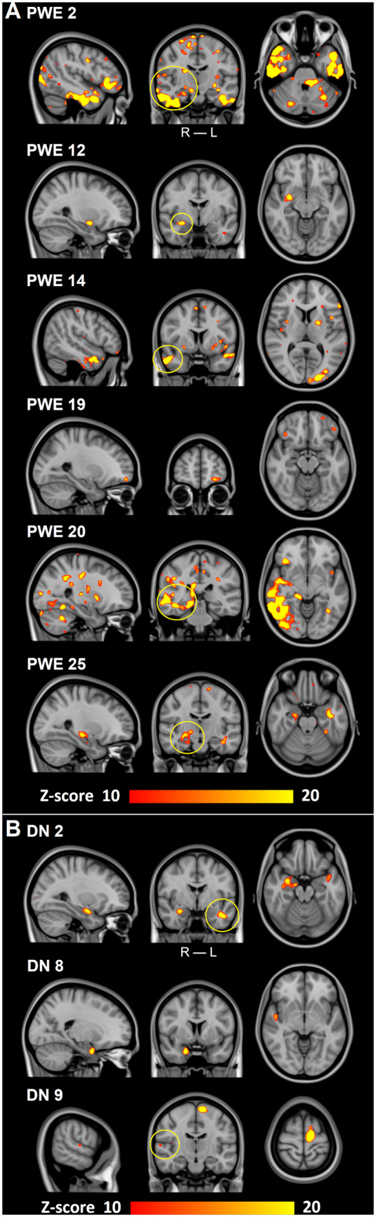
Resting-state functional MRI has shown potential for detecting changes in cerebral blood oxygen level-dependent signal in patients with epilepsy, even in the absence of epileptiform activity. Furthermore, it has been suggested that coefficient of variation mapping of fast functional MRI signal may provide a powerful tool for the identification of intrinsic brain pulsations in neurological diseases such as dementia, stroke and epilepsy. In this study, we used fast functional MRI sequence (magnetic resonance encephalography) to acquire ten whole-brain images per second. We used the functional MRI data to compare physiological brain pulsations between healthy controls (n = 102) and patients with epilepsy (n = 33) and furthermore to drug-naive seizure patients (n = 9). Analyses were performed by calculating coefficient of variation and spectral power in full band and filtered sub-bands. Brain pulsations in the respiratory-related frequency sub-band (0.11-0.51 Hz) were significantly (P < 0.05) increased in patients with epilepsy, with an increase in both signal variance and power. At the individual level, over 80% of medicated and drug-naive seizure patients exhibited areas of abnormal brain signal power that correlated well with the known clinical diagnosis, while none of the controls showed signs of abnormality with the same threshold. The differences were most apparent in the basal brain structures, respiratory centres of brain stem, midbrain and temporal lobes. Notably, full-band, very low frequency (0.01-0.1 Hz) and cardiovascular (0.8-1.76 Hz) brain pulses showed no differences between groups. This study extends and confirms our previous results of abnormal fast functional MRI signal variance in epilepsy patients. Only respiratory-related brain pulsations were clearly increased with no changes in either physiological cardiorespiratory rates or head motion between the subjects. The regional alterations in brain pulsations suggest that mechanisms driving the cerebrospinal fluid homeostasis may be altered in epilepsy. Magnetic resonance encephalography has both increased sensitivity and high specificity for detecting the increased brain pulsations, particularly in times when other tools for locating epileptogenic areas remain inconclusive.

Keywords: brain physiology; brain pulsations; epilepsy; fast fMRI; respiration.


Figures

Graphical Abstract
Graphical Abstract
Figure 1
Figure 1
Group-level differences in respiration-related brain pulsation. (A) Variation in respiration-related brain pulsation (CVresp) was significantly (P <0.05) increased in PWE compared to HC. (B) Power of respiration-related brain pulsation (SPresp) was significantly (P <0.05) increased in PWE compared to HC. In both A and B, significant increases are located in basal structures of the brain, upper brain stems respiratory pneumotaxic centre, midbrain and temporal lobes, including also amygdalae, hippocampi, pallida and putamina.
Figure 2
Figure 2
Mean spectral power of brain pulsations between HC and PWE groups. (A) Group mean maps in respiration-related power, transformed into a logarithmic scale. PWE group mean values are larger in the whole brain compared to HC. (B) Distributions of mean power in the respiratory sub-band voxel values in each group calculated from maps of section (A). Despite an overlap between the two similar distributions (HC: red, PWE: black), difference between the groups is highly significant (P <0.001).
Figure 3
Figure 3
Differences in the respiratory bandpass filtered signal within the brainstem ROI. (A) The selected ROI in the upper brainstem area and example individual bandpass filtered signals from the PWE (black) and HC (red) groups and their respective frequency spectra. (B) Individual time-domain signals shown in A and corresponding individual lower and upper signal envelopes (control: red, patient: black). (C) Difference of envelopes (max−min) i.e. amplitude of signal for the example subjects (control: red, patient: black). (D) Violin plot of mean envelope differences between groups (HC: red, PWE: black). Groups differ significantly (P =0.0035). ROI = region-of-interest.
Figure 4
Figure 4
Individual changes in respiratory-related brain pulsations power. (A) Six examples of individual findings after thresholding (Z-score > 10) from PWE. All PWE show different areas of increased spectral power that correlate with the clinical diagnosis (yellow circles in coronal plane) in Table 2. PWE 19 has a TLE, although our method found affected areas in the frontal parts of the brain. First three PWE are from Freiburg and last three from Oulu. (B) Three DN examples of individual findings after thresholding (Z-score > 10). All DN have different areas influenced similarly as in A and which correlate with the known clinical diagnoses (yellow circles in coronal plane) in Table 3. DN 8 had a clear affected area, even though no diagnosis could be set with standard diagnostic tools. ROI = region-of-interest; TLE = temporal lobe epilepsy.

Similar articles

  • Human NREM Sleep Promotes Brain-Wide Vasomotor and Respiratory Pulsations.
    Helakari H, Korhonen V, Holst SC, Piispala J, Kallio M, Väyrynen T, Huotari N, Raitamaa L, Tuunanen J, Kananen J, Järvelä M, Tuovinen T, Raatikainen V, Borchardt V, Kinnunen H, Nedergaard M, Kiviniemi V. Helakari H, et al. J Neurosci. 2022 Mar 23;42(12):2503-2515. doi: 10.1523/JNEUROSCI.0934-21.2022. Epub 2022 Feb 8. J Neurosci. 2022. PMID: 35135852 Free PMC article.
  • Increased interictal synchronicity of respiratory related brain pulsations in epilepsy.
    Kananen J, Järvelä M, Korhonen V, Tuovinen T, Huotari N, Raitamaa L, Helakari H, Väyrynen T, Raatikainen V, Nedergaard M, Ansakorpi H, Jacobs J, LeVan P, Kiviniemi V. Kananen J, et al. J Cereb Blood Flow Metab. 2022 Oct;42(10):1840-1853. doi: 10.1177/0271678X221099703. Epub 2022 May 14. J Cereb Blood Flow Metab. 2022. PMID: 35570730 Free PMC article.
  • Altered physiological brain variation in drug-resistant epilepsy.
    Kananen J, Tuovinen T, Ansakorpi H, Rytky S, Helakari H, Huotari N, Raitamaa L, Raatikainen V, Rasila A, Borchardt V, Korhonen V, LeVan P, Nedergaard M, Kiviniemi V. Kananen J, et al. Brain Behav. 2018 Sep;8(9):e01090. doi: 10.1002/brb3.1090. Epub 2018 Aug 15. Brain Behav. 2018. PMID: 30112813 Free PMC article.
  • Ultra-fast magnetic resonance encephalography of physiological brain activity - Glymphatic pulsation mechanisms?
    Kiviniemi V, Wang X, Korhonen V, Keinänen T, Tuovinen T, Autio J, LeVan P, Keilholz S, Zang YF, Hennig J, Nedergaard M. Kiviniemi V, et al. J Cereb Blood Flow Metab. 2016 Jun;36(6):1033-45. doi: 10.1177/0271678X15622047. Epub 2015 Dec 21. J Cereb Blood Flow Metab. 2016. PMID: 26690495 Free PMC article.
  • Surgery for epilepsy.
    West S, Nevitt SJ, Cotton J, Gandhi S, Weston J, Sudan A, Ramirez R, Newton R. West S, et al. Cochrane Database Syst Rev. 2019 Jun 25;6(6):CD010541. doi: 10.1002/14651858.CD010541.pub3. Cochrane Database Syst Rev. 2019. PMID: 31237346 Free PMC article.

Cited by

References

    1. Abbott NJ, Pizzo ME, Preston JE, Janigro D, Thorne RG.. The role of brain barriers in fluid movement in the CNS: is there a ‘glymphatic’ system? Acta Neuropathol 2018; 135: 387–407. - PubMed
    1. Andrews JP, Gummadavelli A, Farooque P, Bonito J, Arencibia C, Blumenfeld H, et al.Association of seizure spread with surgical failure in epilepsy. JAMA Neurol 2019; 76: 462–9. - PMC - PubMed
    1. Aroniadou-Anderjaska V, Fritsch B, Qashu F, Braga M.. Pathology and pathophysiology of the amygdala in epileptogenesis and epilepsy. Epilepsy Res 2008; 78: 102–16. - PMC - PubMed
    1. Aslan S, Hocke L, Schwarz N, Frederick B.. Extraction of the cardiac waveform from simultaneous multislice fMRI data using slice sorted averaging and a deep learning reconstruction filter. Neuroimage 2019; 198: 303–16. - PMC - PubMed
    1. Assländer J, Zahneisen B, Hugger T, Reisert M, Lee H-L, LeVan P, et al.Single shot whole brain imaging using spherical stack of spirals trajectories. Neuroimage 2013; 73: 59–70. - PubMed

LinkOut - more resources