. 2021 Jan 1;31(1):48-61.
doi: 10.1093/cercor/bhaa203.

Sulcal Depth in the Medial Ventral Temporal Cortex Predicts the Location of a Place-Selective Region in Macaques, Children, and Adults

Affiliations

Sulcal Depth in the Medial Ventral Temporal Cortex Predicts the Location of a Place-Selective Region in Macaques, Children, and Adults

Vaidehi S Natu et al. Cereb Cortex. .

Abstract

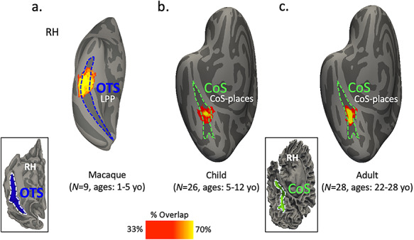
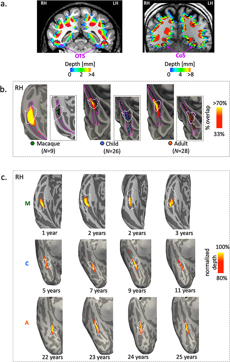
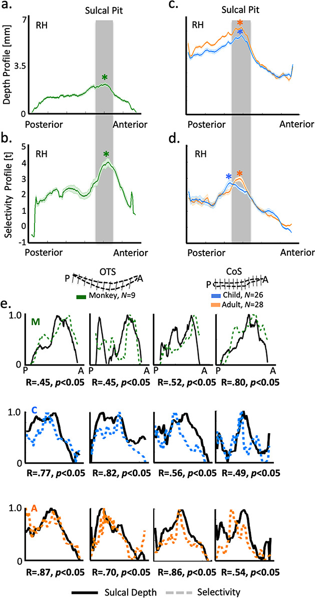
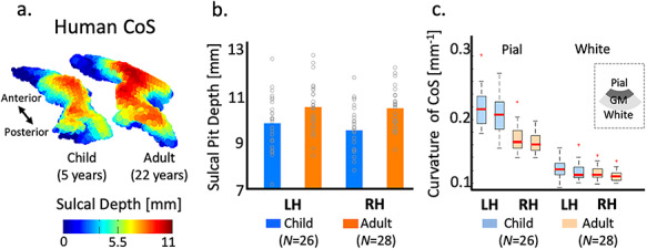
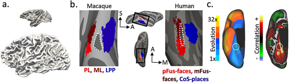
The evolution and development of anatomical-functional relationships in the cerebral cortex is of major interest in neuroscience. Here, we leveraged the fact that a functional region selective for visual scenes is located within a sulcus in the medial ventral temporal cortex (VTC) in both humans and macaques to examine the relationship between sulcal depth and place selectivity in the medial VTC across species and age groups. To do so, we acquired anatomical and functional magnetic resonance imaging scans in 9 macaques, 26 human children, and 28 human adults. Our results revealed a strong structural-functional coupling between sulcal depth and place selectivity across age groups and species in which selectivity was strongest near the deepest sulcal point (the sulcal pit). Interestingly, this coupling between sulcal depth and place selectivity strengthens from childhood to adulthood in humans. Morphological analyses suggest that the stabilization of sulcal-functional coupling in adulthood may be due to sulcal deepening and areal expansion with age as well as developmental differences in cortical curvature at the pial, but not the white matter surfaces. Our results implicate sulcal features as functional landmarks in high-level visual cortex and highlight that sulcal-functional relationships in the medial VTC are preserved between macaques and humans despite differences in cortical folding.

Keywords: functional magnetic resonance imaging (fMRI); human development; macaque; sulcal depth; sulcal pits.


Figures

Figure 1
Figure 1
Probabilistic maps of a functionally defined place-selective region in macaques and humans are located within a sulcus in the medial ventral temporal cortex (VTC) across species. (a) Inflated cortical surface reconstruction of the right hemisphere of a macaque (NMT; Seidlitz et al. 2018). The map indicates the most probable location of the lateral place patch (LPP) in nine macaques. The most probable location (yellow) is within the occipitotemporal sulcus (OTS; blue). (b) Inflated cortical surface reconstruction of the right hemisphere of a human anatomical template (the FreeSurfer average brain from 39 independent adult brains (Fischl, Sereno, Dale, et al. 1999a, Fischl, Sereno, Tootell, et al. 1999b) showing the probabilistic location of CoS-places (also known as the PPA; Epstein and Kanwisher 1998) in 26 children. The most probable location (yellow) of CoS-places is within the collateral sulcus (CoS; green). (c) Same as (b), but for 28 human adults. Like children, the most probable location (yellow) of CoS-places is within the CoS (green). Warmer colors in the probabilistic maps represent greater overlap of place selectivity across participants within a group. Abbreviations: RH, right hemisphere; CoS, collateral sulcus; OTS, occipital temporal sulcus. Brain sizes not to scale.
Figure 2
Figure 2
A place-selective region in the medial VTC overlaps the deepest sulcal points in macaques, children, and adults. (a) Coronal slices showing depth maps obtained from FreeSurfer’s auto-segmentation algorithm in sample macaque and human adult brains (warmer colors represent deeper). For simplicity, the map has been thresholded to only show positive depth values representing sulcal folds, walls, and pits. (b) Inflated cortical surfaces of a right hemisphere showing probabilistic maps (percent overlap) of sulcal depth across participants within each group (macaque, children, and adults) at each point of the occipitotemporal sulcus (OTS) in macaques and the collateral sulcus (CoS) in humans (magenta dotted outlines). The locations of the sulcal pit from each individual subject (colored circles) is shown in the inset on the right of each group surface. In each group, deep sulcal points (warmer colors) within the middle of the sulcus overlap the most across individuals. Additionally, probabilistic locations of the place-selective region (dotted white lines obtained from maps in Fig. 1) overlapped with the probabilistic sulcal depth maps (warm colors) in macaques, children, and adults. (c) Four sample macaque (top row), child (middle row), and adult (bottom row) ventral temporal surfaces, showing the overlap between the functionally defined place-selective region (white dotted lines) and sulcal depth maps (warm colors). To visualize the deep points of the sulcus, the depth maps in each individual subject are thresholded at 80% depth (100% is the deepest point within the sulcus in each individual). Warmer colors indicate deeper sulcal points. All individual surfaces are shown in Supplementary Figures 2–7. Brain sizes are not to scale.
Figure 3
Figure 3
Sulcal depth and place selectivity in the medial VTC are correlated in both humans and macaques. (a, c) Mean (dark line) and standard deviation (lighter shading) of sulcal depth along the anterior–posterior length of the right OTS in nine macaques (a) and the right CoS in humans (c). In (c), data are averaged across 26 children (blue) and 28 adults (orange). Star (*)represents deepest sulcal point. (b, d) Mean place selectivity (t-value) in macaques (b, green) and humans (d, blue/orange). Across macaques and humans, the profiles are well-matched along the anterior–posterior axis in which there is a functional peak (represented by * in b, d) around the deepest sulcal points (represented by gray bar). Left hemisphere data in Supplementary Fig. 8. (e) Normalized depth (black lines) and selectivity (colored dotted lines) values as a function of distance along the posterior (P) to anterior (A) axis in four sample macaques (top row), human children (middle row), and human adults (bottom row). Correlation (R) and significance values below each subplot relate the depth and selectivity curves to one another. All individual profiles and their respective correlations are in Supplementary Figures 9–11.
Figure 4
Figure 4
Morphological differences in the CoS between children and adults. (a) Three-dimensional sulcal depth maps of the CoS in an example child (5 years old) and adult (22 years old) showing differences in depth (represented by increasing height of the 3D maps). (b) Mean depth of place-selective sulcal pits in children (blue) and adults (orange). Gray circles: individual subjects. (c) Box plots showing median (thick red line), 25th and 75th percentile (box), and range (whiskers) of curvature of the CoS at the pial and white matter surfaces in children (blue) and adults (orange). GM, gray matter. LH, left hemisphere. RH, right hemisphere.
Figure 5
Figure 5
The topology of place- and face-selective regions relative to shallow and deep sulci is relatively preserved in the VTC despite significant differences in brain size and macroanatomy between macaques and humans. (a) Sagittal views of a sample macaque and a human brain (to scale) showing that human brains are ~15 times bigger than macaque brains. (b) Middle, top: lateral view of the macaque cortical surface (NMT; Seidlitz et al. 2018). Black rectangle: zoomed portion illustrated in the left image showing the macaque face-selective patches ML (maroon) and PL (red) relative to the lateral place-selective patch (LPP; blue). Each patch is the probabilistic location across nine macaques. The zoomed image has been rotated (superior: left of the image; anterior: top of the image). A shallow sulcus (posterior middle temporal sulcus, pmts; dotted white outline) divides ML and PL from LPP. Middle, bottom: ventral view of the human cortical surface (FreeSurfer average template from 39 adults; Fischl, Sereno, Dale, et al. 1999a, Fischl, Sereno, Tootell, et al. 1999b). Black rectangle: zoomed portion illustrated in the right image showing the human face-selective regions (mFus-faces (maroon) and pFus-faces (red)) relative to the place-selective region, CoS-places (blue). Each region is the probabilistic definition across 12 adults from Weiner et al. (2018). A shallow sulcus (mid-fusiform sulcus, MFS; dotted white outline) divides mFus-faces and pFus-faces from CoS-places. (c) Left: image of the human brain (from the PALS-B12 atlas) adapted from Hill et al. (2010), with permission. Map shows regional evolutionary cortical expansion between an adult macaque and the average human adult on the right ventral surface. White circle has been added to the original images to indicate the approximate location of CoS-places examined in humans in the present study. Regions in the medial VTC show minimal cortical expansion compared to the remainder of cortex, while the anterior–lateral VTC (warm colors) exhibits much more expansion compared to the medial VTC. Right: the same cortical reconstruction of the right hemisphere with a correlation map between evolutionary and postnatal cortical expansion (adapted from Hill et al. (2010)), with permission. Regions in the medial VTC show a positive correlation across evolutionary and postnatal expansion patterns, while regions in the anterior–lateral VTC show little or a negative correlation. A, anterior; S, superior; M, medial.

Similar articles

Cited by

References

    1. Abbasi N, Duncan J, Rajimehr R. 2020. Genetic influence is linked to cortical morphology in category-selective areas of visual cortex. Nat Commun. 11:709. - PMC - PubMed
    1. Amiez C, Petrides M. 2014. Neuroimaging evidence of the anatomo-functional organization of the human cingulate motor areas. Cereb Cortex. 24:563–578. - PubMed
    1. Amiez C, Sallet J, Hopkins WD, Meguerditchian A, Hadj-Bouziane F, Ben Hamed S, Wilson CRE, Procyk E, Petrides M. 2019. Sulcal organization in the medial frontal cortex provides insights into primate brain evolution. Nat Commun. 10:3437. - PMC - PubMed
    1. Amiez C, Wilson CRE, Procyk E. 2018. Variations of cingulate sulcal organization and link with cognitive performance. Sci Rep. 8:13988. - PMC - PubMed
    1. Arcaro MJ, Kastner S. 2015. Topographic organization of areas V3 and V4 and its relation to supra-areal organization of the primate visual system. Vis Neurosci. 32:E014. - PMC - PubMed

Publication types