

Idiopathic middle meningeal artery and middle meningeal vein fistula presenting as temporal intraparenchymal hemorrhage
- PMID: 32954912
- PMCID: PMC8050525
- DOI: 10.1177/1591019920960248
Idiopathic middle meningeal artery and middle meningeal vein fistula presenting as temporal intraparenchymal hemorrhage
Abstract
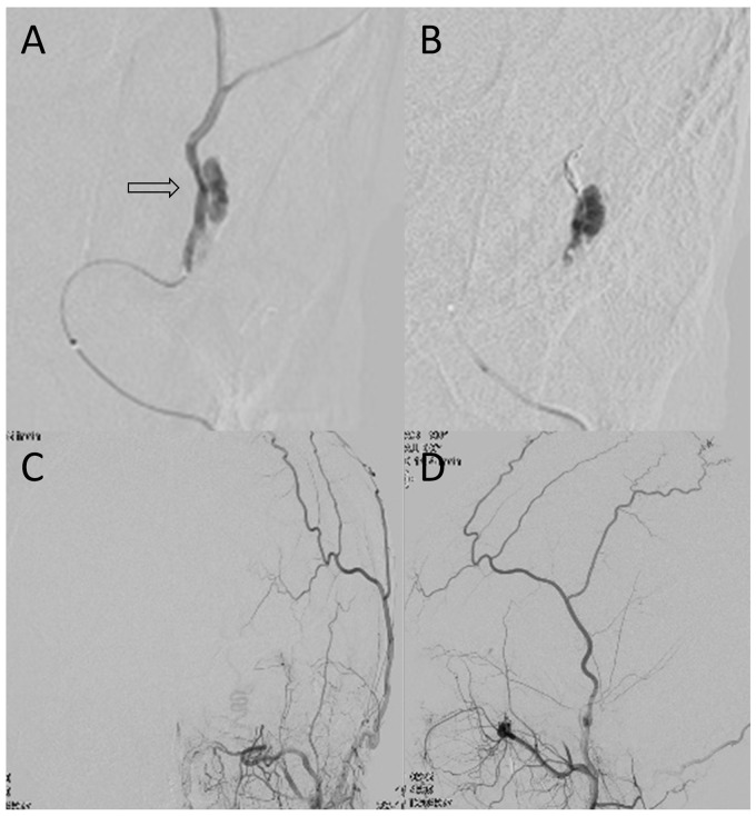
Middle meningeal artery (MMA) and middle meningeal vein (MMV) fistula is a rare lesion. Hemorrhagic propensity for this pathology has not been widely recognized. We encountered a case of idiopathic MMA-MMV fistula presenting as intracerebral hemorrhage. MMA and MMV showed a single fistula and the enlarged MMV coiled around the MMA, resembling a varix. We treated the case using endovascular techniques. This case highlights the anatomy along the course of the MMV and is unique, since no backward leptomeningeal drainage was observed. A bone defect in the meningeal groove on computed tomography and a "green caviar appearance" on angiography may be characteristic of this rare pathology.
Keywords: MM-AVF; MMA-MMV fistula; intraparenchymal hemorrhage; transarterial embolization.
Conflict of interest statement
Figures


Similar articles
-
Embolization of a traumatic arteriovenous fistula between the middle meningeal artery and middle meningeal vein in a child with pulsatile tinnitus.Childs Nerv Syst. 2018 Mar;34(3):571-575. doi: 10.1007/s00381-017-3665-x. Epub 2017 Nov 23. Childs Nerv Syst. 2018. PMID: 29170838
-
Traumatic Arteriovenous Fistula Between Extracranial Middle Meningeal Artery and Petrosal Vein.World Neurosurg. 2019 Oct;130:50-53. doi: 10.1016/j.wneu.2019.06.171. Epub 2019 Jun 29. World Neurosurg. 2019. PMID: 31265928
-
Intracerebral hemorrhage from a middle meningeal arteriovenous fistula with a giant venous varix.Surg Neurol. 1992 Jun;37(6):460-3. doi: 10.1016/0090-3019(92)90136-b. Surg Neurol. 1992. PMID: 1595052
-
Traumatic arteriovenous fistula between the extracranial middle meningeal artery and the pterygoid plexus: A case report and literature review.Interv Neuroradiol. 2017 Feb;23(1):90-96. doi: 10.1177/1591019916673584. Epub 2016 Oct 27. Interv Neuroradiol. 2017. PMID: 27798326 Free PMC article. Review.
-
Iatrogenic Middle Meningeal Arteriovenous Fistula During Embolization: Two Case Reports and Literature Review.J Stroke Cerebrovasc Dis. 2021 Aug;30(8):105909. doi: 10.1016/j.jstrokecerebrovasdis.2021.105909. Epub 2021 Jun 11. J Stroke Cerebrovasc Dis. 2021. PMID: 34119750 Review.
Cited by
-
Spontaneous middle meningeal arteriovenous fistula without cortical venous reflux presenting with acute subdural hematoma: illustrative case.J Neurosurg Case Lessons. 2023 Aug 7;6(6):CASE23306. doi: 10.3171/CASE23306. Print 2023 Aug 7. J Neurosurg Case Lessons. 2023. PMID: 37581592 Free PMC article.
-
Spontaneous Middle Meningeal Arteriovenous Fistula Caused by Aneurysm Rupture: A Case Report.NMC Case Rep J. 2023 Mar 24;10:81-85. doi: 10.2176/jns-nmc.2022-0376. eCollection 2023. NMC Case Rep J. 2023. PMID: 37065874 Free PMC article.
References
-
- Tokairin K, Osanai T, Kazumata K, et al.. Contrecoup injury-induced middle meningeal arteriovenous fistula detected by time-of-flight magnetic resonance angiography and magnetic resonance arterial spin labeling: case report and review of the literature. World Neurosurg 2019; 127: 79–84. - PubMed
-
- Roski RA, Owen M, White RJ, et al.. Middle meningeal artery trauma. Surg Neurol 1982; 17: 200–203. - PubMed
-
- Sakata H, Nishimura S, Mino M, et al.. Serial angiography of dynamic changes of traumatic middle meningeal arteriovenous fistula: case report. Neurol Med Chir (Tokyo) 2009; 49: 462–464. - PubMed
-
- Abla AA, Albuquerque FC, Theodore N, et al.. Delayed presentation of traumatic cerebral and dural arteriovenous fistulae after a BB gun accident in a pediatric patient: case report. Neurosurgery 2011; 68: E1750–E1754. discussion E4–E5. - PubMed
-
- Pritz MB, Pribram HF. Intracerebral hemorrhage from a middle meningeal arteriovenous fistula with a giant venous varix. Surg Neurol 1992; 37: 460–463. - PubMed
Publication types
MeSH terms
LinkOut - more resources
Full Text Sources
