Review
. 2020 Oct 20;9(19):e018506.
doi: 10.1161/JAHA.120.018506. Epub 2020 Sep 21.

Degeneration of Bioprosthetic Heart Valves: Update 2020

Affiliations
Review

Degeneration of Bioprosthetic Heart Valves: Update 2020

Alexander E Kostyunin et al. J Am Heart Assoc. .

Abstract

The implantation of bioprosthetic heart valves (BHVs) is increasingly becoming the treatment of choice in patients requiring heart valve replacement surgery. Unlike mechanical heart valves, BHVs are less thrombogenic and exhibit superior hemodynamic properties. However, BHVs are prone to structural valve degeneration (SVD), an unavoidable condition limiting graft durability. Mechanisms underlying SVD are incompletely understood, and early concepts suggesting the purely degenerative nature of this process are now considered oversimplified. Recent studies implicate the host immune response as a major modality of SVD pathogenesis, manifested by a combination of processes phenocopying the long-term transplant rejection, atherosclerosis, and calcification of native aortic valves. In this review, we summarize and critically analyze relevant studies on (1) SVD triggers and pathogenesis, (2) current approaches to protect BHVs from calcification, (3) obtaining low immunogenic BHV tissue from genetically modified animals, and (4) potential strategies for SVD prevention in the clinical setting.

Keywords: bioprosthesis; calcification; genetically modified animals; immune rejection; inflammation; structural valve degeneration; valve replacement.


Conflict of interest statement

None.

Figures

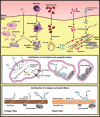
Figure 1
Figure 1. The importance of red blood cells for structural valve degeneration development.
Red blood cells may penetrate the bioprosthetic heart valves through the extracellular matrix disintegration (A) or capillary‐like tubes (B) formed as a result of mechanical stress or chronic inflammation. Red blood cell demise causes iron deposition and further oxidation‐driven degradation of the prosthetic tissue.
Figure 2
Figure 2. Mechanisms driving dystrophic calcification responsible for structural valve degeneration.
Both implant‐related (residual donor cells and their debris, loss of glycosaminoglycans, and damage of collagen/elastin fibers during chemical treatment and storage) and recipient‐related (immune cell and red blood cell penetration, serum proteolytic enzymes, and calcium‐binding proteins) factors promote dystrophic calcification of bioprosthetic heart valves (BHVs). ALP indicates alkaline phosphatase; and MMP, matrix metalloproteinase.
Figure 3
Figure 3. Mechanical stress and structural valve degeneration.
Mechanical load and stress distribution in systolic phase in native mitral and aortic valves (A) and in the case of heart valve implantation into mitral and aortic positions (B).
Figure 4
Figure 4. The molecular basis of chronic inflammation in relation to structural valve degeneration.
Xenogeneic glycans and thrombi adhered to bioprosthetic heart valves (BHVs) recruit monocytes, which can penetrate the tissue with the subsequent differentiation into macrophages and multinucleated giant cells. Immune cells internalize disintegrated fragments of collagen fibers and release reactive oxygen species, proteolytic enzymes, and calcium‐binding extracellular vesicles, altogether promoting degradation and calcification of the extracellular matrix. In addition, immune cells produce proteoglycans, which bind low‐density lipoprotein (LDL) from the plasma. Macrophages engulfing LDL are then transformed into the foam cells reminiscent of the pathophysiological scenarios observed in atherosclerotic plaques and calcific aortic valve disease. MMP indicates matrix metalloproteinase; and oxLDL, oxidized LDL.
Figure 5
Figure 5. Chemical modifications of collagen induced by various fixatives.
A, Aldehyde groups of glutaraldehyde (GA) interact with amino group of lysine (Lys) or hydroxylysine (Hyl) residues within the collagen, thereby forming a stable chemical bond (Schiff base) for a stable cross‐linking. B, One of GA aldehyde groups interacts with a collagen amino group, resulting in a cross‐linking, whereas the second aldehyde group remains free to other chemical interactions, including calcium binding. C, Polymerization of GA is performed through aldol condensation. Despite collagen molecules that are cross‐linked, free aldehyde groups still remain. D, All epoxy groups of ethylene glycol diglycidyl ether (EGDE) interact with amino group of Lys or Hyl residues within the collagen, forming a stable covalent bond for a stable cross‐linking. E, Collagen fixation with 1‐ethyl‐3‐(3‐dimethylaminopropyl)carbodiimide (EDC) and N‐hydroxysuccinimide (NHS) is conducted via the activation of carboxyl groups of aspartic acid/glutamic acid residues in the peptide chain and through the formation of intermediate compound, which is able to interact with free amino groups of lysine or hydroxylysine.
Figure 6
Figure 6. Key factors of structural valve degeneration (SVD) development and strategies to retard SVD.
 

References

    1. Baumgartner H, Falk V, Bax JJ, De Bonis M, Hamm C, Holm PJ, Iung B, Lancellotti P, Lansac E, Rodriguez Muñoz D, et al. 2017 ESC/EACTS guidelines for the management of valvular heart disease. Eur Heart J. 2017;38:2739–2791. - PubMed
    1. Nishimura RA, Otto CM, Bonow RO, Carabello BA, Erwin JP III, Fleisher LA, Jneid H, Mack MJ, McLeod CJ, O'Gara PT, et al. 2017 AHA/ACC focused update of the 2014 AHA/ACC guideline for the management of patients with valvular heart disease: a report of the American College of Cardiology American Heart Association task force on clinical practice guidelines. Circulation. 2017;135:e1159–e1195. - PubMed
    1. Head SJ, Çelik M, Kappetein AP. Mechanical versus bioprosthetic aortic valve replacement. Eur Heart J. 2017;38:2183–2191. - PubMed
    1. Capodanno D, Petronio AS, Prendergast B, Eltchaninoff H, Vahanian A, Modine T, Lancellotti P, Sondergaard L, Ludman PF, Tamburino C, et al. Standardized definitions of structural deterioration and valve failure in assessing long‐term durability of transcatheter and surgical aortic bioprosthetic valves: a consensus statement from the European Association of Percutaneous Cardiovascular Interventions (EAPCI) endorsed by the European Society of Cardiology (ESC) and the European Association for Cardio‐Thoracic Surgery (EACTS). Eur Heart J. 2017;38:3382–3390. - PubMed
    1. Dvir D, Bourguignon T, Otto CM, Hahn RT, Rosenhek R, Webb JG, Treede H, Sarano ME, Feldman T, Wijeysundera HC, et al. Standardized definition of structural valve degeneration for surgical and transcatheter bioprosthetic aortic valves. Circulation. 2018;137:388–399. - PubMed

Publication types

MeSH terms