. 2020 Sep 2:(163):10.3791/61620.
doi: 10.3791/61620.

A Model Membrane Platform for Reconstituting Mitochondrial Membrane Dynamics

Affiliations

A Model Membrane Platform for Reconstituting Mitochondrial Membrane Dynamics

Yifan Ge et al. J Vis Exp. .

Abstract

Mitochondrial dynamics is essential for the organelle's diverse functions and cellular responses. The crowded, spatially complex, mitochondrial membrane is a challenging environment to distinguish regulatory factors. Experimental control of protein and lipid components can help answer specific questions of regulation. Yet, quantitative manipulation of these factors is challenging in cellular assays. To investigate the molecular mechanism of mitochondria inner-membrane fusion, we introduced an in vitro reconstitution platform that mimics the lipid environment of the mitochondrial inner-membrane. Here we describe detailed steps for preparing lipid bilayers and reconstituting mitochondrial membrane proteins. The platform allowed analysis of intermediates in mitochondrial inner-membrane fusion, and the kinetics for individual transitions, in a quantitative manner. This protocol describes the fabrication of bilayers with asymmetric lipid composition and describes general considerations for reconstituting transmembrane proteins into a cushioned bilayer. The method may be applied to study other membrane systems.


Figures

Figure 1:
Figure 1:. Monitoring mitochondrial membrane fusion.
(A) Organelles are cellular membrane compartments. (B) Sequential steps of mitochondrial membrane fusion. Fusion of the outer membrane of mitochondria is catalyzed by Mfn1 and/or Mfn2, while inner-membrane fusion is mediated by Opa1. (C-F) Schematic of the in vitro reconstitution platform to study mitochondrial membrane fusion. The platform includes two parts: a proteoliposome and a polymer-tethered lipid bilayer, both with reconstituted l-Opa1. Fluorescent labels, including two different fluorescent membrane dyes and a content marker, help distinguish steps during membrane fusion. The two membrane markers (Cy5-PE (red) and TexasRed PE (orange) make a FRET pair, which can report on close membrane docking. Diffusion of TexasRed-PE that labels proteoliposome is an indicator of lipid demixing (hemifusion). Content release is monitored through the dequenching of the calcein signal (shown in green). Panels A and B created using Biorender. Please click here to view a larger version of this figure.
Figure 2:
Figure 2:. Steps in making a polymer-tethered lipid bilayer.
Steps of making lipid bilayers using Langmuir-Blodgett dipping (A-C) and Langmuir-Schaefer transfer (D) techniques. (E) The final “sandwich” containing the lipid bilayer. Please click here to view a larger version of this figure.
Figure 3:
Figure 3:. Procedure for reconstituting l-Opa1 into a polymer-tethered lipid bilayer.
Please click here to view a larger version of this figure.
Figure 4:
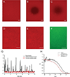
Figure 4:. Distribution of lipid and reconstituted protein in the model membrane.
(A-C) Example images of a lipid bilayer and its lipid fluidity validated by epifluorescence microscopy. (A) Homogeneous lipid distribution in bilayer prior to photobleaching. (B) Snapshot immediately after photobleaching. (C) Bilayer imaged after fluorescence recovery indicates good lipid fluidity of the membrane following reconstitution. (D,E) Representative images of lipid distribution before (D), and after (E) l-Opa1 reconstitution indicate the reconstitution process did not create defects in the bilayer. Representative TIRF image of l-Opa1 labeled with Alexa 488 conjugated antibody (F) showing a homogeneous distribution of Opa1 upon reconstitution. G. Representative raw photon counts of l-Opa1 signal by fluorescent correlation spectroscopy. In the control, no l-Opa1 was reconstituted in the bilayer, while antibody was added and rinsed. The diffusion of l-Opa1 is significantly slower than lipids in the membrane, consistent with successful reconstitution of transmembrane l-Opa1 (H). Scale bar: 10 μm. Please click here to view a larger version of this figure.
Figure 5:
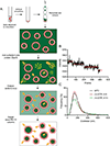
Figure 5:. Fabrication and characterization of proteoliposomes.
(A) Steps in fabricating proteoliposomes with encapsulated, quenched calcein. (B) Representative data of fluorescent step-bleaching show an average of 2–3 copies of l-Opa1 embedded in the liposome. (C) Representative size distributions of proteoliposomes (red) without any nucleotide 1 h after GTP incubation (green). Please click here to view a larger version of this figure.
Figure 6:
Figure 6:. Representative results showing particle tethering (A, scale bar 10 μm), hemifusion (B, scale bar 0.5 μm), and fusion (C, scale bar 0.5 μm).
(A) Proteoliposomes tethered to Opa1-reconstituted lipid bilayer before GTP addition. (B) An example of hemifusion. The upper row in B shows lipid demixing (TexasRed signal, red), lower row in B shows no content release (calcein signal, green) under these conditions. (C) A representative trace of proteoliposome fusing with the lipid bilayer. Content release can be observed from images in the lower row of showing dequenching of calcein (lower row, green). Please click here to view a larger version of this figure.

References

    1. Sackmann E, Biological membranes architecture and function. In Structure and Dynamics of Membranes. Ed: Lipowsky R, Sackmann E. Elsiever. 1, 1–63 (1995).
    1. Rajendran L, Simons K, Lipid rafts and membrane dynamics. Journal of Cell Science. 118 (Pt 6), 1099–1102 (2005). - PubMed
    1. Schink KO, Tan KW, Stenmark H Phosphoinositides in Control of Membrane Dynamics. Annual Review of Cell and Developmental Biology. 32, 143–171 (2016). - PubMed
    1. Gu RX, Ingolfsson HI, de Vries AH, Marrink SJ, Tieleman DP Ganglioside-Lipid and Ganglioside-Protein Interactions Revealed by Coarse-Grained and Atomistic Molecular Dynamics Simulations. Journal of Physical Chemistry B. 121 (15), 3262–3275 (2017). - PMC - PubMed
    1. Schafer DA Coupling actin dynamics and membrane dynamics during endocytosis. Current Opinion in Cell Biology. 14 (1), 76–81 (2002). - PubMed

Publication types

MeSH terms

LinkOut - more resources