Characterizing Enzyme Reactions in Microcrystals for Effective Mix-and-Inject Experiments using X-ray Free-Electron Lasers
- PMID: 32955854
- PMCID: PMC8367009
- DOI: 10.1021/acs.analchem.0c02569
Characterizing Enzyme Reactions in Microcrystals for Effective Mix-and-Inject Experiments using X-ray Free-Electron Lasers
Abstract
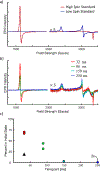
Mix-and-inject serial crystallography is an emerging technique that utilizes X-ray free-electron lasers (XFELs) and microcrystalline samples to capture atomically detailed snapshots of biomolecules as they function. Early experiments have yielded exciting results; however, there are limited options to characterize reactions in crystallo in advance of the beamtime. Complementary measurements are needed to identify the best conditions and timescales for observing structural intermediates. Here, we describe the interface of XFEL compatible mixing injectors with rapid freeze-quenching and X-band EPR spectroscopy, permitting characterization of reactions in crystals under the same conditions as an XFEL experiment. We demonstrate this technology by tracking the reaction of azide with microcrystalline myoglobin, using only a fraction of the sample required for a mix-and-inject experiment. This spectroscopic method enables optimization of sample and mixer conditions to maximize the populations of intermediate states, eliminating the guesswork of current mix-and-inject experiments.
Conflict of interest statement
The authors declare no competing financial interest.
Figures


Similar articles
-
Microfluidic Mixing Injector Holder Enables Routine Structural Enzymology Measurements with Mix-and-Inject Serial Crystallography Using X-ray Free Electron Lasers.Anal Chem. 2019 Jun 4;91(11):7139-7144. doi: 10.1021/acs.analchem.9b00311. Epub 2019 May 22. Anal Chem. 2019. PMID: 31060352
-
Effective coupling of rapid freeze-quench to high-frequency electron paramagnetic resonance.PLoS One. 2020 May 11;15(5):e0232555. doi: 10.1371/journal.pone.0232555. eCollection 2020. PLoS One. 2020. PMID: 32392255 Free PMC article.
-
Mix-and-Inject Serial Femtosecond Crystallography to Capture RNA Riboswitch Intermediates.Methods Mol Biol. 2023;2568:243-249. doi: 10.1007/978-1-0716-2687-0_16. Methods Mol Biol. 2023. PMID: 36227573
-
Virus Structures by X-Ray Free-Electron Lasers.Annu Rev Virol. 2019 Sep 29;6(1):161-176. doi: 10.1146/annurev-virology-092818-015724. Annu Rev Virol. 2019. PMID: 31567066 Review.
-
Microsecond freeze-hyperquenching: development of a new ultrafast micro-mixing and sampling technology and application to enzyme catalysis.Biochim Biophys Acta. 2004 May 12;1656(1):1-31. doi: 10.1016/j.bbabio.2004.02.006. Biochim Biophys Acta. 2004. PMID: 15136155 Review.
Cited by
-
Chaotic advection mixer for capturing transient states of diverse biological macromolecular systems with time-resolved small-angle X-ray scattering.IUCrJ. 2023 May 1;10(Pt 3):363-375. doi: 10.1107/S2052252523003482. IUCrJ. 2023. PMID: 37144817 Free PMC article.
-
Analysis of Covarine Particle in Toothpaste Through Microfluidic Simulation, Experimental Validation, and Electrical Impedance Spectroscopy.ACS Omega. 2024 Feb 23;9(9):10539-10555. doi: 10.1021/acsomega.3c08799. eCollection 2024 Mar 5. ACS Omega. 2024. PMID: 38463280 Free PMC article.
-
Emerging Time-Resolved X-Ray Diffraction Approaches for Protein Dynamics.Annu Rev Biophys. 2023 May 9;52:255-274. doi: 10.1146/annurev-biophys-111622-091155. Annu Rev Biophys. 2023. PMID: 37159292 Free PMC article. Review.
-
Aspartate or arginine? Validated redox state X-ray structures elucidate mechanistic subtleties of FeIV = O formation in bacterial dye-decolorizing peroxidases.J Biol Inorg Chem. 2021 Oct;26(7):743-761. doi: 10.1007/s00775-021-01896-2. Epub 2021 Sep 3. J Biol Inorg Chem. 2021. PMID: 34477969 Free PMC article. Review.
-
Time-resolved cryogenic electron tomography for the study of transient cellular processes.Mol Biol Cell. 2024 Jul 1;35(7):mr4. doi: 10.1091/mbc.E24-01-0042. Epub 2024 May 8. Mol Biol Cell. 2024. PMID: 38717434 Free PMC article.
References
-
- Kupitz C; Olmos JL Jr.; Holl M; Tremblay L; Pande K; Pandey S; Oberthür D; Hunter M; Liang M; Aquila A; Tenboer J; Calvey G; Katz A; Chen Y; Wiedorn MO; Knoska J; Meents A; Majriani V; Norwood T; Poudyal I; Grant T; Miller MD; Xu W; Tolstikova A; Morgan A; Metz M; Martin-Gracia J; Zook JD; Roy-Chowdhury S; Coe J; Nagaratnam N; Meza D; Fromme R; Basu S; Frank M; White T; Barty A; Bajt S; Yefanov O; Chapman HN; Zatsepin N; Nelson G; Weierstall U; Spence J; Schwander P; Pollack L; Fromme P; Ourmazd A; Phillips GN; Schmidt M Struct. Dyn 2017, 4, No. 044003. - PMC - PubMed
-
- Stagno JR; Liu Y; Bhandari YR; Conrad CE; Panja S; Swain M; Fan L; Nelson G; Li C; Wendel DR; White TA; Coe JD; Wiedorn MO; Knoska J; Oberthuer D; Tuckey RA; Yu P; Dyba M; Tarasov SG; Weierstall U; Grant TD; Schwieters CD; Zhang J; Ferré-D’Amaré AR; Fromme P; Draper DE; Liang M; Hunter MS; Boutet S; Tan K; Zuo X; Ji X; Barty A; Zatsepin NA; Chapman HN; Spence JCH; Woodson SA; Wang Y-X Nature 2017, 541, 242–246. - PMC - PubMed
-
- Olmos JL Jr.; Pandey S; Martin-Garcia JM; Calvey G; Katz A; Knoska J; Kupitz C; Hunter MS; Liang M; Oberthuer D; Yefanov O; Wiedorn M; Heyman M; Holl M; Pande K; Barty A; Miller MD; Stern S; Roy-Chowdhury S; Coe J; Nagaratnam N; Zook J; Verburgt J; Norwood T; Poudyal I; Xu D; Koglin J; Seaberg MH; Zhao Y; Bajt S; Grant T; Mariani V; Nelson G; Subramanian G; Bae E; Fromme R; Fung R; Schwander P; Frank M; White TA; Weierstall U; Zatsepin N; Spence J; Fromme P; Chapman HN; Pollack L; Tremblay L; Ourmazd A; Phillips GN; Schmidt M BMC Biol 2018, 16, 1–15. - PMC - PubMed
-
- Dasgupta M; Budday D; de Oliveira SHP; Madzelan P; Marchany-Rivera D; Seravalli J; Hayes B; Sierra RG; Boutet S; Hunter MS; Alonso-Mori R; Batyuk A; Wierman J; Lyubimov A; Brewster AS; Sauter NK; Applegate GA; Tiwari VK; Berkowitz DB; Thompson MC; Cohen AE; Fraser JS; Wall ME; van den Bedem H; Wilson MA Proc. Natl. Acad. Sci. U. S. A 2019, 116, 25634–25640. - PMC - PubMed
-
- Schmidt M Adv. Condens. Matter Phys 2013, 2013, 1–10.
Publication types
MeSH terms
Substances
Grants and funding
LinkOut - more resources
Full Text Sources

