. 2020 Sep 21;16(9):e1008850.
doi: 10.1371/journal.ppat.1008850. eCollection 2020 Sep.

The interplays between Crimean-Congo hemorrhagic fever virus (CCHFV) M segment-encoded accessory proteins and structural proteins promote virus assembly and infectivity

Affiliations

The interplays between Crimean-Congo hemorrhagic fever virus (CCHFV) M segment-encoded accessory proteins and structural proteins promote virus assembly and infectivity

Natalia Freitas et al. PLoS Pathog. .

Abstract

Crimean-Congo hemorrhagic fever virus (CCHFV) is a tick-borne orthonairovirus that has become a serious threat to the public health. CCHFV has a single-stranded, tripartite RNA genome composed of L, M, and S segments. Cleavage of the M polyprotein precursor generates the two envelope glycoproteins (GPs) as well as three secreted nonstructural proteins GP38 and GP85 or GP160, representing GP38 only or GP38 linked to a mucin-like protein (MLD), and a double-membrane-spanning protein called NSm. Here, we examined the relevance of each M-segment non-structural proteins in virus assembly, egress and infectivity using a well-established CCHFV virus-like-particle system (tc-VLP). Deletion of MLD protein had no impact on infectivity although it reduced by 60% incorporation of GPs into particles. Additional deletion of GP38 abolished production of infectious tc-VLPs. The loss of infectivity was associated with impaired Gc maturation and exclusion from the Golgi, showing that Gn is not sufficient to target CCHFV GPs to the site of assembly. Consistent with this, efficient complementation was achieved in cells expressing MLD-GP38 in trans with increased levels of preGc to Gc conversion, co-targeting to the Golgi, resulting in particle incorporation and restored infectivity. Contrastingly, a MLD-GP38 variant retained in the ER allowed preGc cleavage but failed to rescue miss-localization or infectivity. NSm deletion, conversely, did not affect trafficking of Gc but interfered with Gc processing, particle formation and secretion. NSm expression affected N-glycosylation of different viral proteins most likely due to increased speed of trafficking through the secretory pathway. This highlights a potential role of NSm in overcoming Golgi retention and facilitating CCHFV egress. Thus, deletions of GP38 or NSm demonstrate their important role on CCHFV particle production and infectivity. GP85 is an essential viral factor for preGc cleavage, trafficking and Gc incorporation into particles, whereas NSm protein is involved in CCHFV assembly and virion secretion.


Conflict of interest statement

The authors have declared that no competing interests exist.

Figures
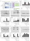
Fig 1
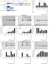
Fig 1. Effect of deleting the MLD, GP38 and NSm proteins from CCHFV M-segment on production of infectious CCHFV tc-VLPs.
(A) Schematic representation of the polyproteins encoded by CCHFV wt-M cDNA and mutant cDNA clones: three internal deletion mutants (ΔMLD-M, ΔMLDΔGP38-M and ΔMLDΔGP38ΔSKI-M), two C-terminal truncation mutants (preGn999 and preGn856) and one N-terminal deletion mutant (SP-preGc). The polyprotein precursor organization and positions of the first amino-acid residues after cleavage marking protein boundaries (MLD, GP38, Gn, NSm, and Gc) within the CCHFV polyprotein are shown. The N-terminal signal peptide (T0) and putative transmembrane domains (T1 to T5) are shown as grey boxes, signal peptidase cleavage sites are indicated by black arrows and other host protein convertase cleavage sites are indicated by red and orange arrows. (B) Infectivity titers of intracellular and extracellular CCHFV tc-VLPs. At 72h post-transfection, clarified supernatants and cell-associated tc-VLPs were used to infect Huh7 cells pre-transfected with L and N, and titers were determined by FACS analysis 24h post-infection. (C-E) Intracellular levels of CCHFV proteins expression and processing. Cell lysates of tc-VLP-producing cells were analyzed by Western blotting with antibodies against the indicated proteins including Gn, Gc, NP and host actin. Intracellular protein band intensities were quantified and normalized relative to actin and expressed as fold change compared to wt-M. Representative western blot analysis and relative quantification of intracellular Gn (C), preGc and Gc (D), and NP (E) protein levels. (F-H) tc-VLP containing supernatants were concentrated by ultracentrifugation through 20% sucrose cushions, resuspended in Opti-MEM medium and analyzed by western blot. Representative blot analysis and relative quantification of Gn (F), Gc (G), and NP (H) expressed as fold change compared to wt-M. Molecular weight markers are marked on the left (kDa). Arrow correspond to Gn precursor raised from ΔMLDΔGP38-M construct and asterisks depict the shift in Gc migration. Statistical significance was determined using non-parametric two-tailed Mann–Whitney test. Average number of repeats for intracellular CCHFV proteins: Gn = 12 (6≤Gn≤19), Gc = 14 (6≤Gc≤22), NP = 11 (7≤NP≤16). Average number of repeats for CCHFV proteins in pellets: Gn = 11 (6≤Gn≤18), Gc = 13 (7≤Gc≤20), NP = 10 (6≤NP≤16). Average number of repeats for extracellular infectivity (E) was 10 (5≤E≤16) and intracellular infectivity (I) was 6 (3≤I≤9).
Fig 2
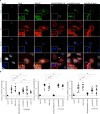
Fig 2. Golgi localization of CCHFV glycoprotein Gc is dependent on MLDGP38.
(A) Confocal microscopy analysis of Huh7 cells transfected with pUC19-empty vector, wt-M, ΔMLD-M, ΔMLDΔGP38-M, a 1:1 mix of preGn999 or preGn856 (ΔNSm) with SP-preGc. At 48h post-transfection, cells were fixed, permeabilized with Triton X-100, and stained for GP38 (6B12, green channel), Gc (11E7, red channel), Golgi (anti-GM130, blue channel) and nuclei (Hoechst, grey channel). Magnification of the squared area is shown at the bottom of each condition, zooms from squared area represent 10μm. (B) Pearson’s coefficients were calculated using FIJI (JACoP) on 5 cells from 3 separated experiments (15 cells in total) and expressed as means ± SEM. Statistical significance was determined using non-parametric two-tailed Mann–Whitney test. Scale bars represent 10 μm.
Fig 3
Fig 3. Rescue of defective tc-VLP infectivity by trans-complementation with wt-MLDGP38 or GP38.
(A) Schematic representation of wt-M, ΔMLDΔGP38ΔSKI-M segment, MLD, MLD-GP38, MLD-GP38-KDEL and GP38 expressing constructs. CCHFV tc-VLPs were generated using constructs encoding either wt-M polyprotein, or ΔMLDΔGP38ΔSKI-M deletion mutant complemented in trans with either pUC19, or with MLD, MLD-GP38, MLD-GP38-HA-KDEL and GP38 expression vectors. Infectivity, CCHFV protein expression and tc-VLP secretion were analyzed at 72h post-transfection. (B) Clarified supernatants were inoculated on L and N pre-transfected Huh7 cells and infectious titers were determined by FACS 24h post-infection. (C-E) Intracellular levels of CCHFV proteins expression and processing. Cell lysates of tc-VLP-producing cells were analyzed by Western blotting as described in Fig 1. Representative western blot analysis and quantification of intracellular Gn (C), Gc (D) and NP (E) protein levels. (F-H) tc-VLP secretion. Western blot analysis of tc-VLP-associated proteins purified by ultracentrifugation through 20% sucrose cushion. Representative blot analysis and relative quantification of Gn (F), Gc (G) and NP (H) expressed as fold change relative to wt-M. Molecular weight markers are marked on the left. Statistical significance was determined using non-parametric two-tailed Mann–Whitney test. Average number of repeats for intracellular CCHFV proteins: Gn = 8 (3≤Gn≤19), Gc = 9 (3≤Gc≤22), NP = 8 (3≤NP≤16). Average number of repeats for CCHFV proteins in pellets: Gn = 8 (4≤Gn≤18), Gc = 9 (3≤Gc≤20), NP = 8 (4≤NP≤16). Average number of repeats for extracellular infectivity (E) was 8 (3≤E≤16) and intracellular infectivity (I) was 5 (3≤I≤9).
Fig 4
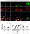
Fig 4. GP38 and Gc co-localization in the Golgi is required for rescue of tc-VLP production with ΔMLDΔGP38ΔSKI-M segment.
(A) Confocal microscopy analysis of Huh7 cells transfected with different expression plasmids as indicated. At 48h post-transfection, cells were fixed, permeabilized with Triton X-100, and stained for GP38 (6B12, green channel), Gc (11E7, red channel), Golgi (anti-GM130, blue channel) and nuclei (Hoechst, grey channel). Magnification of the squared areas are shown at the left bottom of each condition. (B) Pearson’s coefficients were calculated using FIJI (JACoP) on 5 cells from 3 separated experiments (15 cells in total) and expressed as mean (and SEM). Statistical significance was determined using non-parametric two-tailed Mann–Whitney test. Scale bars represent 10μm.
Fig 5
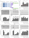
Fig 5. Expression of NSm in trans restores the assembly and secretion of tc-VLPs originated with preGn/Nsm C-terminal truncation mutants.
(A) The polyprotein precursor layout encoded by the wt-M segment is shown at the top. Below are shown schematics of i) preGn999, ii) three preGn/NSm truncation mutants with progressively shorter NSm sequence ending at amino-acid position 961, 881 and 856, iv) NSm and SP-preGc expression constructs. Infectivity, CCHFV protein expression and tc-VLP secretion was analyzed at 72h post-transfection. (B) Infectious titers of intra and extracellular CCHFV tc-VLPs. Clarified supernatants and cell-associated tc-VLPs isolated after three freeze-thaw cycles were used to infect Huh7 cells pre-transfected with L and N, and titers were determined by FACS analysis 24h post-infection. (C-E) Intracellular levels of CCHFV proteins expression and processing. Cell lysates of tc-VLP-producing cells were analyzed by Western blotting as described in Fig 1. Representative western blot analysis and relative quantification of intracellular Gn (C), preGc and Gc (D), and NP (E) protein levels. (F-H) tc-VLP secretion. tc-VLP containing supernatants were first concentrated by ultracentrifugation through 20% sucrose cushions, and analyzed by western blot. Representative blot analysis and relative quantification of Gn (F), Gc (G) and NP (H) expressed as fold change compared to wt-M. Asterisks depict the shift in Gc migration. Statistical significance was determined using non-parametric two-tailed Mann–Whitney test. Average number of repeats for intracellular CCHFV proteins: Gn = 9 (3≤Gn≤19), Gc = 9 (3≤Gc≤22), NP = 8 (3≤NP≤16). Average number of repeats for CCHFV proteins in pellets: Gn = 9 (4≤Gn≤18), Gc = 10 (4≤Gc≤20), NP = 8 (3≤NP≤16). Average number of repeats for extracellular infectivity (E) was 10 (7≤E≤16) and intracellular infectivity (I) was 7 (4≤I≤9).
Fig 6
Fig 6. CCHFV NSm alters the glycosylation of viral glycoproteins and accelerates VSV-G trafficking in a dose dependent fashion.
(A-C) Mobility shift of hemagglutinin (HA) of H7N1 induced by co-expression of NSm. Cell lysates and supernatants of H7N1pp-producer cells were analyzed by western blot after treatment with PNGase F (enzyme free and PNGase F-treated samples were migrated on the same SDS-PAGE gel), using antibodies against HA and host actin (A-B). Infectious titers of H7N1pp titers (C). (D-F) Impact of NSm on VSV-Gts endoH resistance and intracellular trafficking kinetics. Huh7 were transfected with plasmids encoding for VSV-Gts and different doses of NSm (100 ng, 500 ng and 1,000 ng) or HCV p7 when specified. Representative western blot and quantification analysis of cell lysates digested with endoH (D-E). Cell surface expression of VSV-G 1h at 32°C by flow cytometry, using the 41A1 mAb directed against VSV-G ectodomain (F). The values represent the variations of the mean fluorescence intensity (delta MFI) of cell surface-expressed VSV-Gts relative to time 0h at 32°C and relative to control condition. Data represent mean values ± SEM. Significance values were calculated by applying the unpaired one sample t-test using the GraphPad Prism 6 software (GraphPad Software, USA).
Fig 7
Fig 7. Working model of GP38 and NSm proteins functions in CCHFV glycoprotein processing, virion assembly and secretion.
(A) CCHFV M segment encoded polyprotein is co-translationally cleaved at three sites by the ER SPase releasing Gn and Gc precursors (preGn and preGc) and the membrane associated NSm protein. PreGc to Gc conversion and Golgi targeting requires association with either preGn or mature Gn released by SKI-1/S1P in complex with GP85/GP38. Accumulation of GPs in the Golgi and interactions with the virus RNPs leads to assembly and budding of progeny virions into the Golgi cisternae and SVPs. (B) Deletion of GP38 likely prevents Gn and preGc association resulting in impairment of preGc processing, intracellular trafficking and consequently abrogation of virion and SVP assembly. (C) Efficient particle secretion involves manipulation of the secretory pathway by NSm, which increases the rate of protein trafficking and alteration in protein N-glycosylation profiles. Deletion of NSm results in defects in virion and SVP assembly and/or incomplete particle morphogenesis due to retention of progeny virions in the Golgi cisternae. Formation and secretion of NP-VLP as in B) and C) is unchanged because it is independent of CCHFV GPs biogenesis and trafficking.

Similar articles

Cited by

References

    1. Abudurexiti A, Adkins S, Alioto D, Alkhovsky SV, Avsic-Zupanc T, Ballinger MJ, et al. Taxonomy of the order Bunyavirales: update 2019. Arch Virol. 2019;164(7):1949–65. 10.1007/s00705-019-04253-6 - DOI - PMC - PubMed
    1. Kalunda M, Mukwaya LG, Mukuye A, Lule M, Sekyalo E, Wright J, et al. Kasokero virus: a new human pathogen from bats (Rousettus aegyptiacus) in Uganda. Am J Trop Med Hyg. 1986;35(2):387–92. 10.4269/ajtmh.1986.35.387 . - DOI - PubMed
    1. Lasecka L, Baron MD. The molecular biology of nairoviruses, an emerging group of tick-borne arboviruses. Arch Virol. 2014;159(6):1249–65. 10.1007/s00705-013-1940-z . - DOI - PMC - PubMed
    1. Walker PJ, Widen SG, Firth C, Blasdell KR, Wood TG, Travassos da Rosa AP, et al. Genomic Characterization of Yogue, Kasokero, Issyk-Kul, Keterah, Gossas, and Thiafora Viruses: Nairoviruses Naturally Infecting Bats, Shrews, and Ticks. Am J Trop Med Hyg. 2015;93(5):1041–51. 10.4269/ajtmh.15-0344 - DOI - PMC - PubMed
    1. Gargili A, Estrada-Pena A, Spengler JR, Lukashev A, Nuttall PA, Bente DA. The role of ticks in the maintenance and transmission of Crimean-Congo hemorrhagic fever virus: A review of published field and laboratory studies. Antiviral Res. 2017;144:93–119. 10.1016/j.antiviral.2017.05.010 - DOI - PMC - PubMed

Publication types

MeSH terms

Substances