. 2020 Sep 21;15(9):e0239152.
doi: 10.1371/journal.pone.0239152. eCollection 2020.

A tissue engineering approach for repairing craniofacial volumetric muscle loss in a sheep following a 2, 4, and 6-month recovery

Affiliations

A tissue engineering approach for repairing craniofacial volumetric muscle loss in a sheep following a 2, 4, and 6-month recovery

Brittany L Rodriguez et al. PLoS One. .

Abstract

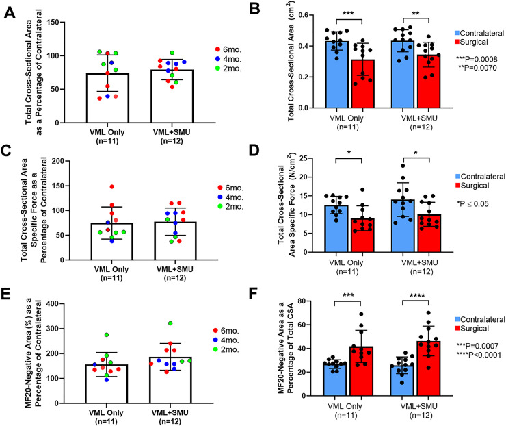
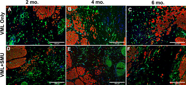
Volumetric muscle loss (VML) is the loss of skeletal muscle that results in significant and persistent impairment of function. The unique characteristics of craniofacial muscle compared trunk and limb skeletal muscle, including differences in gene expression, satellite cell phenotype, and regenerative capacity, suggest that VML injuries may affect craniofacial muscle more severely. However, despite these notable differences, there are currently no animal models of craniofacial VML. In a previous sheep hindlimb VML study, we showed that our lab's tissue engineered skeletal muscle units (SMUs) were able to restore muscle force production to a level that was statistically indistinguishable from the uninjured contralateral muscle. Thus, the goals of this study were to: 1) develop a model of craniofacial VML in a large animal model and 2) to evaluate the efficacy of our SMUs in repairing a 30% VML in the ovine zygomaticus major muscle. Overall, there was no significant difference in functional recovery between the SMU-treated group and the unrepaired control. Despite the use of the same injury and repair model used in our previous study, results showed differences in pathophysiology between craniofacial and hindlimb VML. Specifically, the craniofacial model was affected by concomitant denervation and ischemia injuries that were not exhibited in the hindlimb model. While clinically realistic, the additional ischemia and denervation likely created an injury that was too severe for our SMUs to repair. This study highlights the importance of balancing the use of a clinically realistic model while also maintaining control over variables related to the severity of the injury. These variables include the volume of muscle removed, the location of the VML injury, and the geometry of the injury, as these affect both the muscle's ability to self-regenerate as well as the probability of success of the treatment.


Conflict of interest statement

The authors have declared that no competing interests exist.

Figures

Fig 1
Fig 1. Experimental groups.
(A) A full-thickness longitudinal portion of the ZM constituting 30% of the total mass was dissected to simulate a VML injury. (B) The VML Only group (negative control) received the injury and nerve re-route without any additional repair. (C) In the VML+SMU group, the injury was immediately repaired by placing n = 2 single SMUs within the defect (white arrow). The re-routed nerve was splayed and sutured to the SMU. (D) The SMU was then sutured into defect site. Black arrows in B-C represent re-routed nerve.
Fig 2
Fig 2. Characterization of In vitro SMUs.
(A) The final modular assembly of n = 2 SMUs was 7cm long. (B) SMU monolayers prior to delamination showed abundant, networking myotubes. (C) Tetanic forces produced by sentinel SMUs were 72.0 ± 42.1μN on average. Red data points indicate the SMUs whose force peaked below the maximum current and/or frequency allowed by our force testing system. Masson’s trichrome staining of a single SMU cross-section (D) and modularly fused SMU cross-section (G) revealed an extracellular matrix composed of collagen. Immunohistochemical staining of a single SMU cross-section (E) and a modularly fused SMU (F) revealed the presence of muscle fibers (MF20, green) and laminin (red), as well as the presence of DAPI-stained nuclei (blue) throughout the thickness of the construct. (H) Immunostaining of a longitudinal section of a single SMU for desmin (red), α-smooth muscle actin (green), and DAPI (blue) revealed a parallel, linear arrangement of these proteins. (I, J) DAPI-stained nuclei (white) of the area denoted by the (I) dotted line box and the (J) hash line box in image (F). Scale bars on B, D-H = 500μm. Scale bars on I, J = 200μm.
Fig 3
Fig 3. VML injury and size of SMUs.
(A) The average percentage of the VML injury was 31.8 ± 7.1% in the VML Only group and 30.41 ± 6.1% in the VML+SMU group. There were no significant differences in the size of the VML injuries between timepoints or between experimental groups (two-way ANOVA: P = 0.5207 and P = 0.1424, respectively). (B) The modular SMUs implanted into the 6mo. animals were significantly larger than those in the 2mo. (P = 0.0006) or 4mo. (P = 0.0003) groups and (C) contributed significantly more mass to the defect those in the 2mo. (P = 0.0006) or 4mo. (P = 0.0003) groups. *** indicates P ≤ 0.001; **** indicates P ≤ 0.0001.
Fig 4
Fig 4. Animal body weight.
We measured the animals’ body weight at the time of implantation and the time of explant for the (A) 2-month, (B) 4-month, and (C) 6-month recovery groups. A paired two-way ANOVA performed for each recovery timepoint showed that the experimental group did not significantly affect the animals’ body weight (P = 0.6224 for the 2mo. group, P = 0.4439 for the 4mo. group, P = 0.3054 for the 6mo. group, n = 10 per group). However, there was a significant difference in body weight between the time of implantation and the time of explant in the 4mo. (P = 0.0003) and 6mo. (P<0.0001) groups, but not the 2mo. group (P = 0.6206).
Fig 5
Fig 5. Mechanical properties of explanted muscles.
(A) A two-way ANOVA showed there was no significant difference in force of the injured ZMs as a percentage of the contralalteral between timepoints (P = 0.3576) or between experimental groups (P = 0.3041). (B) In both experimental groups, the maximum tetanic force of the surgical ZM was significantly lower than the uninjured contralateral (P<0.0001 in both groups). (C) As a percentage of the contralateral, there was no difference in the optimal lengths of the muscle between recovery timepoints (P = 0.2270) or experimental groups (P = 0.1905). (D) There was no siginificant difference in the optimal length of the VML Only group realtive to the contralateral (P = 0.8424), but the optimal length of the VML+SMU group was significantly lower than the contralateral (P = 0.0318). (E) Interestingly, there was no correlation (r = 0.1082) between the SMU weight (as a percentage of the mass deficit) and the percentage of force recovery (P = 0.7379).
Fig 6
Fig 6. MF20+ area, specific force, and fiber counts.
(A) A two-way ANOVA showed there was no significant difference in normalized MF20+ area between recovery timepoints (P = 0.3462) or between experimental groups (P = 0.7293). (B) In both experimental groups, the MF20+ area of the surgical ZM was significantly lower than the uninjured contralateral (P<0.0001 in both groups, n = 12 for VML Only, n = 13 for VML+SMU). (C) There was no significant difference in normalized specific force between recovery timepoints (P = 0.2247) or between experimental groups (P = 0.3608). (D) Interestingly, in both experimental groups, there was no significant difference in specific force between the contralateral and surgical ZMs (P = 0.7653, n = 11 for VML Only; P = 0.9529, n = 12 for VML+SMU). (E) A two-way ANOVA showed there was no significant difference in normalized muscle fiber counts between recovery timepoints (P = 0.4046) or between experimental groups (P = 0.7197). (F) In both experimental groups, the total number of muscle fibers in the surgical ZM was significantly lower than the uninjured contralateral (P<0.0001 in both groups, n = 11 for VML Only, n = 12 for VML+SMU).
Fig 7
Fig 7. Total cross-sectional area and specific force.
(A) A two-way ANOVA revealed that there was no significant difference in the total cross-sectional area as a percentage of the contralateral between experimental groups (P = 0.6753). (B) In both experimental groups, the total cross-sectional area of the surgical ZM was significantly lower than the uninjured contralateral (P = 0.0008, n = 11 for VML Only; P = 0.0070, n = 12 for VML+SMU). (C) There was no significant difference in normalized total cross-sectional area specific force between experimental groups (P = 0.6753). (D) In both experimental groups, the total cross-sectional area specific force was significantly lower in the surgical ZM than the uninjured contralateral (P = 0.0384, n = 11 for VML Only; P = 0.0165, n = 12 for VML+SMU). (E) There was no significant difference in the MF20-negative area as a percentage of the contralateral between recovery timepoints (P = 0.1138) or between experimental groups (P = 0.1016). (F) In both experimental groups the percentage of the total cross-sectional area that did not stain for MF20 was significantly higher than the uninjured contralateral (P = 0.0007, n = 11 for VML Only; P<0.0001, n = 12 for VML+SMU).
Fig 8
Fig 8. Histology at the 6-month recovery timepoint.
Cross sections of explants from contralateral (A,D,G), VML only (B,E,H), and VML+SMU (C,F,I) groups were taken from the midbelly of the muscle. Tissues stained with H&E (A-C) show that the surgical site is characterized by the presence of disorganized, hypercellular tissue. Serial sections stained with Masson’s trichrome (D-F) indicates that there are large fibrotic regions in the injury site, evidenced by positive collagen staining (blue) that spans between the native muscle (red) in the surgical groups (E,F). Immunostaining for myosin heavy chain (MF20, red) and laminin (green) (G-I) show that muscle is absent in a large portion of the surgical site.
Fig 9
Fig 9. Muscle fibers in the injury site.
Immunohistochemical staining for myosin heavy chain (MF20, red), laminin (green), and DAPI (blue) revealed the presence of small muscle fibers within the injury site in both the VML only (A-C) and VML+SMU (D-F) groups. These smaller fibers were noted at the 2-month (A,D), 4-month (B,E), and 6-month (C,F) recovery timepoints.
Fig 10
Fig 10. Neuromuscular junctions in the injury site.
Immunostaining of longitudinal sections for acetylcholine receptors (red), synaptic vesicle protein-2 (green), and neurofilament (green) was performed to identify the presence of neuromuscular junctions. We noted the presence of neuromuscular junctions (i.e. overlap of the pre and post-synaptic structures) in the contralateral muscles (A,D,G) as well as the injury site of the (H) VML Only group and (I) VML+SMU group at the 6mo. timpeoint. The small muscle fibers in the injury site do not appear to be fully innvervated at the (B-C) 2mo. and (E-F) 4mo. timpeoints. Scale bars = 50μm.
Fig 11
Fig 11. Fiber type analysis at the 6-month recovery timepoint.
To note any changes to the fiber type of the msucle at the 6-month recovery timepoint, we performed immunostaining for fast myosin isoform (type II fibers, green) and slow myosin isoform (type I fibers, red) in the (A) contralateral, (B) VML Only, and (C) VML+SMU groups. Scale bars on A-C = 1000μm. (D) There was no significant difference in the percentage of the number of slow fibers between groups (P = 0.0807). (E) There was no significant difference in the percentage of the total area made up by slow fibers between groups (P = 0.3852).
Fig 12
Fig 12. Correlation between fiber grouping and force in the 6-month recovery group.
(A) There was no significant correlation between the number of grouped fibers and the percentage of force recovery in the 6-month recovery group as a whole (r = -0.2488, P = 0.4883, n = 10). (B) In the VML Only group, however, there was a significant negative correlation between the number of grouped fibers and the percentage of force recovery (r = -0.9506, P = 0.0131, n = 5). (C) In contrast, there was no significant correlation in the VML+SMU group (r = -0.1515, P = 0.8079, n = 5).
Fig 13
Fig 13. Intramuscular fat.
(A-I) Immunostaining for neurofilament (SMI312, green) and fat (perilipin, red) in muscle cross-sections showed the presence of intramuscular fat and nerve in the explanted muscles. Scale bars = 1000μm. (J) There was a significantly higher amount of intramuscular fat in the VML+SMU group compared to the contralateral muscle (P = 0.0334), while there was no significant difference in the amount of intramuscular fat between the VML Only group and the contralateral (P = 0.9666).

Similar articles

Cited by

References

    1. Garland C, Pomerantz J. Regenerative Strategies for Craniofacial Disorders. Frontiers in Physiology. 2012;3(453). 10.3389/fphys.2012.00453 - DOI - PMC - PubMed
    1. Grogan BF, Hsu JR, Consortium STR. Volumetric muscle loss. J Am Acad Orthop Surg. 2011;19 Suppl 1:S35–7. 10.5435/00124635-201102001-00007 . - DOI - PubMed
    1. Garg K, Ward CL, Hurtgen BJ, Wilken JM, Stinner DJ, Wenke JC, et al. Volumetric muscle loss: persistent functional deficits beyond frank loss of tissue. J Orthop Res. 2015;33(1):40–6. Epub 2014/09/18. 10.1002/jor.22730 . - DOI - PubMed
    1. Mase VJ, Hsu JR, Wolf SE, Wenke JC, Baer DG, Owens J, et al. Clinical application of an acellular biologic scaffold for surgical repair of a large, traumatic quadriceps femoris muscle defect. Orthopedics. 2010;33(7):511 10.3928/01477447-20100526-24 . - DOI - PubMed
    1. Surgeons ASoP. 2018 National Plastic Surgery Statistics. 2018.

Publication types