Review
. 2020 Dec:182:114225.
doi: 10.1016/j.bcp.2020.114225. Epub 2020 Sep 19.

Role of proteolytic enzymes in the COVID-19 infection and promising therapeutic approaches

Affiliations
Review

Role of proteolytic enzymes in the COVID-19 infection and promising therapeutic approaches

Magda Gioia et al. Biochem Pharmacol. 2020 Dec.

Abstract

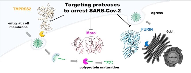
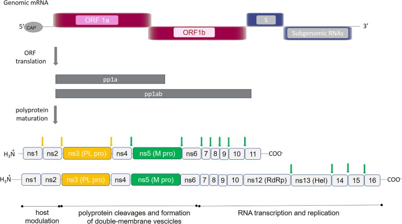
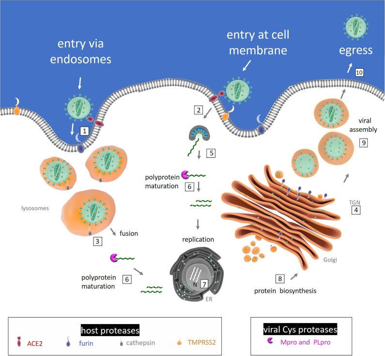
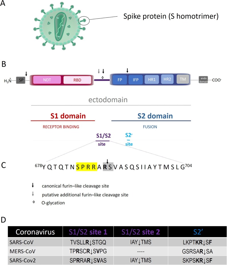
In the Fall of 2019 a sudden and dramatic outbreak of a pulmonary disease (Coronavirus Disease COVID-19), due to a new Coronavirus strain (i.e., SARS-CoV-2), emerged in the continental Chinese area of Wuhan and quickly diffused throughout the world, causing up to now several hundreds of thousand deaths. As for common viral infections, the crucial event for the viral life cycle is the entry of genetic material inside the host cell, realized by the spike protein of the virus through its binding to host receptors and its activation by host proteases; this is followed by translation of the viral RNA into a polyprotein, exploiting the host cell machinery. The production of individual mature viral proteins is pivotal for replication and release of new virions. Several proteolytic enzymes either of the host and of the virus act in a concerted fashion to regulate and coordinate specific steps of the viral replication and assembly, such as (i) the entry of the virus, (ii) the maturation of the polyprotein and (iii) the assembly of the secreted virions for further diffusion. Therefore, proteases involved in these three steps are important targets, envisaging that molecules which interfere with their activity are promising therapeutic compounds. In this review, we will survey what is known up to now on the role of specific proteolytic enzymes in these three steps and of most promising compounds designed to impair this vicious cycle.


Figures

None
Graphical abstract
Fig. 1
Fig. 1
SARS-CoV-2 polyproteins encoded by ORF1a and ORF1ab. Schematic representation of the open reading frames 1a and 1ab, which encode for polyproteins pp1a and pp1ab. Proteins composing each polyprotein are shown: (ns) indicates non-structural proteins; RNA dependent RNA polymerase and Helicase are indicated by (RdRp) and (Hel), respectively. Proteolytic sites cleaved by PLpro and Mpro are reported in yellow and green arrows, respectively. (For interpretation of the references to color in this figure legend, the reader is referred to the web version of this article.)
Fig. 2
Fig. 2
Diagram of the involvement of host and viral proteases in SARS-CoV-2 life cycle. Activation of coronavirus spike proteins by host cell proteases occurs at different stages in the viral life cycle and in different cell localizations. The ACE2-dependent infectious entry at the cell membrane is triggered through the S protein cleavage by host proteases: furin (1) and/or TMPRSS2 (2). Intracellular activation of S protein is mediated by cathepsin in lysosomes (3) and/or by Furin in trans-Golgi network (TGN) (4). After the receptor recognition, the viral genome is released into the cytoplasm of the host cell (5), RNA attaches directly to the host ribosome for translation of two polyproteins (not shown). Polyprotein (pp) maturation into mature fragments is catalysed by viral Cys proteases (Mpro and PL pro) (6). RNA is translated into DNA and inside the nucleus (N) replication amplifies the number of virus genome copies (7). The viral genome produces pps, which help to take command over host ribosomes for their own translation process; protein biosynthesis starts at the endoplasmic reticulum (ER) and follows the constitutive secretory pathway along Golgi compartments (8). The virion assembly occurs (9) and the newly packed viral particles can egress (10).
Fig. 3
Fig. 3
(A): Schematic representation of coronavirus particle. Spike proteins are highly glycosylated type I transmembrane protein, which assemble into trimers on the virion surface to form the distinctive “corona” (crown-like) appearance. (B): Domain organization and cleavage sites of the coronavirus Spike monomer (S). The ectodomain of all CoV spike proteins share the same organization in two domains, that is a N-terminal domain, named S1 and responsible for receptor binding, and a C-terminal S2 domain responsible for fusion. The domain organization of the S monomer consists of a signal peptide (SP), the N-terminal domain (NTD), the receptor-binding domain (RBD), the fusion peptide (FP), the internal fusion peptide (IFP), the heptad repeat 1/2 (HR1/2), and the transmembrane domain (TM).The region between the two domain is termed S1/S2 site. (C): Sequence of S1/S2 cleavage site of S protein from SARS-CoV-2. The four amino acid insertion (SPRRs), unique to SARS-CoV-2, is marked in yellow, the conserved S1/S2 cleavage site is marked in grey. (D): Comparative sequences of S protein cleavage sites. (For interpretation of the references to color in this figure legend, the reader is referred to the web version of this article.)
Fig. 4
Fig. 4
Schematic representation of the furin structure. (A): Schematic representation of the domains of furin. Each domain is represented by different shape and color and is defined by arabic numbers listed above them. Asp153 (D), His194 (H), Ser368 (S), are the amino acid residues that form the catalytic triad of furin; Asn295 contributes to the oxyanion hole. (B): Crystal structure of human furin (PDB ID: 5JXG) . The catalytic domain of furin is shown with its surface in gold and the P-domain in green. The amino acid residues of the catalytic triad and of the binding sites of furin to the viral S protein are displayed. (C): Crystal structure of mouse furin in complex with the inhibitor Dec-Arg-Val-Lys-Arg-CMK (PDB ID: 1P8J) . Furin is shown in light blue. The inhibitor is displayed in black sticks with nitrogen in blue and oxygen in red. The figures of panels B and C were drawn using the UCSF Chimera software . For details, see the text. (For interpretation of the references to color in this figure legend, the reader is referred to the web version of this article.)
Fig. 5
Fig. 5
Schematic representation of the TMPRSS2 structure. (A): Schematic representation of the domains of TMPRSS2. Each domain is represented by different shape and color and is defined by arabic numbers listed above them. His296 (H), Asp345 (D) and Ser441 (S) are the amino acid residues that form the catalytic triad of TMPRSS2. (B): The three-dimensional model of TMPRSS2 was built according to . The catalytic domain of TMPRSS2 is shown with its surface in gold, the SRCR domain in cyan and the LDL domain in pink. The amino acid residues of the catalytic triad and of the predicted active site are displayed. (C): Three-dimensional model of TMPRSS2 in complex with the standard inhibitor camostat mesylate . The catalytic triad (i.e., His296, Asp345, Ser441) and the predicted interactions of camostat with the active site residues (i.e., Asp187, Asn346, Cys348, and Asn450) of the human serine protease are shown. TMPRSS2 is shown in tan. The inhibitor is displayed in black sticks with nitrogen in blue, and oxygen in red. The figures of panels B and C were drawn using the UCSF Chimera software . For details, see the text. (For interpretation of the references to color in this figure legend, the reader is referred to the web version of this article.)
Fig. 6
Fig. 6
Analysis of sequence and structure of Mpro (3CL) protease. (A): Multi-alignment of Mpro homologues from the seven known human coronaviruses: SARS-CoV-2 (Uniprot ID: P0DTD1), SARS-CoV (P0C6X7), HCoV-OC43 (P0C6U7), HCoV-KU1 (P0C6U3), MERS-CoV (V9TU05), HCoV-NL63 (P0C6U6), HCoV-229E (P0C6U2). Secondary structure references were taken from SARS-CoV-2 Mpro (PDB: 6M2Q). (B): Three-dimensional structure of apo SARS-CoV-2 Mpro (PDB: 6M2Q). The structural domains I, II and III of Mpro are colored in light blue, orange and green, respectively. The localization of the protease active site (i.e., residues Cys41 and His145) and the “N-finger” are indicated by arrows. (C): SARS-CoV-2 Mpro complexed with N3 peptide (PDB: 6LU7). Mpro structure is represented by molecular surface colored with the same scheme used in panel B. Subsite pockets S1 and S2 are explicitly indicated. (For interpretation of the references to color in this figure legend, the reader is referred to the web version of this article.)
Fig. 7
Fig. 7
Ligands docked in Mpro active site. Examples of ligands docked in the active site: (A) Amodiaquine; (B) Bonducellpin D; (C) Heptafuhalol; (D) Simeprevir; (E) Pitavastatin; (F) Eszopiclone. Ligands were docked using Autodock Vina with the protocol developed elsewhere . The structural domains I, II and III of Mpro are colored in light blue, orange and green, respectively. (For interpretation of the references to color in this figure legend, the reader is referred to the web version of this article.)

References

    1. Morse J.S., Lalonde T., Xu S., Liu W.R. Learning from the past: possible urgent prevention and treatment options for severe acute respiratory infections caused by 2019-nCoV. ChemBioChem. 2020;21:730–738. doi: 10.1002/cbic.202000047. - DOI - PMC - PubMed
    1. Chen Y., Liu Q., Guo D. Emerging Coronaviruses: genome structure, replication, and pathogenesis. J. Med. Virol. 2020;92:418–423. doi: 10.1002/jmv.25681. - DOI - PMC - PubMed
    1. Bredenbeek P.J., Pachuk C.J., Noten A.F., Charite J., Luytjes W., Weiss S.R., Spaan W.J. The primary structure and expression of the second open reading frame of the polymerase gene of the coronavirus MHV-A59: a highly conserved polymerase is expressed by an efficient ribosomal frameshifting mechanism. Nucl. Acids Res. 1990;18:1825–1832. doi: 10.1093/nar/18.7.1825. - DOI - PMC - PubMed
    1. Lee H.J., Shieh C.K., Gorbalenya A.E., Koonin E.V., La Monica N., Tuler J., Bagdzhadzhyan A., Lai M.M. The complete sequence (22 kilobases) of murine coronavirus gene 1 encoding the putative proteases and RNA polymerase. Virology. 1991;180:567–582. doi: 10.1016/0042-6822(91)90071-i. - DOI - PMC - PubMed
    1. Simmons G., Zmora P., Gierer S., Heurich A., Pöhlmann S. Proteolytic activation of the SARS-Coronavirus Spike protein: cutting enzymes at the cutting edge of antiviral research. Antivir. Res. 2013;100:605–614. doi: 10.1016/j.antiviral.2013.09.028. - DOI - PMC - PubMed

Publication types