. 2020 Sep 17;21(18):6815.
doi: 10.3390/ijms21186815.

Cooperativity between the Ribosome-Associated Chaperone Ssb/RAC and the Ubiquitin Ligase Ltn1 in Ubiquitination of Nascent Polypeptides

Affiliations

Cooperativity between the Ribosome-Associated Chaperone Ssb/RAC and the Ubiquitin Ligase Ltn1 in Ubiquitination of Nascent Polypeptides

Arnab Ghosh et al. Int J Mol Sci. .

Abstract

Eukaryotic cells have evolved multiple mechanisms to detect and eliminate aberrant polypeptides. Co-translational protein surveillance systems play an important role in these mechanisms. These systems include ribosome-associated protein quality control (RQC) that detects aberrant nascent chains stalled on ribosomes and promotes their ubiquitination and degradation by the proteasome, and ribosome-associated chaperone Ssb/RAC, which ensures correct nascent chain folding. Despite the known function of RQC and Ssb/ribosome-associated complex (RAC) in monitoring the quality of newly generated polypeptides, whether they cooperate during initial stages of protein synthesis remains unexplored. Here, we provide evidence that Ssb/RAC and the ubiquitin ligase Ltn1, the major component of RQC, display genetic and functional cooperativity. Overexpression of Ltn1 rescues growth suppression of the yeast strain-bearing deletions of SSB genes during proteotoxic stress. Moreover, Ssb/RAC promotes Ltn1-dependent ubiquitination of nascent chains associated with 80S ribosomal particles but not with translating ribosomes. Consistent with this finding, quantitative western blot analysis revealed lower levels of Ltn1 associated with 80S ribosomes and with free 60S ribosomal subunits in the absence of Ssb/RAC. We propose a mechanism in which Ssb/RAC facilitates recruitment of Ltn1 to ribosomes, likely by detecting aberrations in nascent chains and leading to their ubiquitination and degradation.

Keywords: Ssb/RAC triad; r-protein; rRNA; ribosome; ribosome-associated chaperones; ribosome-associated protein quality control (RQC); ribosome-bound nascent chains (RNCs); ubiquitin; ubiquitin ligase Ltn1; ubiquitination of polypeptides bound to a ribosome.


Conflict of interest statement

The authors declare no conflict of interest.

Figures
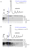
Figure A1
Figure A1
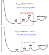
(Related to Figure 2). Ectopically expressed FLAG-tagged Ltn1 is catalytically active and ubiquitinates polypeptides associated with 60S and 80S ribosomal species. Results of the experiment depicted in Figure 2 is shown with absorbance curves for gradients derived from cells transformed with empty vector control (A) and Ltn1FLAG (B). Absorbance was measured at 254 nm during sucrose gradients fractionation.
Figure A2
Figure A2
(Related to Figure 4). Complete curves of absorbance measured at 254 nm during fractionation of sucrose gradients shown in Figure 4. Fractions 5–12 analyzed in Figure 4 for levels of Ltn1FLAG, Rpl3, and poly-Ub species are boxed.
Figure A3
Figure A3
(Related to Figure 5). (AD) Entire absorbance curves measured at 254 nm during fractionation of sucrose gradients shown in Figure 5 Fractions containing 60S and 80S based on absorbance and co-sedimentation of Rpl3 are boxed in red or green, respectively.
Figure 1
Figure 1
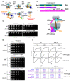
Schematic representation of (A) RQC (details are in the text), and (B) the functional domains of Ssb/ribosome-associated complex (RAC) and interaction of Ssb/RAC with the 80S ribosome. Ssb1 is in green, Ssz1 is in purple, and Zuo1 is in pink. Functional domains are marked in yellow. Cartoon at the bottom depicts positioning of the Ssb/RAC complex on the 80S ribosome. (A,B) tRNAs located at A and P sites of the ribosome are shown in turquoise; ribosome-associated nascent chain (RNC) is shown in orange. Overexpression of Ltn1 in ssb∆∆cells rescues stress-induced growth suppression. (C) Wild-type (BY4741) and ssb1∆ ssb2∆ (ssb∆∆) yeast strains were grown in YPD. Cell cultures were adjusted to the same cell density, and five-fold dilutions were spotted onto YPD agar plates and on YPD plates supplemented with either 25 μg/mL of hygromycin B (HygB) or 0.8 M NaCl. Plates were incubated at 30 °C or at 20 °C for 48 h, as indicated. (D) Wild-type and ssb∆∆ cells were transformed with empty vector control (V) or Ltn1FLAG-expressing constructs. Cells were grown in SC ura- medium and adjusted to the same cell density. Serial dilutions were spotted onto plates as described in (B). (E) Cell cultures from (C) were adjusted to OD600nm ~0.2 and grown in liquid cultures in YPD, YPD + HygB (25 μg/mL), or YPD + NaCl (0.8 M) medium for 24 h with continued shaking at 30 °C. OD600nm were measured every 5 min. The representative growth curves are shown on the top, arrows indicate the end of lag phase; lag and doubling time parameters are shown at the bottom.
Figure 2
Figure 2
Ectopically expressed Ltn1FLAG is functionally active: it rescues hygromycin-induced lethality in ltn1∆ cells and ubiquitinates polypeptides associated with 60S and 80S ribosomal species. (A) Ltn1FLAG cloned under control of the constitutive ADH promoter in 2 μc plasmid was expressed in PTET-07-CDC48 cells grown in SC leu- medium. Expression was detected by western blotting using anti-FLAG primary and HRP-fused secondary antibodies. Empty vector (V) was used as a negative control. (B) ltn1∆ cells and their parental strain BY4741 (wild-type, WT) were transformed with empty vector control (V) or with 2 μc plasmid constitutively expressing Ltn1FLAG. Transformants were grown in SC leu- medium and adjusted to the same cell density. Five-fold dilutions were spotted onto SC leu control agar plates and onto SC leu plates supplemented with 100 μg/mL HygB. Plates were grown at 30 °C for 3 days. (C,D) PTET-0-CDC48 ltn1∆ cells transformed with Ltn1FLAG-expressing construct (C) or empty vector control (D) were grown in SC leu- medium in the presence of Dox for 16 h, diluted to OD600nm~0.3, and grown for 4 h in YPD+Dox. Before harvesting, cells were treated with cycloheximide (CHX) for 5 min. Whole cellular lysates were centrifuged through a 15–42% sucrose gradient (10.8 mL) and fractionated. Total protein extracts were isolated from 23 individual fractions (fractions 5–27; ~390 μL each). Proteins were separated on 10% SDS-polyacrylamide gels in duplicate and transferred onto nitrocellulose membranes. One membrane was probed with anti-FLAG antibodies to detect Ltn1FLAG (top panel). The bottom part of the second membrane was probed with anti-Rpl3 antibodies (middle panel), while the top part of the same membrane was probed with anti-Ub antibodies (bottom panels; two exposures are shown). We used HRP-fused secondary antibodies and ECL system to visualize protein signals. The experiment was repeated 3 times; representative images are shown. Peak levels of ribosomal species detected by absorbance measurements at 254 nm are shown at the top of the figure. The blue vertical line indicates visual separation between 40S–60S and 80S-polysome fractions of the gradient, while 60S- and 80S-containing fractions are boxed in red.
Figure 3
Figure 3
Lack of Ssb/RAC components significantly decreases ubiquitination of polypeptides associated with the 80S monosome. Sucrose gradient sedimentation analysis of ribosomes extracted from: (A) PTET-07-CDC48 cells and their derivative strains containing additional deletions of (B) SSB1 and SSB2, or ZUO1, or SSZ1, or (C) SSZ1 and LTN1. Cells were grown in YPD media in the presence of Dox for 16 h, diluted to OD600nm~0.3, and grown for 4 h in YPD+Dox. Lysates were centrifuged through 15–42% sucrose gradients (10.8 mL) and fractionated into 14 fractions (~780 μL). Total protein was isolated from each fraction and analyzed as described in Figure 2C,D. Western blotting images of 7 critical fractions containing 60S (1–3) and 80S (4–7) ribosomal species are shown. The experiment was repeated 3 separate times; representative images are shown.
Figure 4
Figure 4
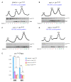
Lack of Ssz1 co-chaperone of the Ssb/RAC complex results in reduced levels of polyubiquitinated nascent chains on 80S ribosomes due to decreased levels of Ltn1FLAG. (A,B) PTET-07 -CDC48 and PTET-07-CDC48 ssz∆ cells transformed with Ltn1FLAG-expressing construct were grown in SC leu- medium in the presence of Dox for 16 h, diluted to OD600nm ~0.3, and grown for additional 4 h in YPD+Dox. Whole-cell lysates were separated by centrifugation through 15–35% sucrose gradients and fractionated with continuous measurement of absorbance at 254 nm to visualize ribosomal peaks. Fractions containing 60S–80S ribosomal species (5–12) were analyzed further. Proteins extracted from these fractions were separated on SDS-polyacrylamide gels in duplicate and analyzed by quantitative western blotting. One membrane was probed with anti-FLAG antibodies (top panels), while the top part of the second membrane was probed with anti-Ub antibodies and the bottom part with anti-Rpl3 antibodies (middle and bottom panels). We used IR-Dye-680RD secondary antibodies for Rpl3- and Ub-probed blots, and IR-Dye-800CW antibodies for FLAG-probed blots. IR signals were detected on Typhoon imager by scanning membranes at 800 nm and 680 nm; ImageQuant was used to analyze the images. Experiments were repeated 3 independent times; representative images are shown. (C,D) Bands corresponding to Ltn1FLAG, Rpl3, and poly-Ub species were converted to phosphorimager units. To normalize 60S-associated Ltn1FLAG to Rpl3 levels, we used phosphorimager units derived from Ltn1 and Rpl3 signals present in fractions 6 and 7. To normalize 80S-associated Ltn1FLAG to Rpl3, we used units derived from Ltn1 or Rpl3 signals present in fractions 10 and 11. (C) Ltn1FLAG/Rpl3 ratios were plotted as bar graphs. (D) Phosphorimager units derived from poly-Ub signals present in fractions 6 + 7 and 10 + 11 of each gradient (panels A and B) were plotted as bar graphs. p values: ***, <0.001; **, <0.01; *, <0.05; NS: not significant. Two-tailed two-sample unequal variance t-test was used for statistical analysis. Error bars represent SD.
Figure 5
Figure 5
Deleting genes encoding Ssb/RAC leads to decreased association of Ltn1FLAG with 60S and 80S ribosomal species. (AD) Indicated yeast strains transformed with Ltn1FLAG-expressing construct were analyzed as described in Figure 4. Experiment was repeated 3 independent times; representative images are shown. (E) Bands corresponding to Ltn1FLAG and Rpl3 were converted to phosphorimager units. Signal averages were calculated for Ltn1FLAG and Rpl3 present in fractions highlighted in red (for 60S) and green (for 80S). Ltn1FLAG levels were then normalized to Rpl3 in the same fractions and expressed as Ltn1FLAG/Rpl3 ratios plotted as bar graphs. p values: ***, <0.001; **, <0.01; NS: not significant. Two-Tailed two-sample unequal variance t-test was used for statistical analysis. Error bars represent SD.
Figure 6
Figure 6
Model for cooperative function of Ssb/RAC and Ltn1 Ub ligase. See details in the text.

Similar articles

Cited by

References

    1. Ciechanover A., Kwon Y.T. Protein Quality Control by Molecular Chaperones in Neurodegeneration. Front. Neurosci. 2017;11 doi: 10.3389/fnins.2017.00185. - DOI - PMC - PubMed
    1. Shan S.-O. Guiding tail-anchored membrane proteins to the endoplasmic reticulum in a chaperone cascade. J. Biol. Chem. 2019;294:16577–16586. doi: 10.1074/jbc.REV119.006197. - DOI - PMC - PubMed
    1. Joazeiro C.A.P. Mechanisms and functions of ribosome-associated protein quality control. Nat. Rev. Mol. Cell Biol. 2019;20:368–383. doi: 10.1038/s41580-019-0118-2. - DOI - PMC - PubMed
    1. Bengtson M.H., Joazeiro C.A.P. Role of a ribosome-associated E3 ubiquitin ligase in protein quality control. Nature. 2010;467:470–473. doi: 10.1038/nature09371. - DOI - PMC - PubMed
    1. Brandman O., Stewart-Ornstein J., Wong D., Larson A., Williams C.C., Li G.-W., Zhou S., King D., Shen P.S., Weibezahn J., et al. A ribosome-bound quality control complex triggers degradation of nascent peptides and signals translation stress. Cell. 2012;151:1042–1054. doi: 10.1016/j.cell.2012.10.044. - DOI - PMC - PubMed

MeSH terms

Substances

LinkOut - more resources