

Non-Linear Image Distortions in Flexible Fiberoptic Endoscopes and their Effects on Calibrated Horizontal Measurements Using High-Speed Videoendoscopy
- PMID: 32958427
- PMCID: PMC7969477
- DOI: 10.1016/j.jvoice.2020.08.029
Non-Linear Image Distortions in Flexible Fiberoptic Endoscopes and their Effects on Calibrated Horizontal Measurements Using High-Speed Videoendoscopy
Abstract
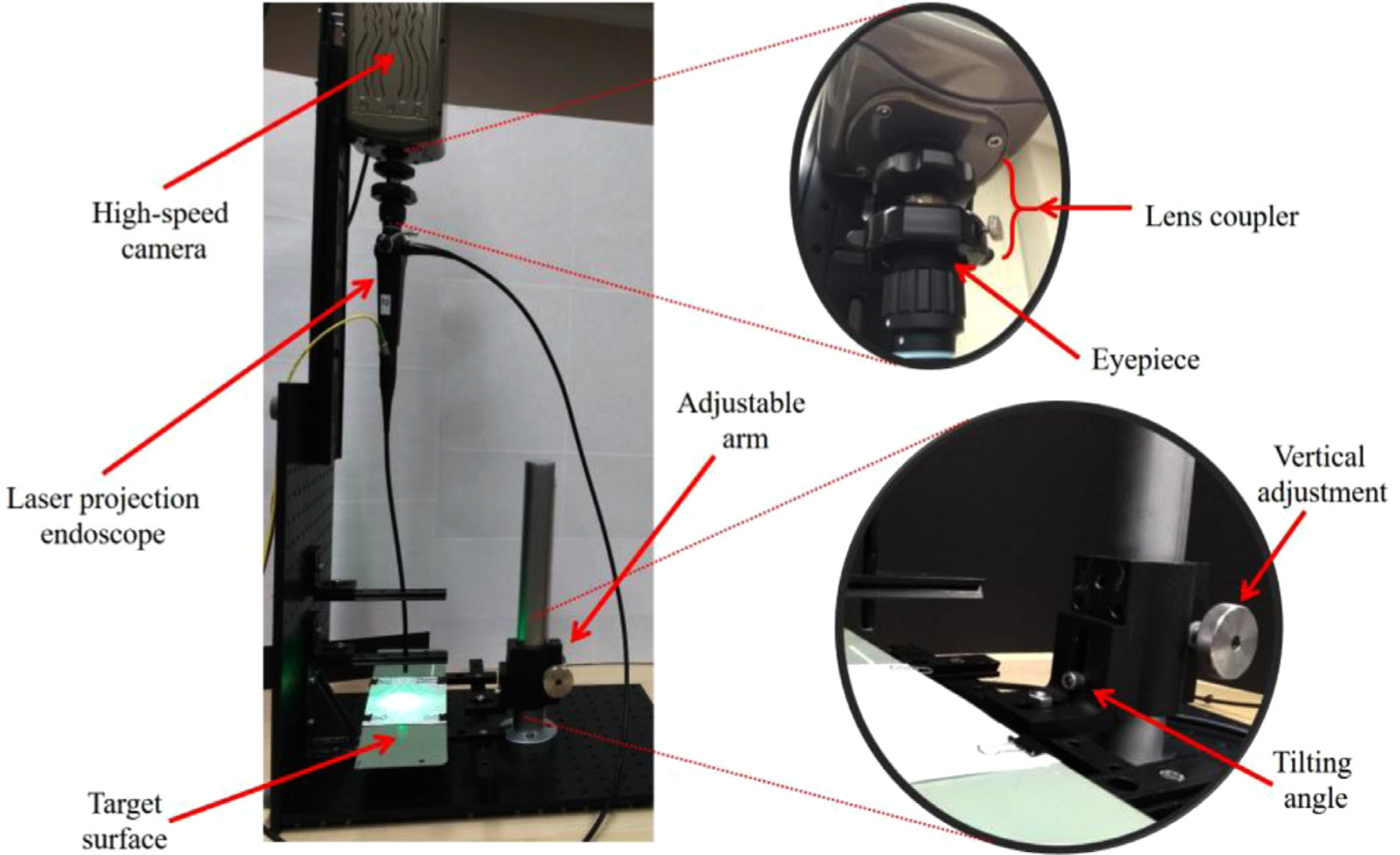
Laryngeal images obtained via high-speed videoendoscopy are an invaluable source of information for the advancement of voice science because they can capture the true cycle-to-cycle vibratory characteristics of the vocal folds in addition to the transient behaviors of the phonatory mechanism, such as onset, offset, and breaks. This information is obtained through relating the spatial and temporal features from acquired images using objective measurements or subjective assessments. While these images are calibrated temporally, a great challenge is the lack of spatial calibration. Recently, a laser-projection system allowing for spatial calibration was developed. However, various sources of optical distortions deviate the images from reflecting the reality. The main purpose of this study was to evaluate the effect of the fiberoptic flexible endoscope distortions on the calibration of images acquired by the laser-projection system. Specifically, it is shown that two sources of nonlinear distortions could deviate captured images from reality. The first distortion stems from the wide-angle lens used in flexible endoscopes. It is shown that endoscopic images have a significantly higher spatial resolution in the center of the field of view than in its periphery. The difference between the two could lead to as high as 26.4% error in calibrated horizontal measurements. The second distortion stems from variation in the imaging angle. It is shown that the disparity between spatial resolution in the center and periphery of endoscopic images increases as the imaging angle deviates from the perpendicular position. Furthermore, it is shown that when the imaging angle varies, the symmetry of the distortion is also affected significantly. The combined distortions could lead to calibrated horizontal measurement errors as high as 65.7%. The implications of the findings on objective measurements and subjective visual assessments are discussed. These findings can contribute to the refinement of the methods for clinical assessment of voice disorders. Considering that the studied phenomena are due to optical principles, the findings of this study, especially those related to the effects of the imaging angle, can provide further insights regarding other endoscopic instruments (eg, distal-chip and rigid endoscopes) and procedures (eg, gastroendoscopy and colonoscopy).
Keywords: Flexible fiberoptic endoscopy; Horizontal calibrated measurements; Image distortion; Imaging angle; Laryngeal imaging; Laser calibrated endoscope; Voice assessment.
Copyright © 2020 The Voice Foundation. Published by Elsevier Inc. All rights reserved.
Figures


Similar articles
-
Method for Vertical Calibration of Laser-Projection Transnasal Fiberoptic High-Speed Videoendoscopy.J Voice. 2020 Nov;34(6):847-861. doi: 10.1016/j.jvoice.2019.04.015. Epub 2019 May 29. J Voice. 2020. PMID: 31151853 Free PMC article.
-
Laser-Calibrated System for Transnasal Fiberoptic Laryngeal High-Speed Videoendoscopy.J Voice. 2021 Jan;35(1):122-128. doi: 10.1016/j.jvoice.2019.07.013. Epub 2019 Aug 2. J Voice. 2021. PMID: 31383516 Free PMC article.
-
Framework for Indirect Spatial Calibration of the Horizontal Plane of Endoscopic Laryngeal Images.J Voice. 2024 May;38(3):595-611. doi: 10.1016/j.jvoice.2021.11.019. Epub 2022 Jan 2. J Voice. 2024. PMID: 34986994 Free PMC article.
-
What have we learned about laryngeal physiology from high-speed digital videoendoscopy?Curr Opin Otolaryngol Head Neck Surg. 2005 Jun;13(3):152-6. doi: 10.1097/01.moo.0000163451.98079.ba. Curr Opin Otolaryngol Head Neck Surg. 2005. PMID: 15908812 Review.
-
State of the art laryngeal imaging: research and clinical implications.Curr Opin Otolaryngol Head Neck Surg. 2010 Jun;18(3):147-52. doi: 10.1097/MOO.0b013e3283395dd4. Curr Opin Otolaryngol Head Neck Surg. 2010. PMID: 20463479 Free PMC article. Review.
Cited by
-
External Validity of Calibrated Measurements from a Laser-Projection Transnasal Fiberoptic High-Speed Videoendoscopy System.J Voice. 2024 Jul;38(4):803-815. doi: 10.1016/j.jvoice.2021.12.017. Epub 2022 Feb 1. J Voice. 2024. PMID: 35115223 Free PMC article.
-
Laser scanner for 3D reconstruction of a wound's edge and topology.Int J Comput Assist Radiol Surg. 2021 Oct;16(10):1761-1773. doi: 10.1007/s11548-021-02459-1. Epub 2021 Aug 23. Int J Comput Assist Radiol Surg. 2021. PMID: 34424457
-
An automatic laryngoscopic image segmentation system based on SAM prompt engineering: from glottis annotation to vocal fold segmentation.Front Mol Biosci. 2025 Jul 10;12:1616271. doi: 10.3389/fmolb.2025.1616271. eCollection 2025. Front Mol Biosci. 2025. PMID: 40708838 Free PMC article.
-
Method for Horizontal Calibration of Laser-Projection Transnasal Fiberoptic High-Speed Videoendoscopy.Appl Sci (Basel). 2021 Jan 2;11(2):822. doi: 10.3390/app11020822. Epub 2021 Jan 17. Appl Sci (Basel). 2021. PMID: 33628469 Free PMC article.
References
-
- Roy N, Barkmeier-Kraemer J, Eadie T, et al. Evidence-based clinical voice assessment: a systematic review. Am J Speech-Language Pathol. 2013;22:212–226. - PubMed
-
- Kendall KA, Leonard RJ, eds. Laryngeal Evaluation: Indirect Laryngoscopy to High-Speed Digital Imaging. Thieme; 2011.
-
- Dejonckere PH, Bradley P, Clemente P, et al. A basic protocol for functional assessment of voice pathology, especially for investigating the efficacy of (phonosurgical) treatments and evaluating new assessment techniques. Eur Arch Oto-rhino-laryngology. 2001;258:77–82. - PubMed
-
- Patel RR, Eadie T, Paul D, et al. Recommended protocols for instrumental assessment of voice: American speech-language-hearing association expert panel to develop a protocol for instrumental assessment of vocal function. Am J Speech-Language Pathol. 2018;27:887–905. 10.1044/2018_ajslp-17-0009. - DOI - PubMed
MeSH terms
Grants and funding
LinkOut - more resources
Full Text Sources
