. 2020 Oct 15;7(5):ENEURO.0426-19.2020.
doi: 10.1523/ENEURO.0426-19.2020. Print 2020 Sep/Oct.

Cortical Presynaptic Boutons Progressively Engulf Spinules as They Mature

Affiliations

Cortical Presynaptic Boutons Progressively Engulf Spinules as They Mature

Charles Campbell et al. eNeuro. .

Abstract

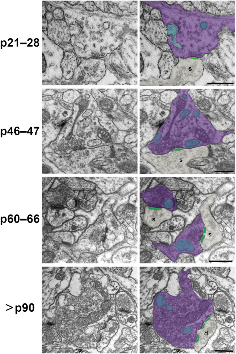
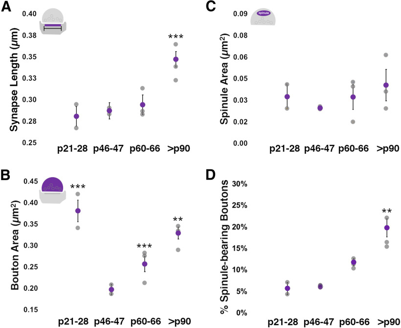
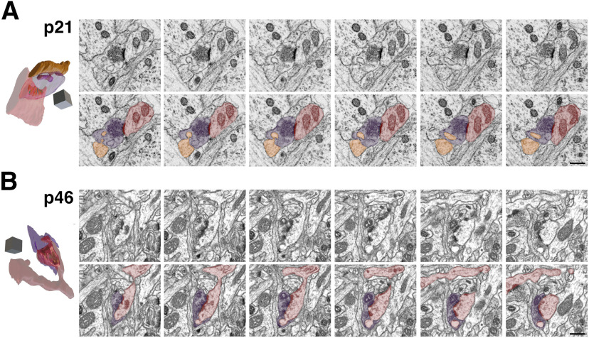
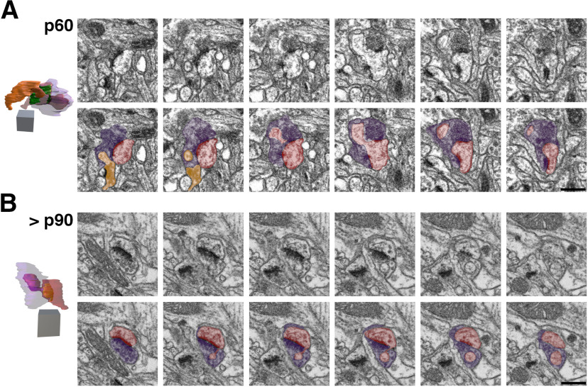
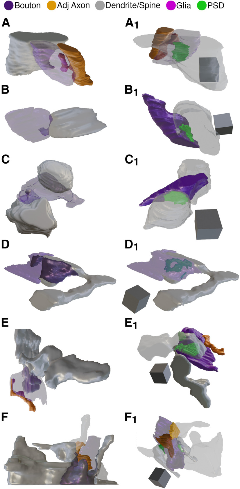
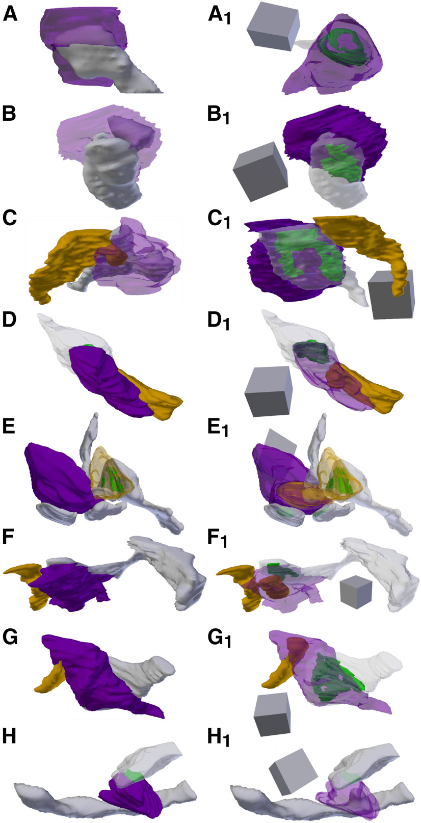
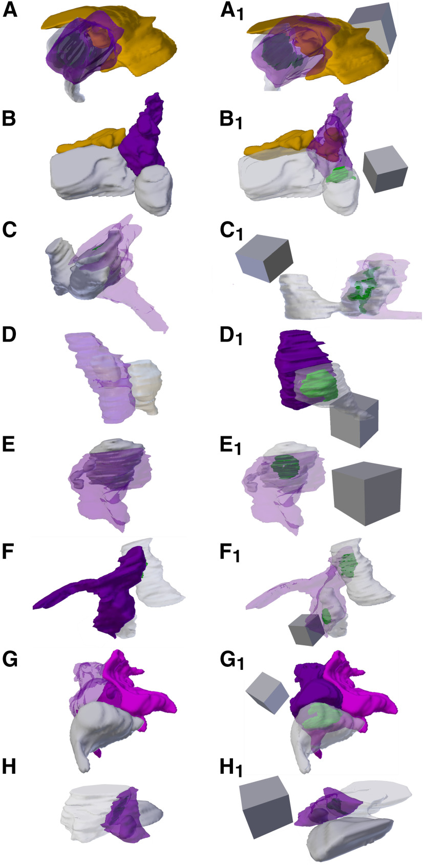
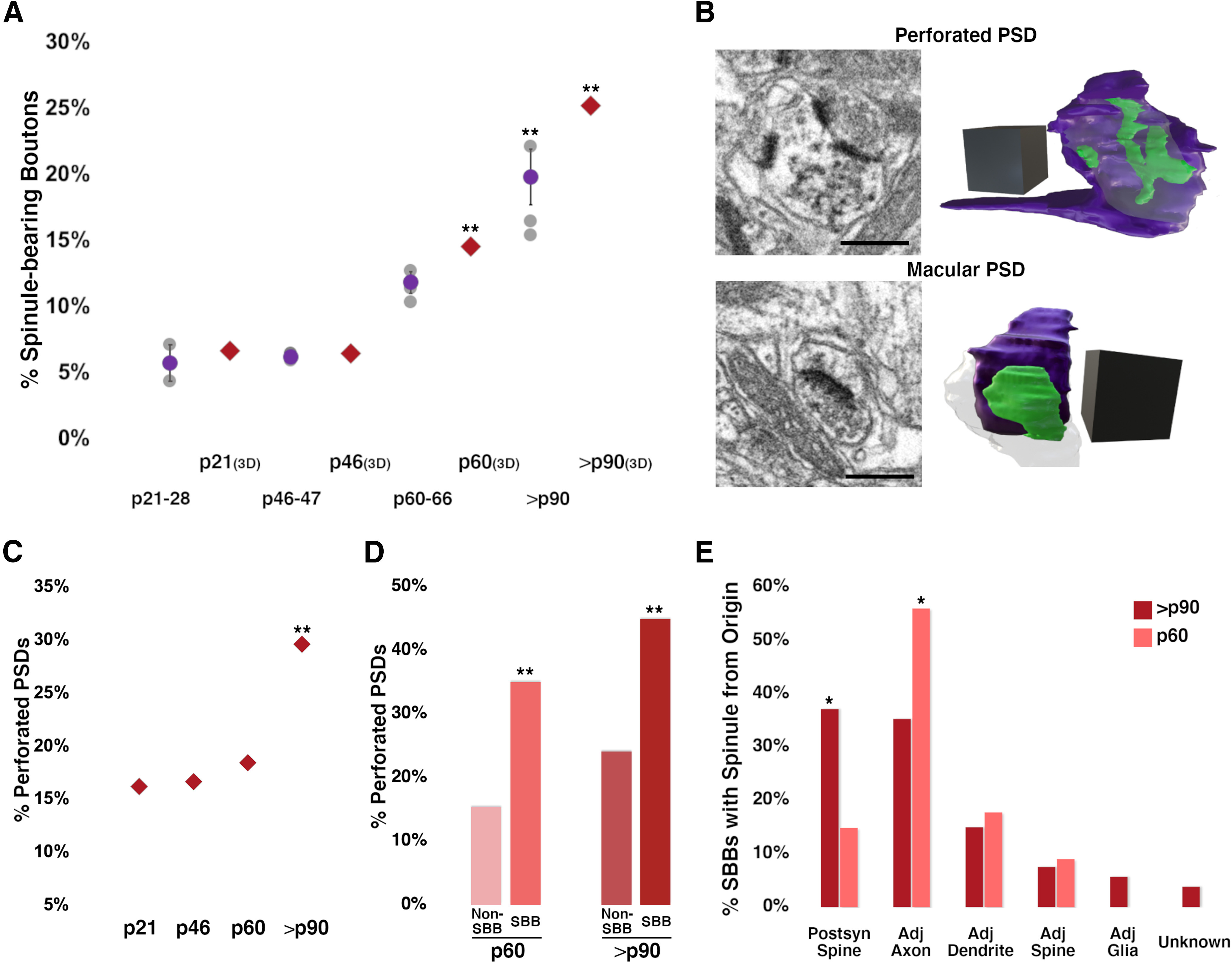
Despite decades of discussion in the neuroanatomical literature, the role of the synaptic "spinule" in synaptic development and function remains elusive. Canonically, spinules are finger-like projections that emerge from postsynaptic spines and can become enveloped by presynaptic boutons. When a presynaptic bouton encapsulates a spinule in this manner, the membrane apposition between the spinule and surrounding bouton can be significantly larger than the membrane interface at the synaptic active zone. Hence, spinules may represent a mechanism for extrasynaptic neuronal communication and/or may function as structural "anchors" that increase the stability of cortical synapses. Yet despite their potential to impact synaptic function, we have little information on the percentages of developing and adult cortical bouton populations that contain spinules, the percentages of these cortical spinule-bearing boutons (SBBs) that contain spinules from distinct neuronal/glial origins, or whether the onset of activity or cortical plasticity are correlated with increased prevalence of cortical SBBs. Here, we employed 2D and 3D electron microscopy to determine the prevalence of spinules in excitatory presynaptic boutons at key developmental time points in the primary visual cortex (V1) of female and male ferrets. We find that the prevalence of SBBs in V1 increases across postnatal development, such that ∼25% of excitatory boutons in late adolescent ferret V1 contain spinules. In addition, we find that a majority of spinules within SBBs at later developmental time points emerge from postsynaptic spines and adjacent boutons/axons, suggesting that synaptic spinules may enhance synaptic stability and allow for axo-axonal communication in mature sensory cortex.

Keywords: critical period; developmental plasticity; electron microscopy; focused ion beam scanning electron microscopy; presynaptic terminal; synaptic plasticity.


Figures

Figure 1.
Figure 1.
Development of excitatory SBB morphology. Spinules (blue) were observed within cortical boutons (purple) at every postnatal day age group examined. In 2D TEM sections, spinules were occasionally observed invaginating into SBB profiles (e.g., p21–p28); however, most spinules appeared as ovoid or circular double membrane-bound structures encapsulated within their “host” bouton. PSDs (green) at SBB synapses sometimes contained perforations (e.g., p60–p66 and >p90 panels). Note that SBBs at times displayed multiple spinule cross-sections (i.e., 2D profiles of putative spinules), yet in our 2D TEM analyses, it was not possible to attribute these to single spinules with complex morphologies, or to multiple spinules protruding into a single SBB. s = postsynaptic spine; d = postsynaptic dendrite. Scale bars = 0.5 μm.
Figure 2.
Figure 2.
Excitatory bouton areas and spinule prevalence increase over development. A, 2D synapse profile length, as defined by the PSD, remains relatively stable from before eye-opening (p28) until the end of the critical period for plasticity in ferret visual cortex (p60). However, PSD length increases substantially from the end of the critical period until at least the cusp of maturity (>p90 ages). B, Excitatory presynaptic bouton areas are largest at a time when most boutons appear to have elongated en passant morphology (p21–p28). Bouton size decreases significantly by p46–p47 and then shows a steady increase until at least p90. C, Average spinule profile areas do not appreciable change across the postnatal ages examined. Note that spinule areas show relatively large interanimal variation attributed to the variation in spinule sizes from distinct parent neurite/glia origins. D, Excitatory SBBs are most prevalent in late adolescence (>p90) versus at the height (p46–p47) or end (p46–p47) of the critical period for plasticity in ferret V1. A trend toward increased SBB prevalence is also seen between p46–p47 and p60 ages. For A–D, statistical comparisons were performed between p21–p28 and p46–p47, p46–p47, and p60–p66, and p60–p66, and >p90 (for details, see Table 3). Gray circles = individual animal means; Purple circles = group means ± SEM; **p < 0.015, ***p < 0.001.
Figure 3.
Figure 3.
FIBSEM images of SBBs in p21 and p46 V1. A, Adjacent FIBSEM images of an SBB in L4 of V1 from a p21 ferret. FIBSEM images are ∼25 nm apart in z (depth) on average, picked to display the progression of spinule invagination and engulfment by the SBB. Top panel, Raw FIBSEM images. Bottom panel, Pseudo-colored to highlight SBB (purple), adjacent axon projecting a spinule (orange), and postsynaptic dendrite (red). Left, Full reconstruction of this SBB showing transparent bouton with engulfed adjacent axon spinule and postsynaptic dendrite. Note the macular shaped PSD (green) formed between the SBB and the red dendrite. Identical SBB as shown in Figure 5A,A1. B, Adjacent FIBSEM images of an SBB in L4 of V1 from a p46 ferret. Top and bottom panels arranged and colored as in A, showing a postsynaptic spine (red) projecting a spinule into its presynaptic SBB partner (purple). Left, Full reconstruction of this p46 SBB showing postsynaptic spine spinule enveloped by its presynaptic bouton. Note the perforated complex shaped PSD (green). Identical SBB as shown in Figure 5D,D1. Scale bars = 0.5 μm; 3D scale cubes = 0.5 μm3.
Figure 4.
Figure 4.
FIBSEM images of SBBs in p60 and >p90 V1. A, Adjacent FIBSEM images of an SBB in L4 of V1 from a p60 ferret. FIBSEM images are ∼25 nm apart in z (depth) on average, picked to display the progression of spinule invagination and engulfment into the SBB. Top panel, Raw FIBSEM images. Bottom panel, Pseudo-colored to highlight SBB (purple), adjacent axon/bouton (orange), and postsynaptic spine (red). Note the adjacent bouton (orange) with a synapse onto a spine that protrudes a spinule into this SBB at the bottom left of the first image in the series, and the spinule from the postsynaptic spine that invaginates into this SBB across the middle of its perforated PSD. Left, Full reconstruction of this SBB showing transparent bouton (purple) with engulfed postsynaptic spine (red) and adjacent bouton (orange) spinules. Note the horseshoe-shaped perforated PSD. Identical SBB as shown in Figure 6C,C1. B, Adjacent FIBSEM images of an SBB in L4 of V1 from a >p90 ferret. Top and bottom panels arranged and colored as in A. Note the postsynaptic spine (red) that sends its spinule into its SBB (purple) partner from the edge of the PSD. Left, Full reconstruction of this SBB (purple), made transparent to show the engulfed anchor-like spinule from its postsynaptic spine partner. Macular-shaped PSD (green) appears yellow within spine. Identical SBB as shown in Figure 7D,D1. 2D scale bars = 0.5 μm; 3D scale cubes = 0.5 μm3.
Figure 5.
Figure 5.
3D Reconstructions of p21 and p46 SBBs. Focused ion beam serial electron microscopy 3D reconstructions of SBBs (purple) and their spinules from axon/boutons (yellow), and spines/dendrites (gray). Object color scheme shown at top. A–C, 3D reconstructions from a p21 ferret showing spinules from an adjacent axon (A, A1), postsynaptic dendrite (B, B1), and adjacent dendrite (C, C1) projecting into L4 SBBs. Note that B1 shows the spinule emerging from the edge (below and left) of the PSD. Reconstruction shown in A is the identical SBB shown in Figure 3A. D–F, 3D reconstructions from a p46 ferret showing hook-like spinule from a postsynaptic spine (D, D1), and spinules from adjacent axons and adjacent dendrites (E, E1, F, F1) projecting into L4 SBBs. Note the complex perforated PSD in D1, the identical SBB shown in Figure 3B. In F, F1, an SBB with synapses (green) onto two postsynaptic spines receives a large adjacent dendrite spinule (F) and a large adjacent axon spinule (F1). A1–F1, Identical SBBs as shown to the left in A–F, but with transparent postsynaptic neurites and/or SBBs to highlight the morphology and locations of the PSDs (green) or spinules. 3D scale cubes = 0.5 μm3.
Figure 6.
Figure 6.
3D Reconstructions of p60 SBBs. Focused ion beam serial electron microscopy 3D reconstructions of SBBs (purple) and their spinules from axon/boutons (yellow), and spines/dendrites (gray). Object color scheme as shown in Figure 5. A–C, Reconstructions of a postsynaptic spine head (A) and postsynaptic spine anchor-like spinules (B, C) projecting into L4 p60 SBBs. Note that in C, a second spinule from a synaptic bouton (orange) projects into the “upper” portion of the SBB (same bouton shown in Fig. 4A). C–G, Reconstructions showing axons/bouton spinules of various sizes engulfed by p60 SBBs. Note that in E, a presynaptic bouton (orange) receives a spinule from its postsynaptic spine partner and a portion of this bouton with its engulfed spinule protrude into a large adjacent SBB (purple) with synapses (green) onto three postsynaptic spines. H, An SBB with a synapse onto a postsynaptic dendrite (top) receives a spinule from an adjacent dendrite (bottom). A1–H1, Identical SBBs as shown to the left in A–H, but with transparent postsynaptic neurites and/or SBBs to highlight the morphology and locations of the PSDs (green) or spinules. 3D scale cubes = 0.5 μm3.
Figure 7.
Figure 7.
3D Reconstructions of >p90 SBBs. Focused ion beam serial electron microscopy 3D reconstructions of SBBs (purple) and their spinules from axon/boutons (yellow), spines/dendrites (gray), and glia (pink) from L4 of V1 of a >p90 ferret. Object color scheme as shown in Figure 5. A, B, Reconstructions showing adjacent axons/bouton spinules engulfed by SBBs with synapses onto postsynaptic spines. C–F, Reconstructions of postsynaptic spine heads (C, F), and anchor-like spinules (D, E) projecting into their SBB partners. Note the horseshoe-shaped perforated PSD (green) in C1, and the SBB shown in F, F1 enveloping approximately two-thirds of its postsynaptic spine partner. Reconstruction shown in D is the identical SBB shown in Figure 4B. G, An SBB with a synapse onto a postsynaptic spine receives a spinule from an adjacent glia. H, An SBB with a synapse onto a postsynaptic dendrite engulfs a spinule from an adjacent dendrite. A1–H1, Identical SBBs as shown to the left in A–H, but with transparent postsynaptic neurites and/or SBBs to highlight the morphology and locations of the PSDs (green) or spinules. 3D scale cubes = 0.5 μm3.
Figure 8.
Figure 8.
SBBs progressively engulf spinules as plasticity wanes in V1. A, Percentages of excitatory SBBs across the developmental ages examined in 2D TEM and FIBSEM (3D) images. 3D FIBSEM data at p21, p46, p60, and >p90 are represented by red diamonds, 2D data as shown in Figure 2D. Statistical comparisons were performed between p21 and p46, p46 and p60, and p60 and >p90 (for details, see Table 3). B, FIBSEM images showing an SBB with a synapse onto a postsynaptic spine containing a perforated PSD (top panel), and an SBB with a with a synapse onto a spine displaying a macular PSD (bottom panel). 3D reconstructions of each SBB and their corresponding PSDs are shown to the right. SBBs shown in purple, PSDs shown in green, postsynaptic spines are made transparent to visualize PSDs. Identical SBBs as shown in Figure 7C,D. C, FIBSEM developmental analysis showing the average percentages of excitatory presynaptic boutons with perforated PSDs in L4. D, FIBSEM analysis showing the percentages of SBBs versus Non-SBBs (i.e., excitatory presynaptic boutons without spinules) with perforated PSDs at p60 and >p90. E, FIBSEM analysis showing the percentages of p60 and >p90 SBBs containing at least one spinule from a defined neurite or glial origin. For example, ∼56% of SBBs at p60 contain at least one spinule from an adjacent axon. Scale bars = 0.5 μm for FIBSEM images in B; 3D scale cubes = 0.5 μm3 for FIBSEM reconstructions in B; *p < 0.05, **p < 0.015.

Similar articles

Cited by

References

    1. Ahmed B, Anderson JC, Douglas RJ, Martin KA, Nelson JC (1994) Polyneuronal innervation of spiny stellate neurons in cat visual cortex. J Comp Neurol 341:39–49. 10.1002/cne.903410105 - DOI - PubMed
    1. Araya R, Vogels TP, Yuste R (2014) Activity-dependent dendritic spine neck changes are correlated with synaptic strength. Proc Natl Acad Sci USA 111:E2895–E2904. 10.1073/pnas.1321869111 - DOI - PMC - PubMed
    1. Bailey CH, Thompson EB, Castellucci VF, Kandel ER (1979) Ultrastructure of the synapses of sensory neurons that mediate the gill-withdrawal reflex in Aplysia. J Neurocytol 8:415–444. 10.1007/BF01214801 - DOI - PubMed
    1. Bopp R, Holler-Rickauer S, Martin KA, Schuhknecht GF (2017) An ultrastructural study of the thalamic input to layer 4 of primary motor and primary somatosensory cortex in the mouse. J Neurosci 37:2435–2448. 10.1523/JNEUROSCI.2557-16.2017 - DOI - PMC - PubMed
    1. Bozhilova-Pastirova A, Ovtscharoff W (1999) Intramembranous structure of synaptic membranes with special reference to spinules in the rat sensorimotor cortex. Eur J Neurosci 11:1843–1846. 10.1046/j.1460-9568.1999.00608.x - DOI - PubMed

LinkOut - more resources