. 2020 Oct 6;117(40):25104-25115.
doi: 10.1073/pnas.2005506117. Epub 2020 Sep 21.

Quality-control mechanisms targeting translationally stalled and C-terminally extended poly(GR) associated with ALS/FTD

Affiliations

Quality-control mechanisms targeting translationally stalled and C-terminally extended poly(GR) associated with ALS/FTD

Shuangxi Li et al. Proc Natl Acad Sci U S A. .

Abstract

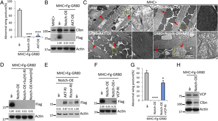
Maintaining the fidelity of nascent peptide chain (NP) synthesis is essential for proteome integrity and cellular health. Ribosome-associated quality control (RQC) serves to resolve stalled translation, during which untemplated Ala/Thr residues are added C terminally to stalled peptide, as shown during C-terminal Ala and Thr addition (CAT-tailing) in yeast. The mechanism and biological effects of CAT-tailing-like activity in metazoans remain unclear. Here we show that CAT-tailing-like modification of poly(GR), a dipeptide repeat derived from amyotrophic lateral sclerosis with frontotemporal dementia (ALS/FTD)-associated GGGGCC (G4C2) repeat expansion in C9ORF72, contributes to disease. We find that poly(GR) can act as a mitochondria-targeting signal, causing some poly(GR) to be cotranslationally imported into mitochondria. However, poly(GR) translation on mitochondrial surface is frequently stalled, triggering RQC and CAT-tailing-like C-terminal extension (CTE). CTE promotes poly(GR) stabilization, aggregation, and toxicity. Our genetic studies in Drosophila uncovered an important role of the mitochondrial protease YME1L in clearing poly(GR), revealing mitochondria as major sites of poly(GR) metabolism. Moreover, the mitochondria-associated noncanonical Notch signaling pathway impinges on the RQC machinery to restrain poly(GR) accumulation, at least in part through the AKT/VCP axis. The conserved actions of YME1L and noncanonical Notch signaling in animal models and patient cells support their fundamental involvement in ALS/FTD.

Keywords: C9-ALS/FTD; CAT-tailing; Notch; YME1L; ribosome-associated quality control.


Conflict of interest statement

The authors declare no competing interest.

Figures

Fig. 1.
Fig. 1.
YME1L negatively regulates poly(GR) expression and toxicity. (AC) Effects of YME1L overexpression (OE) or RNAi (RI) on wing-posture (A) and GR80 protein (B) and mRNA (C) expression in Mhc-Gal4 > Flag-GR80 flies. Examples of normal and droopy wing posture are shown in A. (D) Immunostaining showing increased GR80 accumulation on mitochondria (labeled with mito-GFP) after YME1L-RI. Compared to control flies, Flag-GR80 flies showed regions devoid of mito-GFP (arrows), which was exacerbated by YME1L-RI. (E and F) Immunoblots showing the effect of CRISPR-mediated YME1L knockdown on Flag-GR80 expression in HEK293T cells (E) and rescue with WT or E543Q forms of YME1L (F). Cotransfected GFP serves as control for transfection efficiency in this and subsequent figures. (G and H) Effect of cotransfection of YME1L-GFP, or Flag tagged YME1L, and YME1L-E543Q on Flag-GR80 expression in HEK293T cells. (I and J) In vitro YME1L protease activity assay using hexYME1L as protease and Flag-GR80 or Flag-C-I30 as substrates. **P < 0.01 and ***P < 0.001 in one-way ANOVA test followed by SNK test plus Bonferroni correction (multiple hypotheses correction); N.S., nonsignificant. Values under the Flag-GR80 blots indicate normalized levels relative to control in this and all subsequent figures. Western blots represent at least two biological repeats.
Fig. 2.
Fig. 2.
Stalled translation of poly(GR) mRNA on mitochondria. (A) RT-PCR analysis of Percoll gradient-purified mitochondrial fractions from GR80 transgenic fly muscle showing the presence of GR80 mRNA (Left) and GR80 mRNA levels in total and mitochondrial fractions (Right). Mitochondrial-encoded mtCo-1 and cytosolic tubulin serve as positive and negative controls. (B and C) Release of Flag-GR80 NP by HA (B) or puromycin (C) treatment of mitochondria purified from GR80-expressing HEK293 cells or fly muscle. Untreated samples (control), and the supernatant (HA-release) or mitochondrial pellet (HA-remaining) of HA-treated samples were analyzed. (D) HEK293 cells transfected with MS2-bs or GR60-MS2-bs reporter constructs were costained for the cotransfected MS2-GFP that binds to MS2-bs, and the mitochondrial marker Tom20. (E) Immunoblots showing co-IP between Flag-GR80 and Tom40, and the preferential binding of Flag-GR80 to Rpl3 over Rps6.
Fig. 3.
Fig. 3.
RQC of poly(GR) in fly tissues and mammalian cells. (A) Co-IP assays using extracts treated with or without RNase A to test the role of RNA in facilitating interactions between Flag-GR80 and the RQC factors. (B and C) RNA-IP assays showing binding of NEMF (B) or Clbn (C) to GR80 mRNA in HEK293T (B) or fly muscle (C) samples. Bar graphs show quantification of GR80 mRNA level in IP samples. ***P < 0.001 in Student’s t test. (D) Immunoblots showing co-IP between Flag-GR80 and RQC factors in fly muscle. (EG) Immunoblots showing effects of overexpression or knockdown of RQC factors on Flag-GR80 level in fly muscle. (H) Immunostaining showing effects of overexpressing various RQC factors on Flag-GR80 level on muscle mitochondria. (I) Quantification showing effects of altered activities of RQC factors on GR80-induced wing posture phenotype. *P < 0.05 and **P < 0.01 in one-way ANOVA test followed by SNK test plus Bonferroni correction. Western blots represent at least two biological repeats.
Fig. 4.
Fig. 4.
CAT-tailing–like CTE of ribosome-stalled poly(GR). (A and B) Immunoblots showing effects of Clbn or NEMF RNAi on Flag-GR80 level in fly muscle (A) or HEK293 cells (B). (C) Immunoblots and immunostaining showing effect of anisomycin on Flag-GR80 level in HEK293T cells. (DF) Immunoblots showing effects of Clbn-OE (D) or RNAi of fly Vms1 (E) and various ARSs (F) on Flag-GR80 level in fly muscle. (G) Immunostaining showing effects of RNAi of various ARSs on Flag-GR80 associated with muscle mitochondria. (H) Immunoblots showing effect of IARS RNAi on Flag-GR80 level in fly muscle. (I–K) MS/MS spectra of the parent ion of peptides generated by collision-induced dissociation fragmentation. Matched peptide sequence entries from the custom-built database were shown below each spectrum. The blue and yellow vertical lines in the MS spectra represent “y” and “b” ions, respectively, found in the BY matches. The BY matches are fragment ions found in the high-energy data that suggest a particular identification for a given precursor ion found in the low-energy data.
Fig. 5.
Fig. 5.
Negative regulation of poly(GR) expression and toxicity by noncanonical Notch signaling in fly muscle. (A) Quantification of effect of Notch-OE or AKT-OE on GR80-induced wing posture defect. (B) Immunoblots showing effect of Notch-OE or AKT-OE on Flag-GR80 and Clbn expression. (C) TEM images showing effects of Notch-OE or AKT-OE, or Notch-OE + AKT-RI, on GR80-induced mitochondrial morphology defect. Arrows mark mitochondria. Insets show zoom-in view of selected areas of interest (yellow squares). GR-80 caused swelling and loss of cristae and electron-dense material, phenotypes rescued by Notch-OE or AKT-OE. AKT-RI blocked the Notch-OE effect. Magnification: 5×. (D) Immunoblots showing lack of effect of Su(H) RNAi or OE of dominant-negative Mam [Man(H)] on the inhibition of Flag-GR80 expression by Notch-OE. (E and F) Immunoblots showing the effects of RNAi of AKT or Rictor (E), or VCP RNAi (F) on the inhibition of Flag-GR80 expression by Notch-OE. (G) Quantification of effect of VCP RNAi on the suppression of GR80-induced wing posture defect by Notch-OE. (H) Immunoblots assessing effect of Notch-OE on endogenous VCP or Clbn expression. *P < 0.05; ***P < 0.001.
Fig. 6.
Fig. 6.
The AKT-VCP axis mediates the effects of noncanonical Notch signaling on poly(GR) expression and toxicity. (A) Immunoblots showing co-IP between VCP and AKT. (B) Immunoblots showing the effect of SC79 and AKTi on VCP phosphorylation at consensus AKT target site(s) in HEK293T cells. (C) Immunoblots showing effects of VCP RNAi on the inhibition of Flag-GR80 expression by AKT-OE. (D) Quantification of effect of VCP RNAi on the suppression of GR80-induced wing posture defect by AKT-OE. (E and F) Immunoblots showing the effects of VCP-WT, VCP-S745/747D, and VCP-S745/747A on Flag-GR80 protein level (E), and the effects of VCP-WT and VCP-S745/747D in the presence of AKTi (F). (G and H) Immunostaining showing the effect of Notch, AKT, VCP, and YME1L OE on the levels of Flag-GA80 (G) and Flag-PR80 (H) in fly muscle. Bar graphs show quantification of Flag-GA80 and Flag-PR80 immunofluorescent signals. *P < 0.05, **P < 0.01, and ***P < 0.001 in one-way ANOVA test followed by SNK test plus Bonferroni correction.
Fig. 7.
Fig. 7.
Regulation of poly(GR) expression by noncanonical Notch signaling in C9-ALS/FTD patient fibroblasts. (A) Dot blots showing the effect of genetic manipulation of Notch and VCP on poly(GR) expression in C9-ALS/FTD patient fibroblasts. (B) TEM showing the effect of genetic manipulation or chemical treatment on mitochondrial morphology in patient fibroblasts. Arrows mark mitochondria. Insets show zoom-in view of selected areas of interest (yellow squares). The swelling and loss of electron-dense material and cristae structure was rescued by SC79 treatment or OE of Notch and AKT. Magnification: 5×. (C) Dot blots showing the effect of VCPi and AKTi treatment on poly(GR) level in C9-ALS/FTD patient fibroblasts. (D) Dot blots showing the enrichment of poly(GR) in the mitochondria of patient fibroblasts and the effect of SC79 treatment on poly(GR) level. Dot blots represent at least two biological repeats.

Similar articles

Cited by

References

    1. Klaips C. L., Jayaraj G. G., Hartl F. U., Pathways of cellular proteostasis in aging and disease. J. Cell Biol. 217, 51–63 (2018). - PMC - PubMed
    1. Hou Y. et al. ., Ageing as a risk factor for neurodegenerative disease. Nat. Rev. Neurol. 15, 565–581 (2019). - PubMed
    1. Ciechanover A., Brundin P., The ubiquitin proteasome system in neurodegenerative diseases: Sometimes the chicken, sometimes the egg. Neuron 40, 427–446 (2003). - PubMed
    1. Duttler S., Pechmann S., Frydman J., Principles of cotranslational ubiquitination and quality control at the ribosome. Mol. Cell 50, 379–393 (2013). - PMC - PubMed
    1. Wang F., Durfee L. A., Huibregtse J. M., A cotranslational ubiquitination pathway for quality control of misfolded proteins. Mol. Cell 50, 368–378 (2013). - PMC - PubMed

Publication types

MeSH terms

Supplementary concepts

LinkOut - more resources