. 2020 Sep 21;11(1):4765.
doi: 10.1038/s41467-020-18565-8.

CD36 facilitates fatty acid uptake by dynamic palmitoylation-regulated endocytosis

Affiliations

CD36 facilitates fatty acid uptake by dynamic palmitoylation-regulated endocytosis

Jian-Wei Hao et al. Nat Commun. .

Abstract

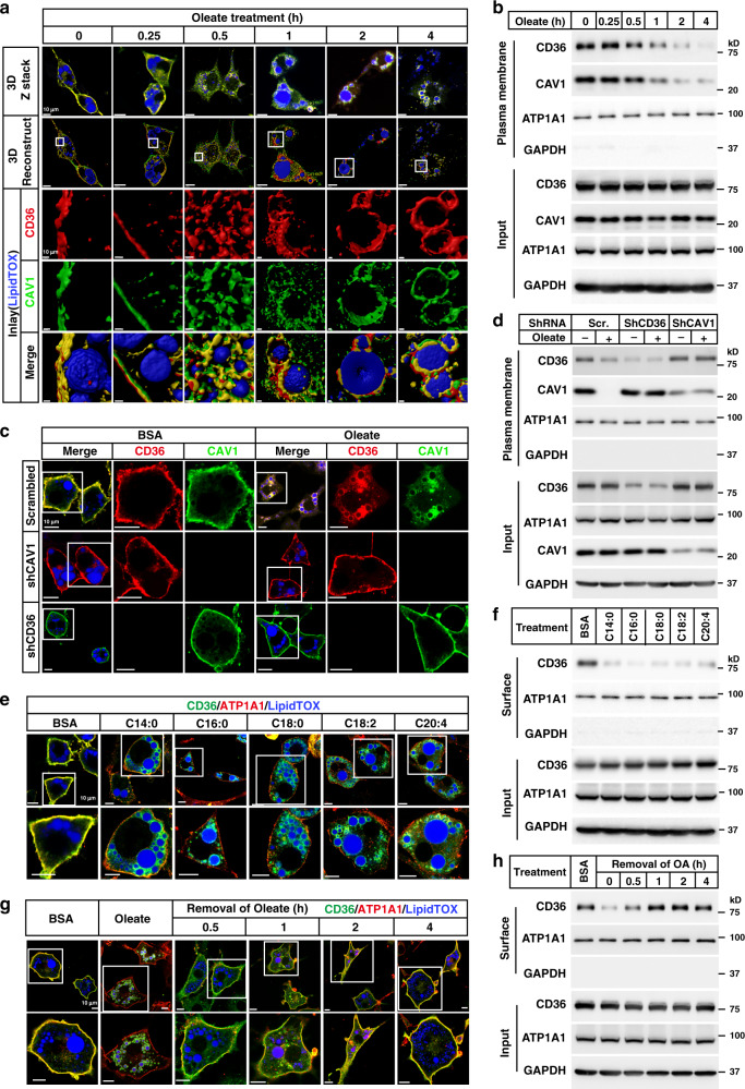
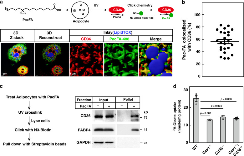
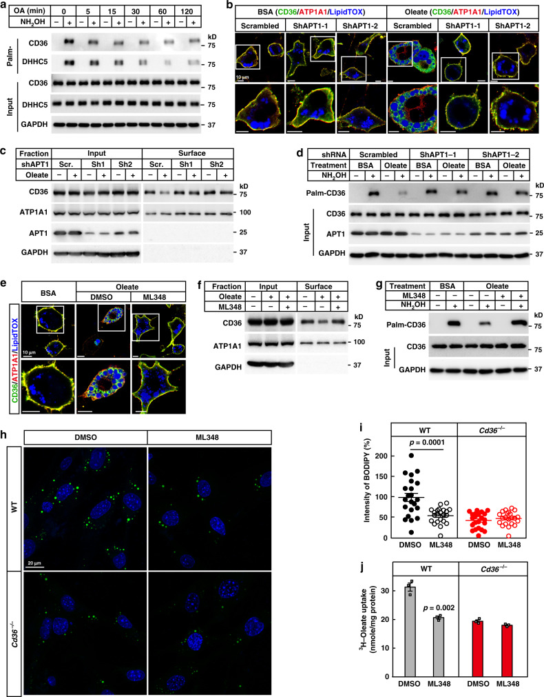
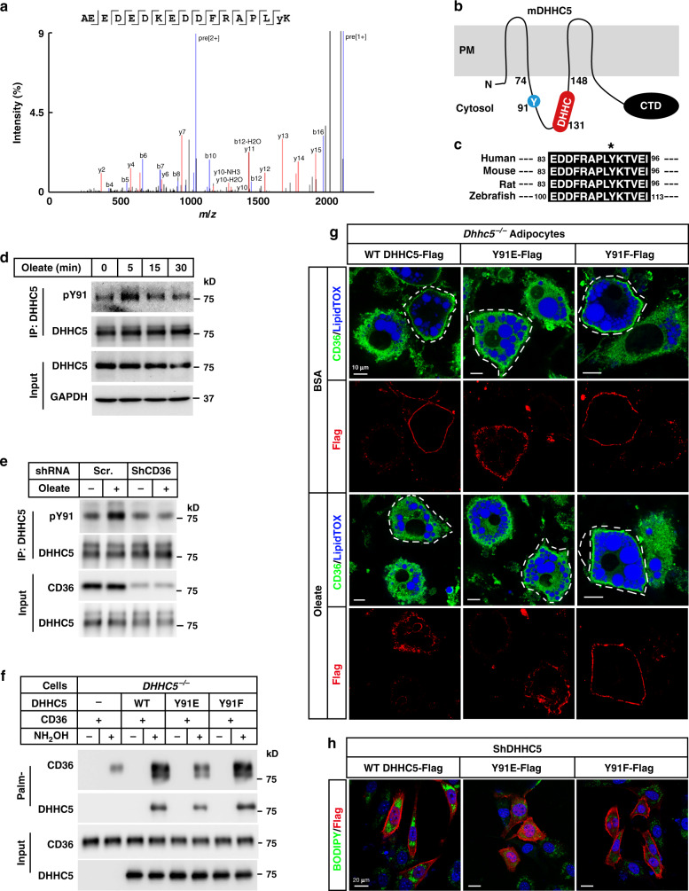
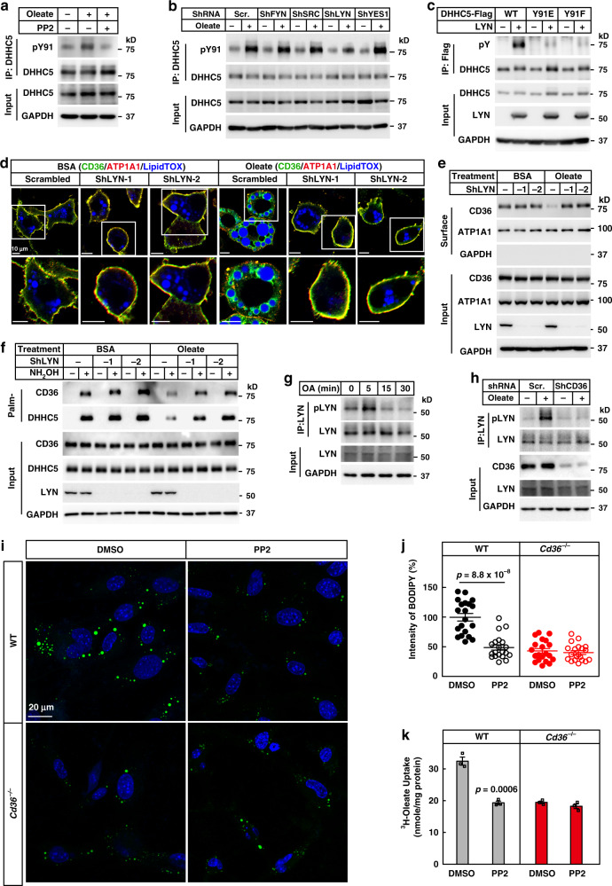
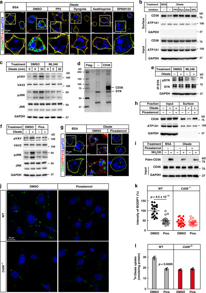
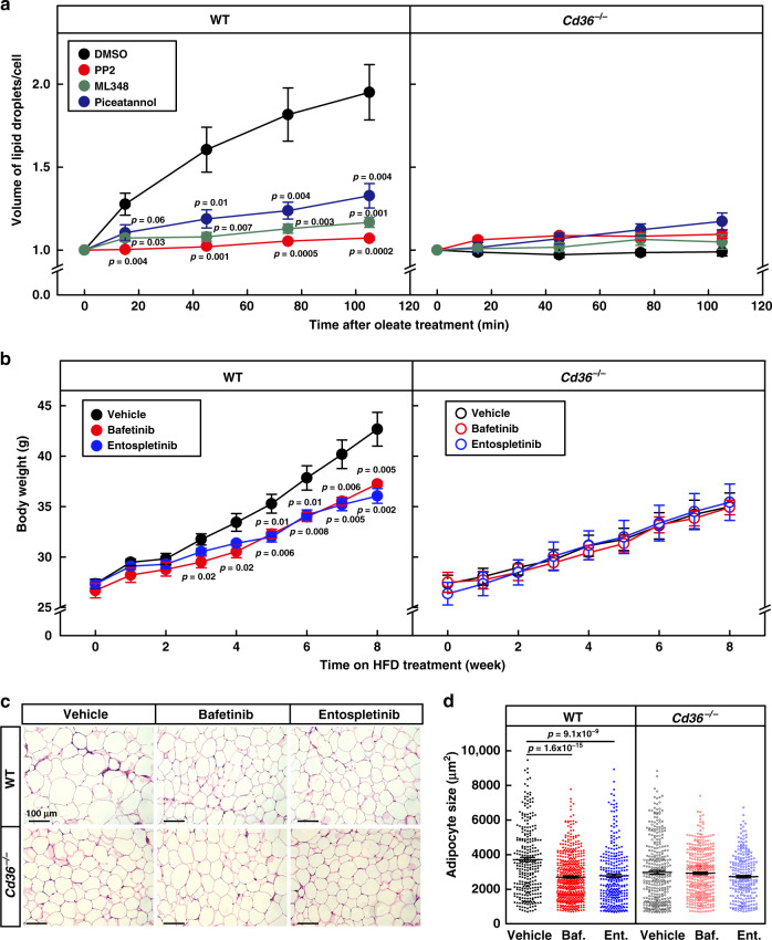
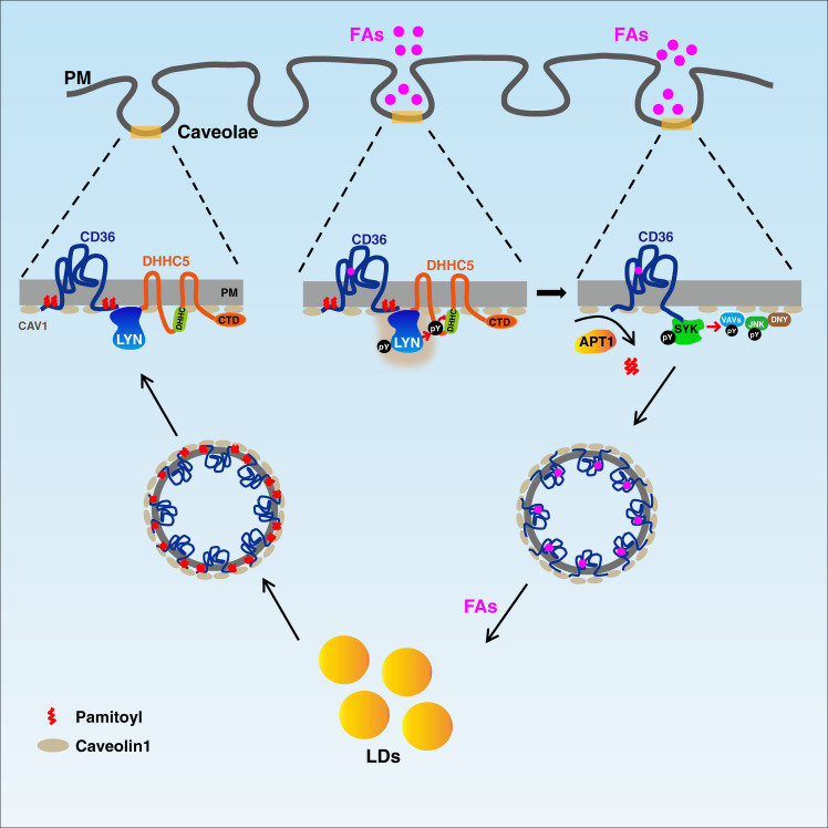
Fatty acids (FAs) are essential nutrients, but how they are transported into cells remains unclear. Here, we show that FAs trigger caveolae-dependent CD36 internalization, which in turn delivers FAs into adipocytes. During the process, binding of FAs to CD36 activates its downstream kinase LYN, which phosphorylates DHHC5, the palmitoyl acyltransferase of CD36, at Tyr91 and inactivates it. CD36 then gets depalmitoylated by APT1 and recruits another tyrosine kinase SYK to phosphorylate JNK and VAVs to initiate endocytic uptake of FAs. Blocking CD36 internalization by inhibiting APT1, LYN or SYK abolishes CD36-dependent FA uptake. Restricting CD36 at either palmitoylated or depalmitoylated state eliminates its FA uptake activity, indicating an essential role of dynamic palmitoylation of CD36. Furthermore, blocking endocytosis by targeting LYN or SYK inhibits CD36-dependent lipid droplet growth in adipocytes and high-fat-diet induced weight gain in mice. Our study has uncovered a dynamic palmitoylation-regulated endocytic pathway to take up FAs.


Conflict of interest statement

The authors declare no competing interests.

Figures

Fig. 1
Fig. 1. FAs trigger internalization of CD36.
a, b On day 8 of differentiation, 3T3-L1 adipocytes were pretreated with serum-free medium for 4 h, and then treated with BSA-conjugated oleate (100 μM) for indicated time. a One set of cells was subjected to immunostaining with anti-CD36 and anti-CAV1 antibodies. LipidTOX was used to label lipid droplets. Images were taken under a Zeiss LSM-780 microscope in a 3D Z-stack mode and reconstructed using Imaris 9.2.0. b The other set of cells was subjected to surface biotinylation assay and blotted with indicated antibodies. c, d On day 4 of differentiation, 3T3-L1 cells were infected with lentivirus encoding scrambled shRNA or shRNAs against CD36 or CAV1. On day 5, cells were selected with 5 μg/ml puromycin. On day 8, cells were pretreated as in (a) and treated with oleate (100 μM) for 4 h, followed by immunostaining with anti-CD36 and anti-CAV1 antibodies (c), or surface biotinylation assay (d). e, f 3T3-L1 adipocytes were pretreated as in (a) and treated with BSA-conjugated FAs with different chain lengths or saturation (100 μM) for 4 h. Cells were subjected to immunostaining with anti-CD36 antibody (e), or surface biotinylation assay (f). After oleate treatment for 4 h, 3T3-L1 adipocytes were switched to serum-free medium for indicated time and harvested for immunostaining (g) and surface biotinylation (h). The scale bars were as indicated in each figure. These experiments were repeated at least three times. Source data are provided as a Source Data file.
Fig. 2
Fig. 2. CD36-mediated caveolar endocytosis transports FAs into cells.
ac 3T3-L1 adipocytes were pretreated with serum-free medium for 4 h, and then treated with BSA-conjugated PacFA (50 μM) for 20 min, followed by UV crosslinking on ice for 30 min. a, b Cells were subjected to click chemistry using an N3-Alexa Fluro 488, and immunostained with anti-CD36 antibody. Colocalization of PacFA and CD36 was quantified from 24 cells over three independent experiments and plotted in (b). The value represents mean ± SEM. c Cells were lysed and subjected to click chemistry assay using N3-biotin. PacFA-labeled proteins were captured with streptavidin beads and subjected to western blot using anti-CD36 and anti-FABP4 antibodies. d WT, Cav1−/−, Cd36−/−, and Cav1−/−;Cd36−/− SVFs were isolated and differentiated into adipocytes and treated with 100 μM 3H-oleate (specific activity, 2268 dpm/nmol) for 1 h. Lipid fractions were extracted from the cells and subjected to scintillation counting. The radioactive counting was normalized to protein content. Each value represents mean ± SEM obtained from three samples. Two-sided Student’s t test was performed between WT and each of the knockout cells. These experiments were repeated twice. Source data are provided as a Source Data file.
Fig. 3
Fig. 3. Depalmitoylation of CD36 is required for the endocytosis of FAs.
a 3T3-L1 adipocytes were pretreated and treated with oleate (100 μM) as in Fig. 1a for indicated time. Cells were harvested for Acyl-RAC assay followed by immunoblotting using indicated antibodies. bd Control (scrambled) and APT1 knockdown 3T3-L1 adipocytes were pretreated and treated with BSA or oleate (100 μM) for 1 h. Cells were then harvested for immunostaining (b), surface biotinylation (c), and Acyl-RAC (d) assays. eg 3T3-L1 adipocytes were pretreated and treated with ML348 (10 μM) for 1 h, followed by BSA or oleate treatment for another 1 h. Cells were then harvested for immunostaining (e), surface biotinylation (f), and Acyl-RAC (g) assays. h, i On day 0, WT and Cd36−/− SVFs were set up at 4 × 104 cells per well in a six-well plate. On day 2, cells were pretreated with serum-free medium for 4 h, and treated with ML348 (10 μM) for 1 h, followed by treatment with oleate (100 μM) and BODIPY 493/503 (0.1 μg/ml) for 4 h. h Cells were then fixed and imaged on a Zeiss LSM-780 confocal microscope. i Quantification of BODIPY fluorescent intensity was performed using ZEN 2010 software. Each value represents mean ± SEM fluorescent intensity per cell from 20 cells. The value in DMSO-treated WT cells was normalized to 1.0. Two-sided Student’s t test was performed between DMSO- and ML348-treated cells. DAPI was used to indicate the nuclei. j WT and Cd36−/− SVFs were differentiated into adipocytes and pretreated with ML348 (10 μM) for 1 h, followed by treatment with 100 μM 3H-oleate (specific activity, 2268 dpm/nmol) for 1 h. Lipid fractions were extracted from the cells and subjected to scintillation counting. The radioactive counting was normalized to protein content. Each value represents mean ± SEM obtained from three samples. Two-sided Student’s t test was performed between DMSO- and ML348-treated cells. These experiments were repeated at least twice. Source data are provided as a Source Data file.
Fig. 4
Fig. 4. Oleate triggers phosphorylation and inactivation of DHHC5.
a On day 8 of differentiation, 3T3-L1 adipocytes were pretreated with serum-free medium for 4 h, and then treated with BSA or BSA-conjugated oleate (100 μM) for 15 min. Cells were harvested and subjected to immunoprecipitation using anti-DHHC5 antibody. The bands of DHHC5 were cut out and sent for mass spectrometry analysis of the phosphorylation sites. The spectrum of pY91-containing peptide was shown. b Indication of Y91 and DHHC motif on a topology of mouse DHHC5, which was predicted at UniProt (www.uniprot.org). c Alignment of the Y91 containing regions of DHHC5 proteins from human, mouse, rat, and zebrafish. Control and CD36 knockdown 3T3-L1 adipocytes were pretreated as in (a), and treated with oleate (100 μM) for indicated time (d) or 5 min (e). Cells were harvested, immunoprecipitated with anti-DHHC5 antibody, and immunoblotted with an anti-pY antibody to detect phosphorylation of DHHC5. f On day 0, DHHC5−/− HEK293T cells were set up at 7.5 × 105 cells per 6-cm dish. On day 2, cells were transfected with 0.5 μg CD36-Flag/pCDH-puro and 0.5 μg of indicated DHHC5 WT, Y91E, or Y91F/pCDH-puro. On day 3, cells were harvested for Acyl-RAC assay and blotted with indicated antibodies. g SVFs were isolated from Rosa-CreERT2;Dhhc5f/f mice, treated with 4-OHT to induce deletion of Dhhc5, and subjected to differentiation. On day 6 of differentiation, cells were electroporated with WT, Y91E, or Y91F of DHHC5-Flag. On day 8, cells pretreated and treated with oleate as in Fig. 1c. Cells were subjected to immunofluorescence using anti-CD36 and anti-Flag antibodies. LipidTOX was used to indicate lipid droplets. Cells expressing WT, Y91E, or Y91F of DHHC5 were outlined as indicated. Scale bar, 10 μm. h DHHC5 knockdown 3T3-L1 preadipocytes were transfected with WT, Y91E, or Y91F of DHHC5-Flag. Cells were treated with oleate (100 μM) and BODIPY 493/503 (0.1 μg/ml) for 8 h, followed by immunostaining with anti-Flag M2 antibody. DAPI was used to indicate the nuclei. These experiments were repeated at least twice. Source data are provided as a Source Data file.
Fig. 5
Fig. 5. LYN phosphorylates DHHC5 and is required for the endocytosis of FAs.
a 3T3-L1 adipocytes were pretreated with serum-free medium for 4 h and PP2 (15 μM) for 1 h, followed by treatment with BSA or oleate (100 μM) for 5 min. Cells were harvested and immunoprecipitated with anti-DHHC5 antibody to detect phosphorylation of Tyr91. b 3T3-L1 adipocytes transduced with indicated shRNA were set up and treated with oleate for 5 min. Cells were harvested to detect Tyr91 phosphorylation as in (a). c On day 0, HEK293T cells were set up at 2.5 × 105 cells per 6-cm dish. On day 2, cells were transfected with 0.5 μg of WT, Y91E, or Y91F of DHHC5-Flag/pCDH-puro and/or 0.5 μg LYN/pCDNA3.3. On day 3, cells were harvested and immunoprecipitated with anti-Flag M2 beads to detect phosphorylation of DHHC5 (pY). Control and LYN knockdown adipocytes were pretreated and treated with BSA or oleate (100 μM) for 1 h, followed by immunostaining (d), surface biotinylation (e), and Acyl-RAC (f) assays. Control and CD36 knockdown 3T3-L1 adipocytes were pretreated and treated with oleate (100 μM) for indicated time (g) or 5 min (h). Cells were harvested and subjected to immunoprecipitation of LYN to detect phosphorylation of LYN (Y396). i, j WT and Cd36−/− SVFs were set up and subjected to FA uptake as in Fig. 3h, i, except that cells were pretreated with PP2 (15 μM). Each value represents mean ± SEM obtained from 20 cells. Two-sided Student’s t test was performed between DMSO and PP2 treated cells. k WT and Cd36−/− adipocytes were set up and subjected to 3H-oleate uptake as in Fig. 3j, except that cells were pretreated with PP2 (15 μM). Each value represents mean ± SEM obtained from three samples. Two-sided Student’s t test was performed between DMSO and PP2 treated cells. These experiments were repeated at least twice. Source data are provided as a Source Data file.
Fig. 6
Fig. 6. Depalmitoylated CD36 recruits SYK to facilitate the endocytosis.
a, b 3T3-L1 adipocytes were pretreated with serum-free medium for 4 h, and DMSO, PP2 (20 μM), Dyngo4a (2 μM), azathioprine (10 μM), or SP600125 (20 μM) for 1 h, followed by treatment with BSA or oleate (100 μM) for 1 h. Cells were harvested for immunostaining (a) and surface biotinylation (b) assays. c 3T3-L1 adipocytes were pretreated with ML348 (10 μM) for 1 h, following by oleate treatment for 0, 5, or 30 min. Cells were harvested and immunoblotted with indicated antibodies. d The interacting proteins of depalmitoylated CD36 were isolated as described in “Methods.” The eluted fractions were separated on SDS-PAGE, and subjected to silver staining and mass spectrometry. e 3T3-L1 adipocytes were pretreated with ML348, followed by BSA or oleate (100 μM) for 5 min. Cells were harvested, subjected to immunoprecipitation with anti-SYK antibody, and blotted with anti-SYK and anti-pSYK (Y525/526). f 3T3-L1 adipocytes were pretreated with serum-free medium for 4 h and piceatannol (Pice., 40 μM) for 1 h, followed by treatment with BSA or BSA-conjugated oleate (100 μM) for 30 min. Cells were harvested for immunoblotting with indicated antibodies. gi 3T3-L1 adipocytes were pretreated with serum-free medium for 4 h and piceatannol (40 μM) for 1 h, followed by treatment with BSA or BSA-conjugated oleate (100 μM) for 1 h. Cells were harvested for immunostaining (g), surface biotinylation (h), and Acyl-RAC (i) assays. j, k WT and Cd36−/− SVFs were set up and subjected to FA uptake as in Fig. 3h, i, except that cells were pretreated with piceatannol (40 μM). Each value represents mean ± SEM obtained from 20 cells. Two-sided Student’s t test was performed between DMSO and piceatannol-treated cells. l WT and Cd36−/− adipocytes were set up and subjected to 3H-oleate uptake as in Fig. 3j, except that cells were pretreated with piceatannol (40 μM). Each value represents mean ± SEM obtained from three samples. Two-sided Student’s t test was performed between DMSO and piceatannol-treated cells. The scale bars were as indicated. These experiments were repeated at least twice. Source data are provided as a Source Data file.
Fig. 7
Fig. 7. Blocking endocytosis inhibits lipid droplet growth and HFD-induced obesity.
a On the day of experiment, WT and Cd36−/− adipocytes were pretreated with PP2 (20 μM), ML348 (10 μM), or piceatannol (40 μM) for 1 h, then treated with oleate (100 μM) and BODIPY 493/503 (0.1 μg/ml), and subjected to live imaging on a Zeiss LSM-780 confocal microscope in a 3D Z-stack mode for 2 h. The number of cells was 12, 9, 8, and 10 for DMSO, PP2, ML348, and piceatannol-treated WT cells, and 8, 8, 8, and 10 for corresponding groups in Cd36−/− cells, respectively. Total volume of lipid droplets in each cell was calculated. Each value represents mean ± SEM, and the volume at 0 min was normalized to 1.0. Two-sided Student’s t test was performed between DMSO- and PP2-, ML348- or piceatannol-treated cells. bd WT and Cd36−/− mice (8-week-old male, n = 6/group) were daily gavaged with vehicle (0.5% methyl cellulose), bafetinib (20 mg/kg), or entospletinib (10 mg/kg) at 7 pm and subjected to HFD feeding for 8 weeks. b Body weight of the mice was monitored every week. c, d Gonadal WAT was subjected to H&E staining, and representative pictures were shown (c). Scale bar, 100 μm. Quantification of the surface area of adipocytes in gWAT by ImageJ. The size of each adipocyte was quantified by ImageJ and plotted as mean ± SEM from 287, 488, 305, 324, 362, and 218 cells, respectively d. Two-sided Student’s t test was performed between vehicle and bafetinib (Baf.), or entospletinib (Ent.) treated group, respectively. These experiments were repeated twice. Source data are provided as a Source Data file.
Fig. 8
Fig. 8. A working model of FA uptake by CD36-mediated caveolar endocytosis.
The plasma membrane of adipocytes is enriched with caveolaes. In a caveolae, CD36 is in the palmitoylated form and its FA binding cavity is on the outer layer, whereas CAV1 is on the inner layer. When FAs bind to CD36, a SRC kinase LYN gets activated, and it phosphorylates DHHC5 at Tyr91 and inactivates DHHC5, resulting in the subsequent depalmitoylation of CD36 by the depalmitoylase APT1. The depalmitoylated CD36 then recruits SYK to phosphorylate VAV and JNK, thereby initiating CD36-mediated caveolar endocytosis. The endocytosed vesicles deliver FAs to lipid droplets for storage. After that, CD36 gets re-palmitoylated and recycled to the plasma membrane for another round of delivery.

Similar articles

Cited by

References

    1. McArthur MJ, et al. Cellular uptake and intracellular trafficking of long chain fatty acids. J. Lipid Res. 1999;40:1371–1383. - PubMed
    1. Hamilton JA. Fatty acid transport: difficult or easy? J. Lipid Res. 1998;39:467–481. - PubMed
    1. Hamilton JA. Fast flip-flop of cholesterol and fatty acids in membranes: implications for membrane transport proteins. Curr. Opin. Lipidol. 2003;14:263–271. - PubMed
    1. Hajri T, Abumrad NA. Fatty acid transport across membranes: relevance to nutrition and metabolic pathology. Annu. Rev. Nutr. 2002;22:383–415. - PubMed
    1. Ehehalt R, et al. Translocation of long chain fatty acids across the plasma membrane–lipid rafts and fatty acid transport proteins. Mol. Cell. Biochem. 2006;284:135–140. - PubMed

Publication types

MeSH terms